
Echocardiographic Monitoring of Cardiac Parameters after Mitral Valve Replacement with the Preservation of Subvalvular Structures
2011-11-22 02:36:32RasulSadirhanovichParpiyevMirdjamalMirumarovichZufarovKhamidullaAmannullaevichAbdumadjivovSayoraAbdullaevaandKhusanGazihanovichKhalikulov
Chinese Medical Sciences Journal 2011年1期
Rasul Sadirhanovich Parpiyev*,Mirdjamal Mirumarovich Zufarov,Khamidulla Amannullaevich Abdumadjivov,Sayora Abdullaeva,and Khusan Gazihanovich Khalikulov
Republic Specialized Center of Surgery Named after Academician V.Vahidov,Tashkent Medical Academy,Tashkent 100115,Republic of Uzbekistan
TO date many monitoring techniques have been used to determine the efficacy of surgical correction of mitral valve disease.The most common non-invasive method in use is echocardiography which can assess the myocardial and mitral valve function changes after mitral valve replacement procedures.In this study,we investigated the five-year follow-up echocardiographic results of 143 patients undergoing mitral valve replacement with preservation of subvalvular apparatus to analyze the recovery of myocardial and mitral valve functions.
PATIENTS AND METHODS
Patients
During a five-year period from 2001 to 2005,a total of 143 patients underwent mitral valve replacement with the preservation of valvular and subvalvular structures for mitral valve diseases at our center.
The patients’ age ranged from 12 to 55 (mean,34.4±11.2) years old.Eighteen (12.6%) patients were in functional class IIId (classification of the New York Heart Association)and the other 125 (87.4%) patients in functional class IV.
Eighty-five (59.4%) patients required surgery for another valve or coronary artery disease during the mitral valve procedure.The concomitant precedures included:left atrial thrombectomy (8.4% patients),tricuspid valve plastic operation (20.3%),3 coronary artery bypass grafting (2.1%),aortic valve replacement (18.9%),aortic valve plastic operation (5.6%),left atrioplasty (2.1%),and subtotal pericardiectomy (2.1%).
Mitral valve replacement with the total preservation of posterior cusp was performed in 91 (63.6%) patients,with the partial preservation of posterior cusp in 52 (36.4%)patients,with the partial preservation of anterior cusp in 13(9.1%) patients,with the total preservation of the both cusps in 5 (3.5%) patients.
There were 68 (47.6%) patients receiving multivalvular surgery.Of those,64 cases underwent the repair of 2 valves and 4 cases received the repair of 3 valves.
Methods
We used mechanical (124 cases) and bioprosthetic valves(19 cases).Transthoracic echocardiography was performed at 7 to 15 days after surgery to evaluate the outcome of surgery.Two-dimensional echocardiography was performed before surgery,at 6 months after surgery thereafter.The following parameters were recorded:left ventricular (LV) dimension and function as well as function of valve prosthesis.
Statistical analysis
RESULTS
Table 1 shows the changes in echocardiographic variables from baseline to follow-up.There was significant improvement in LV systolic function as well as LV chamber dimensions in patients undergoing mitral valve replacement with preservation of subvalvular apparatus 7 to 15 days after surgery (allP<0.05).LV end diastolic diameter was decreased by 11.7% (Р<0.05),LV end diastolic volume decreased by 25.6% (Р<0.01),and LV ejection fraction decreased by 17.3% (Р<0.05).
Totally,45 patients were followed for 6 months,and 29 patients were followed for more than 6 months (Table 1).Echocardiography data of 6-month follow-up showed better preserved end diastolic volume and LV ejection fraction as well as better recovery of cardiac size after surgery (allP<0.05).After 6-month follow-up,echocardiographic data showed the improved LV dimension as well as function.LV end diastolic diameter was decreased by 19.1% (P<0.01) and LV end diastolic volume decreased by 31.7% (Р<0.01).Myocardial contractility was in normal range (ejection fraction:60.00%±0.10%).
Table 2 shows echocardiographic variables before surgery and at follow-up in patients with multivalvular surgery.LV end diastolic diameter was decreased by 11.6% (Р<0.01),LV end diastolic volume decreased by 25.2%% (Р<0.01),and LV ejection fraction decreased by 17.8% (Р<0.01) 7 to 15 days after surgery.And 6 months after surgery,the above three parameters were decreased by 13.8% (Р<0.05),28.7% (Р<0.01),and 12.5% (Р<0.01)respectively compared with those before surgery.After 6-month to 5-year surgery,LV dimension and function came back to near the normal range.LV stroke volume was decreased to 81.75±2.04 mL (normal data:30-89 mL).
Paravalvular fistulae and prosthetic valve endocarditis did not occur in any case.
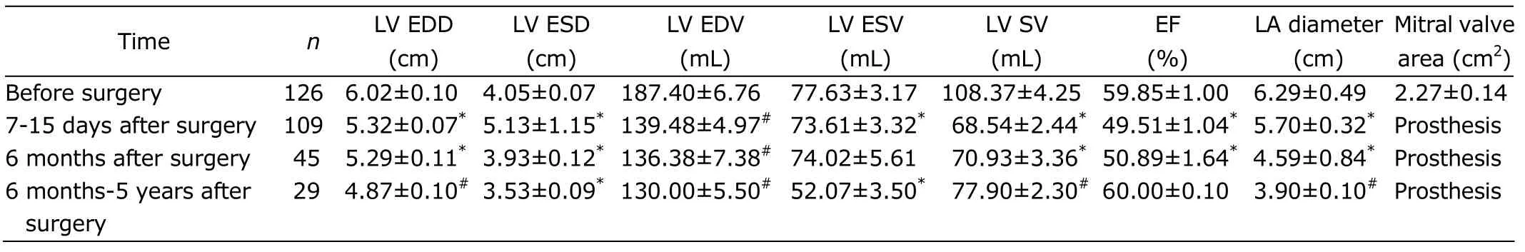
Table 1.Echocardiographic variables before surgery and at follow-up in patients with mitral valve replacement preserving the subvalvular structures§
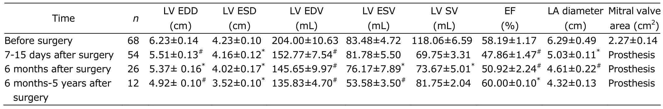
Table 2.Echocardiographic variables before surgery and at follow-up in patients with multivalvular surgery§
CONCLUSIONS
Echocardiography is an important monitoring technique to provide information regarding cardiac size and contractility after mitral valve replacement.LV dimensions and ejection fraction recovered to normal values 6 months after mitral valve replacement with preservation of subvalvular apparatus.Our results confirm that mitral valve replacement surgery is able to restore and maintain normal cardiac and valve function.
 Chinese Medical Sciences Journal2011年1期
- Chinese Medical Sciences Journal的其它文章
- Pathological and High Resolution CT Findings in Churg-Strauss Syndrome
- Observation of Insulin Exocytosis by a Pancreatic β Cell Line with Total Internal Reflection Fluorescence Microscopy
- Changes in Plasma Angiotensin II and Circadian Rhythm of Blood Pressure in Hypertensive Patients with Sleep Apnea Syndrome Before and After Treatment
- Influence of Shenqing Recipe on Morphology and Quantity of Colonic Interstitial Cells of Cajal in Trinitrobenzene Sulfonic Acid Induced Rat Colitis△
- Primary Clinical Evaluation of the Joint Replacement for the Treatment of the First Metatarsophalangeal Arthritis
- Construction of the Subtracted cDNA Library of Striatal Neurons Treated with Long-term Morphine△