Anti-inflammatory activity and qualitative analysis of different extracts of Maytenus obscura (A. Rich.) Cuf. by high performance thin layer chromatography method
Mohamed F. Alajmi, Perwez Alam
Deptartment of Pharmacognosy, College of Pharmacy, King Saud University, Riyadh, Kingdom of Saudi Arabia
Anti-inflammatory activity and qualitative analysis of different extracts of Maytenus obscura (A. Rich.) Cuf. by high performance thin layer chromatography method
Mohamed F. Alajmi*, Perwez Alam
Deptartment of Pharmacognosy, College of Pharmacy, King Saud University, Riyadh, Kingdom of Saudi Arabia
PEER REVIEW
Peer reviewer
Dr. Prawez Alam, Assistant Professor, Salman bin Abdul Aziz University, Al Kharj, Kingdom of Saudia Arabia.
Tel: +96615886014
Fax: +96615886001
E-mail: prawez007@gmail.com
Comments
The research represents advanced methodological qualitative analysis of complex matrix such as herbal medicine where only very limited information can be acquitted using traditional techniques. The manuscript is well written. It has been organized according to well documented researches. The materials and methods are written precisely and well referenced. The results are compatible with what the methods may generate. The results were discussed scientifically according to the results obtained.
Details on Page 156
Objective:To perform aqueous ethanol soluble fraction (AESF) and dichloromethane extract of aerial parts of Maytenus obscura (A. Rich.) Cuf. using high performance thin layer chromatography (HPTLC) and to test anti-inflammatory activity of these extracts.
Maytenus obscura, Phytochemical screening, Finger print, Anti-inflammatory, HPTLC
1. Introduction
Maytenus obscura(A. Rich.) Cuf. (M. obscura) belongs to the genusMaytenuswhich is distributed worldwide, particularly in subtropical and tropical regions, Africa, China, Brazil, Paraguay, Uruguay and Argentina and in southern regions of Saudi Arabia[1]. The genusMaytenusis rich in triterpenes, diterpenes, sesquiterpene alkaloids and spermidine alkaloids[2]. Other secondary metabolites isolate from different species of the genusMaytenus,including flavonoids and flavonoid glycosides[3], phenolic glucoside and agarofurans[4]. Species belonging to this genus are extensively investigated for bioactive compounds as they are widely used in folk medicine such as antiseptic, antiasthmatic, fertility-regulating agents, antitumor and antiulcer[5,6]. SomeMaytenusspecies are marketed in the form of capsules, powders, dried leaves, fresh leaves, aqueous or aqueous-alcoholic preparations[5]. Herbal drugs play major role in treatment of many diseases especially in undeveloped countries[7]. Standardization of plant materials used in traditional medicine is very important in today’s health care system. Pharmacopoeial herbal profiles need to be updated to include the fingerprinting of herbal drugs generated by advanced technology. These modern methods describing the identification and quantification of active constituents in the plant material may be useful for complete standardization of herbals and its formulations[8]. High performance thin layer chromatography (HPTLC) can provide chromatographic fingerprints of herbal drugs that can be visualized, stored as electronic images and used as referene for quality control of herbs. Due to its low cost, high sample throughput and low sample cleanup, HPTLC may become one of standard quality testing techniques. HPTLC has the advantage of simultaneous analysis of large numbers of samples, thus reducing time and cost per analysis[9-13]. The aim of this study is to evaluate and compare the antiinflammatory activity and to develop HPTLC fingerprint profile of alcoholic and dichloromethane extract of aerial parts ofM. obscura.
2. Materials and methods
2.1. Plant material
M. obscurawas collected from Abha, kingdom of Saudi Arabia. The plant was collected and identified by Dr. Mohammad Atiqur Rahman (taxonomist of Medicinal, Aromatic and Poisonous Plants Research Center, College of Pharmacy, King Saud University, Saudi Arabia). Voucher specimens (14295, 13420 and 14309 respectively) were deposited in the herbarium of the College of Pharmacy, King Saud University, Saudi Arabia. Aerial parts were shade dried and coarsely powdered.
2.2. Preparation and extraction of plant material
About 1.3 kg ofM. obscurawas exhaustively extracted at room temperature with 90% ethanol (3×3 L). The ethanol extract was then concentrated under vacuum to yield 182.5 g black waxy residue. A total of 120 g was defatted with petroleum ether (4×200 mL). The remaining alcoholic extract was fractionated with CH2Cl2(5×200 mL) to yield 13.2 g CH2Cl2soluble fraction (SF) and 99.1 g aqueous ethanol soluble fraction (AESF).
2.3. Phytochemical screening
Preliminary phytochemical screening was done following the method of Kokate and Gokhale[14], and Khandelwal[15].
2.4.HPTLCprofile
2.4.1. Chromatographic method
HPTLC studies were carried out following the method of Harborne and Wagneret al[16,17]. The protocol for preparing sample solutions was optimized for high quality fingerprinting. The fingerprinting of dichloromethane and alcoholic extracts ofM. obscurawere executed by spotting 5, 10, 15, 20 μL of suitably diluted sample solution of the SF and AESF on a HPTLC plate. Chromatography was performed on 20 cm×10 cm glass HPTLC plates precoated with 200 μm layers of silica gel 60F254 (E. Merck, Darmstadt, Germany). Samples (5 μL, 10 μL, 15 μL, 20 μL) of each extract were applied as bands 6 mm wide and 8 mm apart by means of Camag (Muttenz, Switzerland) Linomat IV sample applicator equipped with a 25 μL syringe which was programmed through WIN CATS software. The developed plate was dried at 100 °C in hot air oven for 3 min. The plate was kept in photodocumentation chamber (CAMAG Reprostar 3) and captured the images under UV light at 254 and 366 nm, respectively. TheRfvalues and finger print data were recorded by WIN CATS software.
2.4.2. Solvent system development
A number of solvent systems were tried, but the best resolution was obtained in the solvent toluene: ethyacetate: glacial acetic acid (5:3:0.2).
2.4.3. Detection of spots
The developed plate was dried at 100 °C in hot air oven for 3 min to evaporate solvents from the plate. The plate was kept in photo-documentation chamber (CAMAG Reprostar 3) and images captured under UV light at 254 and 366 nm, respectively. TheRfvalues and finger print data were recorded by WIN CATS software.
2.5. Anti-inflammatory activity
Inflammation was induced in the plantar side of the left hind paws of male wistar rats (weighing 100-150 g) by injecting 0.1 mL of 10% aqueous formalin as described by Fuet al[18]. The increase in the paw edema was recorded prior to and 60 min after formalin injection (or 2 h after treatment with extracts) using hydro-plethysmograph (Model 7150, Ugo, Basile, Haly). Instead of the treatment (extracts) (in thetreated group), the control group was administered (i.p.) 0.1 mL of 10% aqueous formalin and the standard group was given (i.p.) injection of Indomethacin (30 mg/kg)[19]. Treated group: the anti-inflammatory activity of the SF and AESF (100 and 200 mg/kg) were tested by injecting the treatment (SF and AESF) solubilized in suitable vehicle as described above 60 min (i.p.) before formalin injection. Then the procedure was continued as outlined above. Effect of the SF and AESF on the formalin-induced paw volume was recorded (2 h after extract treatment or 1 h after formalin injection) as percentage inhibition relative to the control untreated group and were compared to the standard group. Change in volume of edema was quantified using the following formula:
Change in volume of edema=(L2-R2)-(L1-R1)
Where L1is left paw volume before injection of formalin; R1is right paw volume before injecting left paw with formalin; L2is left paw volume 1 h after injecting formalin and 2 h after treatment; R2is right paw volume 1 h after formalin injection in the left paw and 2 h after treatment.
2.6. Statistical analysis
Results were analyzed using one way ANOVA and Dunnett test aspost hoctest (for more than 2 groups) or Kruskal’s wallis ANOVA with Dunn’s test aspost hoctest (unless otherwise stated) and presented as mean±SEM. Only results withP≤0.05 were regarded as significant[20].
3. Results
3.1. Phytochemical screening ofAESFandSF
A preliminary phytochemical screening of AESF and SF showed that both extracts contain protein, lipid, carbohydrate, reducing sugar, phenol, tannin, flavonoid, saponin, triterpenoid, alkaloid and quinone (Table 1).
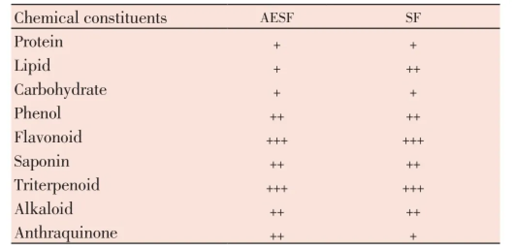
Table 1 Phytochemical contents of AESF and SF.
3.2HPTLCfingerprinting ofAESFandSF
Tables 2 and 3 show HPTLC fingerprinting of AESF and SF, respectively, which revealed several peaks.HPTLC profile of AESF and SF under UV 366 and 254 nm is recorded in the Figures 1 and 2, respectively. The corresponding HPTLC chromatograms are presented in Figures 3A-3H.
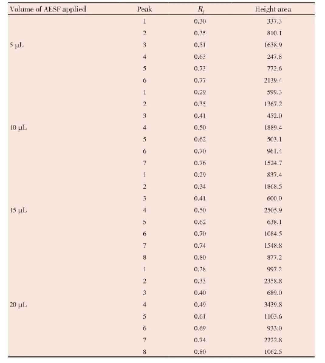
Table 2 HPTLC profile of the AESF.
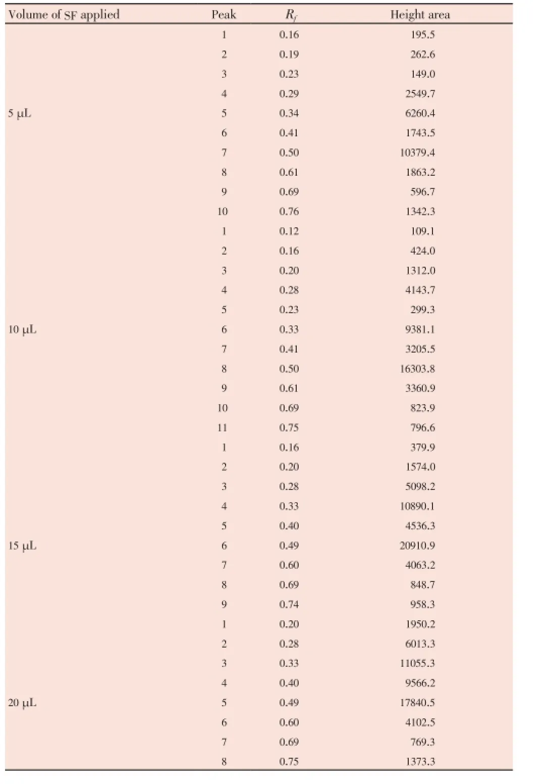
Table 3 HPTLC profile of the SF.

Figure 1. HPTLC picture of the AESF and SF of sample application at UV 254 nm.A: AESF at 5, 10, 15 and 20 μL; B: SF at 5, 10, 15 and 20 μL.
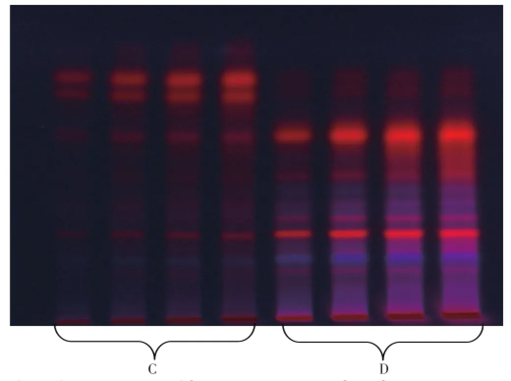
Figure 2. HPTLC picture of the AESF and SF of sample application at 366 nm. C: AESF at 5, 10, 15 and 20 μL; D: SF at 5, 10, 15 and 20 μL.
The AESF revealed 6, 7, 8 and 8 spots withRfvalues in the range of 0.28 to 0.80 for 5, 10, 15 and 20 μL application volume, respectively (Table 2 and Figures 3A-3D). However,SF revealed 10, 11, 9 and 8 spots withRfvalues in the range of 0.12 to 0.76 for 5, 10, 15 and 20 μL application volume, respectively (Table 3 and Figures 3E-3H). The purity of the sample was confirmed by comparing the absorption spectra at start, middle and end position of the band.
3.3. Anti-inflammatory activity ofAESFandSF
Injection of formalin into a rat’s hind paw resulted in swelling as early as 10 min after injection. This swelling reached a plateau by 30 min and was maintained for up to 240 min[21]. Treatment of rats (i.p.) with AESF and SF ofM. obscurain doses of 100 and 200 mg/kg inhibited formalininduced inflammation by 50% and 45.5%, 55.9% and 51.4%, respectively (Table 4).
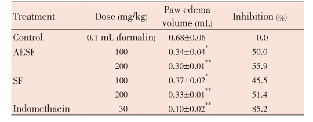
Table 4 Effect of AESF and SF on formalin-induced inflammation in rats.
4. Discussion
The dose-dependent anti-inflammatory activity of both extracts may explain at least partly the rationale beyond using theMaytenusspecies in treatment of inflammatory diseases[1]. Phytochmical screening of both extracts revealed the presence of flavonoids which are known to possess anti-inflammatory and antioxidant activities[22]. Flavonoid content of both extracts are almost equal and hence both extracts have close percentage of inhibition on inflammation. However, it is evident that they are different in polarity and UV absorption. Therefore, the presence of these compounds might be the ultimate cause for their bioactivity. HPTLC analysis of AESF and SF extracts revealed different peaks which are distinct for each extract and are concentration-dependent. Therefore, any additive or coamixed material will definitely affect the HPTLC picture showing new unrelated peaks pointing to the presence of either adulteration or deterioration. HPTLC fingerprinting is proved to be a liner, precise, accurate method for herbal identification and can be used further in authentication and standardization of the medicinally important plant. Such finger printing is useful in quality control of herbal products and checking for the adulterant. Therefore, it can be useful for the evaluation of different marketed pharmaceutical preparations and plant systematic studies[13].
Hence, HPTLC provides great utility for the quality control of these products. It may be utilized to estimate the extent of activity according to the percentage content of these UV active compounds to the present extract’s content. This might form a target for more comprehensive studies. The presence of any adulterant will be easily traced having these profiles developed and stored in a library. Literature review revealed that there is no previous work that has been done on these two extracts. AESF and SF ofM. obscuraproduced dose-dependent significant anti-inflammatory activity in rats. This activity may be partially explained in view of the high content of flavonoids in both extracts. HPTLC profiles of both extrats revealed the presence of eight to nine major spots absorbing in different UV wavelengths, pointing to the difference in these compounds.
Conflict of interest statement
We declare that we have no conflict of interest.
Acknowledgements
The authors are grateful to the research center in College of Pharmacy and the deanship of Scientific Research of the King Saud University (Riyadh, Saudi Arabia) for supporting this project (Grant No. RGP-VPP-150).
Comments
Background
Many countries (including developed ones) suffer a big problem in standardizing and quality control of the herbal plants. This is due to many factors among which are the complex form of these products and the inability of the traditional methods to precisely estimate the quality of the herbs. Regulatory authorities used to follow tedious procedures that may give indication about the quality of the herb but not complete map of the chemical content (which is vital for the biological activity of these plants).
Research frontiers
The research represents an advancement in the quality control of the herbal medicine. It provides new tool for fingerprinting using precise chemical contents mapping which might be utilized in correct identification of not only identity of the plant but even the purity of the plant product and the absence of contaminants. The authors utilized one of the advanced technologiesi.e.HPTLC for that purposes which affords fast, reliable and precise tool for this purpose.
Related reports
No similar reports were found in the literature regarding this plant specifically, since it is being evaluated using this tool for the first time as per literature review availablein our resources.
Innovations and breakthroughs
The previous pharmacopeial methods for testing quality control concentrate on organoleptics, ash, and vague chemical screening which could not give precise chemical contents. Chemical content is the driving force for the medicinal value of the plant. This is why using such new techniques assure not only identity, purity of the plant but even the activity against the claimed diseases.
Applications
It can help to sensitize other groups of researches and adapt similar methodology in quality studies of other plants. It can be utilized even for microbial fermentation products and the presence of the analyte of interest. It may play a good role in food, food supplement screening and quality control.
Peer review
The research represents advanced methodological qualitative analysis of complex matrix such as herbal medicine where only very limited information can be acquitted using traditional techniques. The manuscript is well written. It has been organized according to well documented researches. The materials and methods are written precisely and well referenced. The results are compatible with what the methods may generate. The results were discussed scientifically according to the results obtained.
[1] Rojo de Almeida MT, Rios-Luci C, Pard?n JM, Palermo JA. Antiproliferative terpenoids and alkaloids from the roots of Mayenus vitis-idaea and Maytenus spinosa. Phytochemistry 2010; 71(14-15): 1741-1748.
[2] Gutierrez F, Estévez-Braun A, Ravelo AG, Astudillo L, Zárate R. Terpenoids from the medicinal plant Maytenus ilicifolia. J Nat Prod 2007; 70(6): 1049-1052.
[3] Momtaz S, Hussein AA, Ostad SN, Abbdollahi M, Lall N. Growth inhibition and induction of apoptosis in human cancerous Hela cells by Maytenus procumbens. Food Chem Toxicol 2013; 51: 38-45.
[4] Crestani S, Rattmann YD, Cipriani TR, de Souza LM, Lacomini M, Kassuya CA, et al. A potent and nitiric oxide-dependent hypotensive effect induced in rats by semi-purified fractions from Maytenus ilicifolia. Vascul Pharmacol 2009; 51(1): 57-63.
[5] Costa PM, Ferreira PM, Bolzani VS, Furlan M, de Freitas Formenton Macedo Dos Santos VA, Corsino J, et al. Antiproliferative activity of pristimerin isolated from Maytenus ilicifolia (Celastraceae) in human HL-60 cells. Toxicol In Vitro 2008; 22(4): 854-863.
[6] Bramhachari PV, Ravichand J, Reddy YH, Kotresha D, Viswanatha Chaitanya K, Bobbarala V. Evaluation of hydroxyl radical scavenging activity and HPTLC fingerprint profiling of Aegle marmelos (L.) Correa extracts. J Pharm Res 2011; 4(1): 252.
[7] Kaushik S, Sharma P, Jain A, Sikarwar MS. Preliminary phytochemical screening and HPTLC fingerprinting of Nicotiana tabacum leaf. J Pharm Res 2010; 3(5): 144.
[8] Kaul N, Agrawal H, Patil B, Kakad A, Dhaneshwar SR. Application of stability-indicating HPTLC method for quantitative determination of metadoxine in pharmaceutical dosage form. Farmaco 2005; 60(4): 351-360.
[9] Faiyazuddin Md, Baboota S, Ali J, Ahuja A, Ahmad S, Akhtar J. Validated HPTLC method for simultaneous quantitation of bioactive citral isomers in lemongrass oil encapsulated solid lipid nanoparticle formulation. Int J Essen Oil Ther 2009; 3: 142-146.
[10] Faiyazuddin M, Ali J, Ahmad S, Ahmad N, Akhtar J, Baboota S. Chromatographic analysis of trans and cis-citral in lemongrass oil and in a topical phytonanocosmeceutical formulation, and validation of the method. J Planar Chromat 2010; 23(3): 233-236.
[11] Faiyazuddin Md, Ahmad S, Iqbal Z, Talegaonkar S, Ahmad FJ, Bhatnagar A, et al. Stability indicating HPTLC method for determination of terbutaline sulfate in bulk and from submicronized dry powder inhalers. Anal Sci 2010; 26(4): 467-472.
[12] Faiyazuddin M, Rauf A, Ahmad N, Ahmad S, Iqbal Z, Talegaonkar S, et al. A validated HPTLC method for determination of terbutaline sulfate in biological samples: application to pharmacokinetic study. Saudi Pharm J 2011; 19(3): 185-191.
[13] Yamunadevi M, Wesely EG, Johnson M. Chromatographic finger print analysis of steroids in Aerva lanata L. by HPTLC technique. Asian Pac J Trop Biomed 2011; 1(6): 428-433.
[14] Kokate CK, Gokhale SB. Practical pharmacognosy. Pune: Nirali Prakashan; 2008, p. 42-44.
[15] Khandelwal KR. Practical pharmacognosy. Pune: Pragati Books Pvt. Ltd.; 2008, p. 149-160.
[16] Harborne JB. Phytochemical methods. 3rd ed. London: Chapman and Hall; 1998, p. 44-46.
[17] Wagner H, Baldt S, Zgainski EM. Plant drug analysis. Berlin: Springer; 1996, p. 85.
[18] Fu KY, Light AR, Maixner W. Long-lasting inflammation and long-term hyperalgesia anfter subcutaneous formaline injection into the rat hind paw. J Pain 2001; 2(1): 2-11.
[19] Tavares TG, Spindola H, Longato G, Pintado ME, Caralho JE, Malcata FX. Antinociceptive and anti-inflammatory effects of novel dietary protein hydrolysate produced from whey by proteases of Cynara cardunculus. Int Dairy J 2013; 32(2): 156-162.
[20] Daniel WW. Biostatics: a foundation for analysis in health sciences. 9th ed. New York: Wiley; 2009.
[21] Cannon KE, Leurs R, Hough LB. Activation of peripheral and spinal histamine H3 receptors inhibits formalin-induced inflammation and nociception, respectively. Pharmacol Biochem Behav 2007; 88(1): 122-129.
[22] Mendoza-Wilson AM, Glossman-Mitnik D. Theoretical study of the molecular properties and chemical reactivity of (+)-catechin and (-)-epicatechin related to their antioxidant ability. J Mol Struct 2006; 761(1-3): 97-106.
10.1016/S2221-1691(14)60224-0
*Corresponding author: Mohamed F. Alajmi, Department of Pharmacognosy, College of Pharmacy, King Saud University, Riyadh, Kingdom of Saudi Arabia.
Tel: +966505151846
E-mail: alajmister@gmail.com
Foundation Project: Supported by College of Pharmacy and the deanship of Scientific Research of the King Saud University (Riyadh, Saudi Arabia) (Grant No. RGP-VPP-150).
Article history:
Received 25 Dec 2013
Received in revised form 1 Jan, 2nd revised form 6 Jan, 3rd revised form 10 Jan 2014
Accepted 16 Jan 2014
Available online 28 Feb 2014
Methods:HPTLC studies were carried out using CAMAG HPTLC system equipped with Linomat IV applicator, TLC scanner 3, Reprostar 3, CAMAG ADC 2 and WIN CATS-4 software were used. The anti-inflammatory activity was tested by injecting different groups of rats (6 each) with formalin in hind paw and measuring the edema volume before and 1 h later formalin injection. Control group received saline i.p. The extracts treatment was injected i.p. in doses of 100 and 200 mg/kg 1 h before formalin administration. Indomethacin (30 mg/kg) was used as standard.
Results:The results of preliminary phytochemical studies confirmed the presence of protein, lipid, carbohydrate, phenol, flavonoid, saponin, triterpenoid, alkaloid and anthraquinone in both extracts. Chromatography was performed on glass-backed silica gel 60 F254 HPTLC plates with the green solvents toluene: ethyacetate: glacial acetic acid (5:3:0.2, v/v/v) as mobile phase. HPTLC finger printing of AESF revealed major eight peaks with Rfvalues in the range of 0.28 to 0.80 and the dichloromethane revealed major 11 peaks with Rfvalues in the range of 0.12 to 0.76. The purity of sample was confirmed by comparing the absorption spectra at start, middle and end position of the band. Treatment of rats (i.p.) with AESF and dichloromethane in doses of 100 and 200 mg/kg inhibited singnificantly (P<0.05, n=6) formalin-induced inflammation by 50%, 55.9%, 45.5%, and 51.4%, respectively.
Conclusions:HPTLC finger printing of AESF and dichloromethane of Maytenus obscura revealed eight major spots for alcoholic extracts and nine major spots for dichloromethane extracts. These HPTLC profiles may be of great usefulness in the quality control of herbal products containing these extracts. The anti-inflammatory activity of both extracts also revealed the medicinal importance of these extracts. The plant can be further explored for the isolation of phytoconstituents having anti-inflammatory activity.
 Asian Pacific Journal of Tropical Biomedicine2014年2期
- Asian Pacific Journal of Tropical Biomedicine的其它文章
- Prevalence and antibiogram of bacterial isolates from urinary tract infections at Dessie Health Research Laboratory, Ethiopia
- Inhalation of Shin-I essential oil enhances lactate clearance in treadmill exercise
- Ameliorative effect of alkaloid extract of Cyclea peltata (Poir.) Hook. f. & Thoms. roots (ACP) on APAP/CCl4induced liver toxicity in Wistar rats and in vitro free radical scavenging property
- Investigation of in vivo neuropharmacological effect of Alpinia nigra leaf extract
- A pharmacobotanical study of two medicinal species of Fabaceae
- Pancreatic islet regeneration and some liver biochemical parameters of leaf extracts of Vitex doniana in normal and streptozotocin-induced diabetic albino rats
