Clinico-pathological signi fi cance of extra-nodal spread in special types of breast cancer
Ecmel Isik Kaygusuz, Handan Cetiner, Hulya Yavuz
Department of Pathology, Zeynep Kamil Maternity and Children’s Training and Research Hospital, Istanbul 34668, Turkey
Clinico-pathological signi fi cance of extra-nodal spread in special types of breast cancer
Ecmel Isik Kaygusuz, Handan Cetiner, Hulya Yavuz
Department of Pathology, Zeynep Kamil Maternity and Children’s Training and Research Hospital, Istanbul 34668, Turkey
Objective: To investigate the signi fi cance of extra-nodal spread in special histological sub-types of breast cancer and the relationship of such spread with prognostic parameters.
Breast cancer; extra-nodal spread; prognosis
Introduction
Breast carcinoma is the most common malignant tumor and is the leading cause of cancer death in women, with an incidence of more than 90 new cases per pollution of population 100,000 annually. Breast cancer has speci fi c histopathological types with di ff erent prognostic and clinical characteristics. Histological type, age, tumor diameter, existing lymph node metastasis, the number of the metastatic lymph nodes, the expression of estrogen receptor (ER) and progesterone receptor (PR), proliferative rate (Ki-67), p53 mutation, and c-erbB-2 are oncogenic-prognostic determinants of breast carcinoma1-3. Both the presence and the total number of tumor-positive lymph nodes are of prognostic value. Grouping patients with one to three, four to nine, and ten or more positive lymph nodes metastases is a generally accepted approach and is used to determine the type of chemotherapy to be given after surgery. Previous studies4-8assert that in the identification of prognosis, extra-nodal spread has a significant relationship with complete survival, disease-free survival, and local recurrence. This study examines the significance of extranodal spread in breast cancer cases and its relationship with prognostic parameters.e signi fi cance of the extra-nodal spread in special histological sub-types of breast cancer, as well as its predictive value in guiding treatment, is discussed.
Materials and methods
A total of 303 cases diagnosed between 1993 and 2004 were selected for the study.e study was approved by the institutional ethics commiee. All patients underwent mastectomy and axillary dissection. A total of 100 cases were diagnosed with invasive ductal carcinoma (IDC), 60 with invasive lobular carcinoma (ILC), 37 with pleomorphic lobular carcinoma (PLC), 25 with mucinous carcinoma, 28 with mixed mucinous carcinoma, and 53with micropapillary carcinoma. Aer the 303 cases were classi fi ed according to tumor type, each tumor group was subdivided according to age, tumor diameter, lymph node metastasis, extranodal spread, vascular invasion in the adjacent sotissue, distant metastasis, and immunohistochemical characteristics.
Immunohistochemistry
Tumor specimens were fi xed in 10% neutral bu ff ered formalin for 20 to 28 h before processing and embedding. Sections were cut on a microtome and stained with H&E. ER, PR, c-erbB-2, and Ki67 were determined through immunohistochemistry by using a streptavidine biotine peroxidase system. Staining protocols for each antibody had previously been optimized and standardized. First, 4 mm thick tissue sections were cut from representative blocks and mounted on charged and pre-cleaned slides. Second, sections were dewaxed in xylene and rehydrated through a decreasing concentration of alcohol to water.en, heat-induced antigen retrieval (immersion in citrate buffer, pH 6.0, at 95 °C for 30 min followed by cooling at room temperature for 20 min) or, alternatively, a 10 min treatment with protease XIV was applied. Sections were incubated with 0.3% hydrogen peroxide in methanol for 30 min. to block endogenous peroxidase activity. Immunostaining experiments for the localization of ER and PR, p53, c-erbB-2 protein, and Ki-67 antigen were conducted on consecutive tissue sections from the tumor-containing blocks. The primary antibodies used were the monoclonal antibody (Mab) to ER (clone 1D5, at 1/100 dilution; Dako, Glostrup, Denmark), the Mab to PGR (clone 1A6, at 1/800 dilution; Dako), the MIB-1 Mab to the Ki-67 antigen (at 1/1,200 dilution; Immunotech, Marseille, France), and the polyclonal antibody (at 1/3,200 dilution; Dako) to the c-erbB-2 protein.
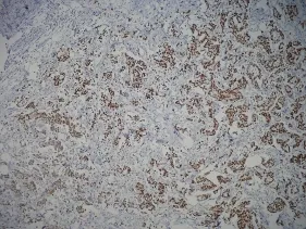
Figure 1 Immunohistochemically ER positive cells (IHC×100). ER, estrogen receptor.
The immunohistochemically stained slides were evaluated by three pathologists who were all very experienced in breast pathology. Observers were asked to select at least three highpower fields to comprise the spectrum of staining seen on the initial overview. The average percentage of positive stained cells across the section were scored. Only nuclear reactivity, irrespective of its intensity, was considered for ER (Figure 1), PR, p53, and Ki-67 antigens, whereas only an intense and complete membrane staining in >10% of the tumor cells quali fi ed for c-erbB-2 overexpression (3+) (Figure 2). Steroid hormone receptors status and p53 were classi fi ed as negative in case of <1% immunoreactive tumor cells (Table 1).e scoring of c-erbB-2 by immunohistochemistry was semiquantitatively evaluated according to the following categories: 0, no membrane staining; 1+, weak and incomplete membrane staining; 2+, weak/moderate heterogeneous complete membrane staining in <10% of tumor cells; or 3+, strong, complete, homogeneous membrane staining in >10% of tumor cells.
Clinical follow-up of 122 cases with extra-nodal spread
Examined data
· Age: under 50 and above 50;
· Lymph node metastasis: no axillary metastasis, metastasis in one to three lymph nodes, metastasis in four to nine lymph nodes, and metastasis in 10 or more lymph nodes;
· Extra-nodal Spread: Extra capsular growth of tumor cells, invasion of perinodal fat and extra-nodal localization of tumor cells;
· Extra-nodal Vascular Invasion: with and without tumor cells in the vascular section in the extra-nodal adipose tissue (Figure 3);
· Prognosis: Patients with extra-nodal spread were reviewed for distant metastases.
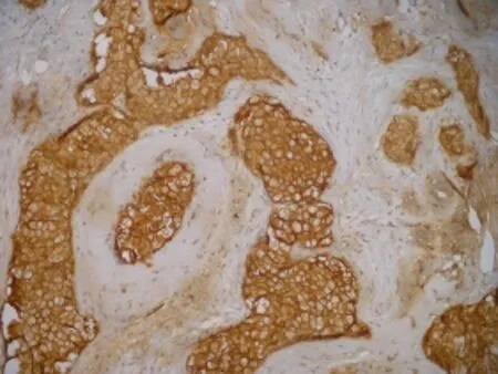
Figure 2 Immunohistochemically c-erbB-2 positive cells (IHC×200).
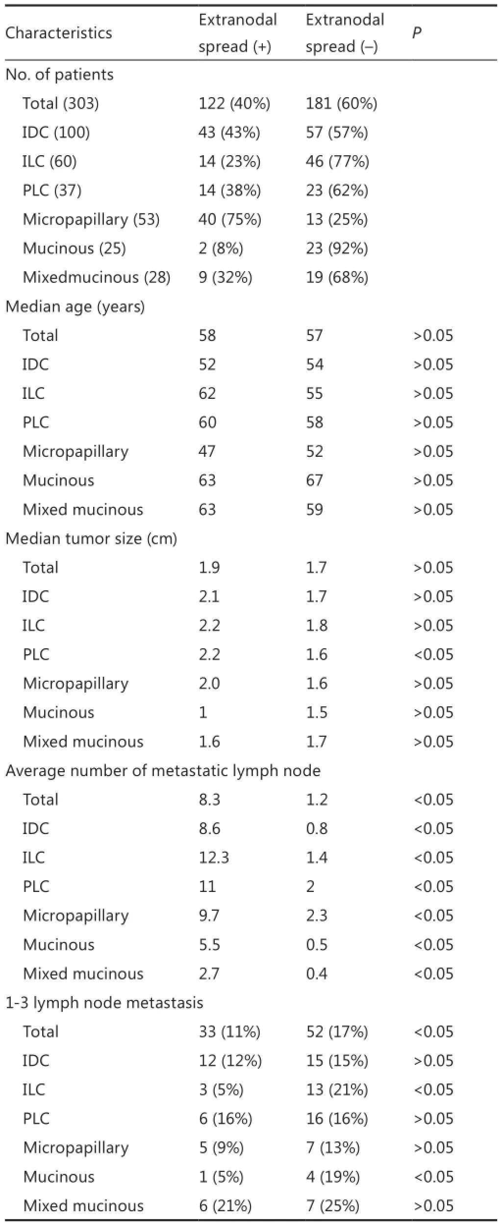
Table 1 Patients and pathologic charecteristics
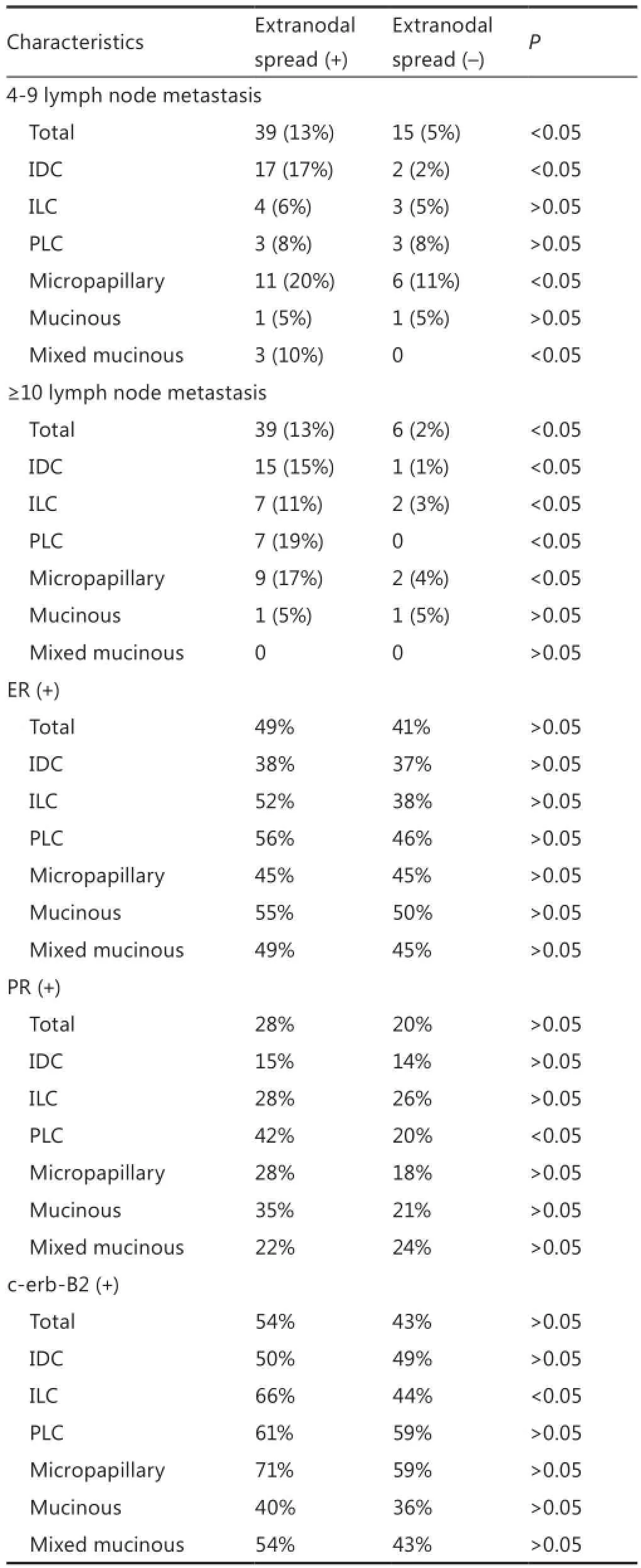
Table 1 (continued)
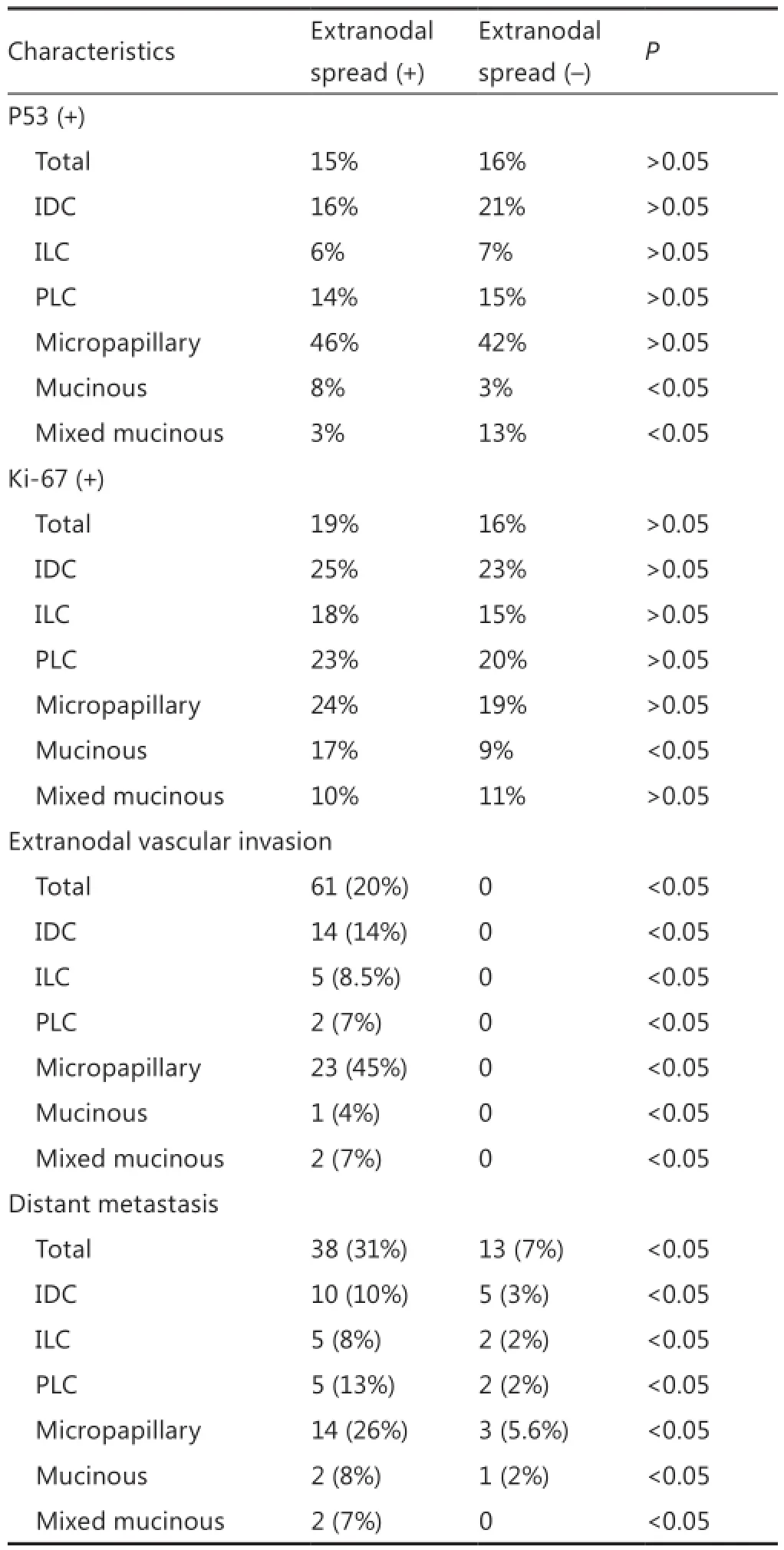
Table 1 (continued)
Statistical analysis
Statistical analysis was performed by using the statistical analysis system. Chi-square and t-tests were used.e cases with P values under 0.05 were considered signi fi cant.

Figure 3 Patients characteristics of with special types breast cancer.
Results
Invasive ductal carcinoma (IDC)
The patients with extra-nodal spread had a significantly higher mean number of metastatic lymph node (8.6) and distant metastasis ratio (10 cases, 10%) compared with the patients without extra-nodal spread (P<0.05). A signi fi cant relationship exists between the extra-nodal spread presence and four or more lymph node metastases (P<0.05) (Figure 3).
Invasive lobular carcinoma (ILC)
The patients with extra-nodal spread had significantly higher mean number of metastatic lymph nodes (12.3) and distant metastasis ratio (5 cases, 8.3%) compared with the patients without extra-nodal spread (P<0.05). A signi fi cant relationship exists between the extra-nodal spread existence and ten or more lymph nodes metastasis (P<0.05). C-erbB-2 was highly positive in patients with extra-nodal spread (66%) compared with patients without such spread (44%) (P<0.05).
Pleomorphic lobular carcinoma (PLC)
Micropapillary carcinoma
Patients with extra-nodal spread had a signi fi cantly higher mean number of metastatic lymph node (9.7) and distant metastasis ratio (14 cases, 26%) compared with the patients without extranodal spread (P<0.05). A signi fi cant relationship exists between the extra-nodal spread and the existence of four or more lymph nodes metastasis (P<0.05). The number of the lymph node metastasis was higher in the axilla in micropapillary carcinoma compared with the cases diagnosed with mucinous and mixed mucinous carcinoma (P<0.05).
Mucinous carcinoma
Patients with extra-nodal spread had a significantly higher mean number of metastatic lymph node (5.5) and the distant metastasis ratio (2 cases, 8%) compared with the patients without extra-nodal spread (P<0.05). In the mucinous carcinoma cases with extra-nodal spread, p53 was significantly more positive than that in cases without extra-nodal spread (P<0.05). The cases diagnosed with mucinous carcinoma had the least ratio (8%) of extra-nodal spread among all groups (P<0.05). Compared with IDC, mucinous carcinoma was observed in the older population (P<0.05).
Mixed mucinous carcinoma
Patients with extra-nodal spread had a significantly higher mean number of metastatic lymph node (2.7%) and distant metastasis ratio (2 cases, 7%) compared with the patients without extra-nodal spread (P <0.05). A signi fi cant relationship exists between extra-nodal spread and the existence of 4-9 lymph node metastases (P<0.05). C-erbB-2 was highly positive in the mixed mucinous tumors with extra-nodal spread, whereas p53 was interestingly high in patients without extra-nodal spread (P<0.05).
Considering all the cases together as a whole
(Table 2)
Extra-nodal spread was found in 40% (122 cases) of 303 breast cancer cases.
While the extra-nodal spread most frequently presents in the histological sub-type of micropapillary carcinoma (40 cases, 75%),it least frequently presents in mucinous carcinoma (2 cases, 8%).
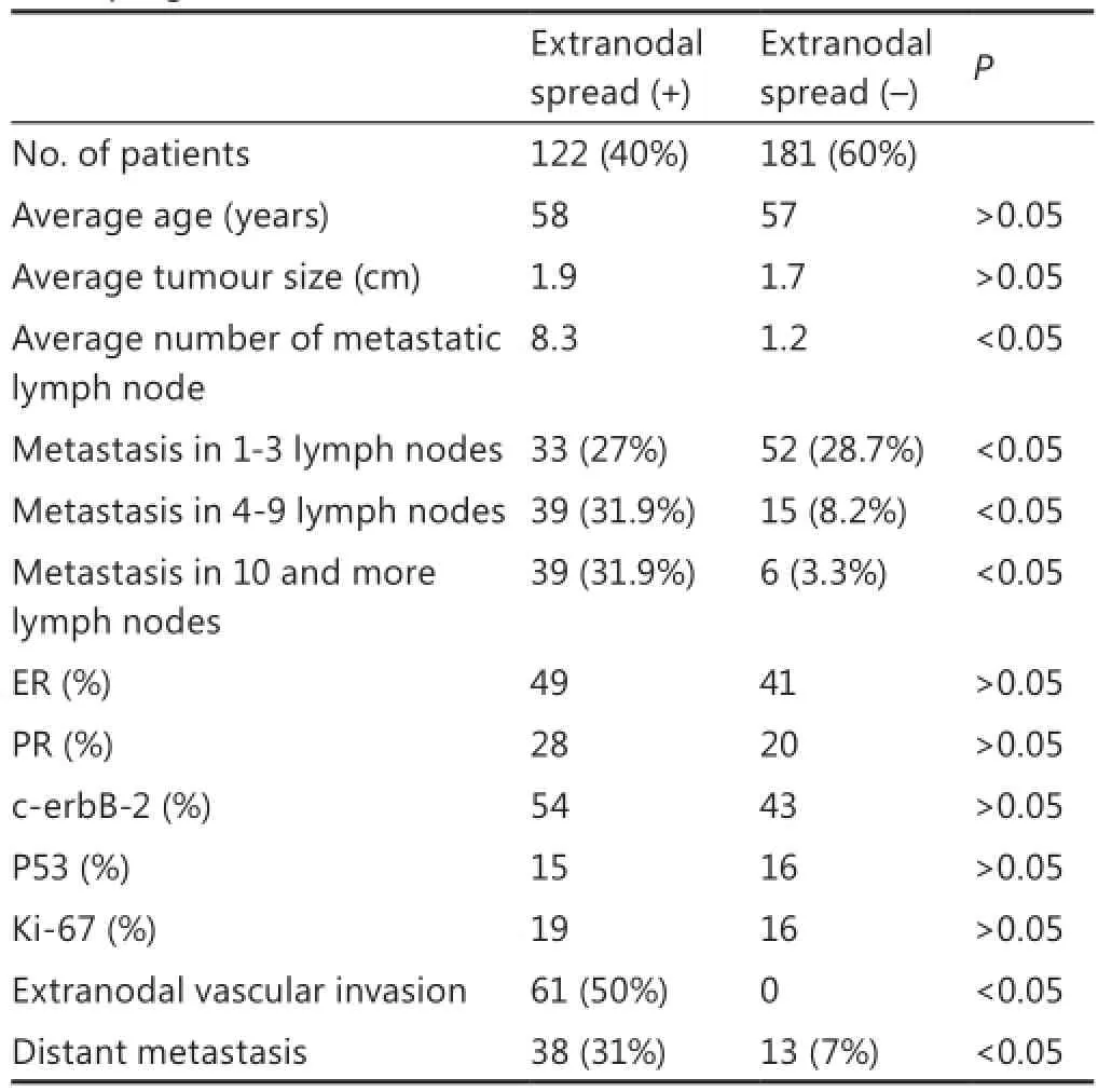
Table 2 Association of the extranodal spread (+) and (-) cases with other prognostic factors
Patients with extra-nodal spreads had higher mean number of metastatic lymph nodes (8.3) and distant metastasis ratio (28 cases, 9%) compared with the patients without extra-nodal spread.
A relationship exists between the extra-nodal spread and the presence of four or more lymph node metastases (P<0.05), as well as between the number of metastatic lymph nodes (≥4) and tumor diameter, extra-nodal spread, distant metastasis, and extra-nodal vascular invasion presence (P<0.05). Apart from the tumor type, the presence of distant metastasis was associated with extra-nodal spread.
The relationship between the extra-nodal vascular invasion and the distant metastasis was investigated. However, the results had a limit value (P=0.061).
Discussion
One of the most important prognostic factors in breast cancer is the regional lymph node status9. Previous studies10-13observed that the extra-nodal spread occurred with disseminated axillary lymph node metastases. Vicini et al.13proved that patients with extra-nodal spread had signi fi cantly more mean metastatic lymph nodes. The mean number of metastatic lymph nodes of thepatients with extra-nodal spread in this study was 8.6 in the IDC group, 12.36 in ILC, 11 in PLC, 9.7 in micropapillary carcinoma, 5.5 in mucinous carcinoma, and 2 in mixed mucinous carcinoma. Meanwhile, the mean number of metastatic lymph nodes was 8.3 in patients with extra-nodal spread and 1.2 in the patients without extra-nodal spread.
Patients were divided into three groups according to the number of metastatic lymph nodes (1-3, 4-9, ≥10). A signi fi cant relationship was found between extra-nodal spread and the presence of four or more lymph nodes metastases (P<0.05) in the IDC, micropapillary, and mixed mucinous carcinoma groups. The ILC, PLC, and mucinous carcinoma groups exhibited a significant relationship between extra-nodal spread and the presence of ten or more lymph node metastases (P<0.05). Previous studies10-13emphasized that the number of metastatic axillary lymph nodes was four or more in patients with extranodal spread. The exclusion of extra-nodal invasion by recent TNM versions seems reasonable, mainly because of its direct association with the number of positive lymph nodes and the lack of independent prognostic signi fi cance.
Although Leonard et al.11, Fisher et al.14, and Mignano et al.4stated that the extra-nodal spread affected survival, whereas Donegan et al.10and Pierce et al.12suggested that it was not.e results of our study show a significant relationship between distant metastases and extra-nodal spread (P<0.05). Notably, distant metastases were observed in 31% of the patients with extra-nodal spread but in only 7% of the patients without extranodal spread.
A significant relationship between the tumor diameter and the extra-nodal spread was only observed in the PLC group (P<0.05).
However, a signi fi cant relationship between tumor diameter and the presence of four or more metastatic lymph nodes was observed in all groups, which suggests that extra-nodal spread might have an indirect relationship with the tumor diameter, which is a prognostic factor.
The cases were also related to the presence of vascular invasion on the extra-nodal area in patients. Vascular invasion on adjacent sotissue was observed in 20% of all patients with extra-nodal spread (61 cases), 14% in IDC (14 cases), 8.5% in ILC (5 cases), 7.1% in PLC (2 cases), 45% in micropapillary carcinoma (23 cases), 4% in mucinous carcinoma (1 case), and 7% in mixed mucinous carcinoma (2 cases) (Table 2). The results were significantly when compared with the findings on patients without extra-nodal spread (no extra-nodal vascular invasion was observed) (P<0.05). The relationship between extra-nodal vein invasion and distant metastases was also researched, but the results had a limit value (P=0.061).
In this study, no axillary recurrence was observed. Previous studies observed axillary recurrence to be 0% (9) or at a very low level of 3% (4 and 10) in cases with extra-nodal spread and no radiotherapy.
Local recurrence was observed in only four (10%) (two IDC, one PLC, one micropapillary carcinoma) of 38 cases that received partial mastectomy. Bucci et al.5also failed to detect local recurrence in any of their 43 patients with extra-nodal spread who received adjuvant radiotherapy. Certainly, the low local recurrence ratio in our cases can be attributed to the adjuvant radiotherapy used on all patients.e current fi ndings show that the presence of extra-nodal spread in the examined breast cancer sub-types has predictive value in forecasting the number of metastatic lymph nodes and in disease prognosis.
Con fl ict of interest statement
No potential con fl icts of interest are disclosed.
1. Prognostic importance of occult axillary lymph node micrometastases from breast cancer. International (Ludwig) Breast Cancer Study Group. Lancet 1990;335:1565-1568.
2. Carter CL, Allen C, Henson DE. Relation of tumor size, lymph node status, and survival in 24,740 breast cancer cases. Cancer 1989;63:181-187.
3. Contesso G, Mouriesse H. Anatomopathologic consensus for de fi ning the prognostic factors of breast cancers. Pathol Biol (Paris) 1990;38:834-835.
4. Mignano JE, Zahurak ML, Chakravarthy A, Piantadosi S, Dooley WC, Gage I. Signi fi cance of axillary lymph node extranodal sotissue extension and indications for postmastectomy irradiation. Cancer 1999;86:1258-1262.
5. Bucci JA, Kennedy CW, Burn J, GilleDJ, Carmalt HL, Donnellan MJ, et al. Implications of extranodal spread in node positive breast cancer: a review of survival and local recurrence. Breast 2001;10:213-219.
6. Palamba HW, Rombouts MC, Ruers TJ, Klinkenbijl JH, Wobbes T. Extranodal extension of axillary metastasis of invasive breast carcinoma as a possible predictor for the total number of positive lymph nodes. Eur J Surg Oncol 2001;27:719-722.
7. Sivridis E, Giatromanolaki A, Galazios G, Koukourakis MI. Noderelated factors and survival in node-positive breast carcinomas. Breast 2006;15:382-389.
8. Altinyollar H, Berbero?lu U, Gülben K, Irkin F.e correlation of extranodal invasion with other prognostic parameters in lymph node positive breast cancer. J Surg Oncol 2007;95:567-571.
9. Alvarenga CA, dos Santos CC, Alvarenga M, Paravidino PI, Morais SS, Brenelli HB, et al. Localization of metastasis within the sentinel lymph node biopsies: a predictor of additional axillary spread of breast cancer? Rev Bras Ginecol Obstet 2013;35:483-499.
10. Donegan WL, Stine SB, Samter TG. Implications of extracapsular nodal metastases for treatment and prognosis of breast cancer. Cancer 1993;72:778-782.
11. Leonard C, Corkill M, Tompkin J, Zhen B, Waitz D, Norton L, et al. Are axillary recurrence and overall survival a ff ected by axillary extranodal tumor extension in breast cancer? Implications for radiation therapy. J Clin Oncol 1995;13:47-53.
12. Pierce LJ, Oberman HA, Strawderman MH, Lichter AS. Microscopic extracapsular extension in the axilla: is this an indication for axillary radiotherapy? Int J Radiat Oncol Biol Phys 1995;33:253-259.
13. Vicini FA, Horwitz EM, Lacerna MD, Brown DM, White J, Dmuchowski CF, et al.e role of regional nodal irradiation in the management of patients with early-stage breast cancer treated with breast-conserving therapy. Int J Radiat Oncol Biol Phys 1997;39:1069-1076.
14. Fisher ER, Gregorio RM, Redmond C, Kim WS, Fisher B. Pathologic findings from the national surgical adjuvant breast project. (Protocol no. 4). III. The significance of extranodal extension of axillary metastases. Am J Clin Pathol 1976;65:439-444.
Cite this article as:Kaygusuz EI, Cetiner H, Yavuz H. Clinico-pathological significance of extra-nodal spread in special types of breast cancer. Cancer Biol Med 2014;11:116-122. doi: 10.7497/j.issn.2095-3941.2014.02.006
Correspondance to: Ecmel Isik Kaygusuz
E-mail: ecmeli@yahoo.com
Received January 30, 2014; accepted May 16, 2014. Available at www.cancerbiomed.org
Copyright ? 2014 by Cancer Biology & Medicine
Methods: A total of 303 breast cancer cases were classi fi ed according to tumor type, and each tumor group was subdivided according to age, tumor diameter, lymph node metastasis, extra-nodal spread, vein invasion in the adjacent soft tissue, distant metastasis, and immunohistochemical characteristics [estrogen receptor (ER), progesterone receptor (PR) existence, p53, c-erbB-2, and proliferative rate (Ki-67)].e 122 cases with extra-nodal spread were clinically followed up.
Results:An extra-nodal spread was observed in 40% (122 cases) of the 303 breast cancer cases.e spread most frequently presented in micro papillary carcinoma histological sub-type (40 cases, 75%), but least frequently presents in mucinous carcinoma (2 cases, 8%). Patients with extra-nodal spread had a high average number of metastatic lymph nodes (8.3) and a high distant metastasis rate (38 cases, 31%) compared with patients without extra-nodal spread.
Conclusion:e existence of extra-nodal spread in the examined breast cancer sub-types has predictive value in forecasting the number of metastatic lymph nodes and the disease prognosis.
 Cancer Biology & Medicine2014年2期
- Cancer Biology & Medicine的其它文章
- Polymeric nanocomposites loaded with fluoridated hydroxyapatite Ln3+ (Ln = Eu or Tb)/iron oxide for magnetic targeted cellular imaging
- An unusual case of aggressive systemic mastocytosis mimicking hepatic cirrhosis
- Spindle cell carcinoma of the breast as complex cystic lesion: a case report
- Effects of postmastectomy radiotherapy on prognosis in different tumor stages of breast cancer patients with positive axillary lymph nodes
- Incidence and mortality of female breast cancer in the Asia-Paci fi c region
- Research progress on the anticarcinogenic actions and mechanisms of ellagic acid
