Impact of venous thromboembolism on the natural history of pancreatic adenocarcinoma
Marseille, France
Impact of venous thromboembolism on the natural history of pancreatic adenocarcinoma
Mehdi Oua?ssi, Cécilia Frasconi, Diane Mege, Laurence Panicot-dubois, Laurence Boiron, Laetitia Dahan, Philippe Debourdeau, Christophe Dubois, Dominique Farge and Igor Sielezneff
Marseille, France
BACKGROUND: Few studies have analyzed the effect of venous thromboembolism (VTE) events on the prognosis of pancreatic cancer, but their results were conficting. The present study was undertaken to determine the effect of VTE on pancreatic adenocarcinoma (PA) outcomes.
METHODS: All consecutive patients diagnosed with PA from May 2004 to January 2012 in a single oncology center were retrospectively studied. Clinical, radiological and histological data at time of diagnosis or within the frst 3 months after surgery, including the presence (+) or absence (-) of VTE were collected. VTE was defned as radiological evidence of either pulmonary embolism (PE), deep venous thrombosis without infection or catheter-related thrombosis. PA with and without PE was compared for survival using the Kaplan-Meier method to estimate overall survival.
RESULTS: Among 162 PA patients with a median follow-up of 15 (3-92) months after diagnosis, 28 demonstrated VTE (+). PA patients with and without PE were similar for age, American Society of Anesthesiologist score, body mass index, and history of treatment. The distribution of cancer stages was similarbetween the two groups VTE (+) and VTE (-). The median duration of survival was signifcantly worse in the VTE (+) group vs VTE (-) (12 vs 18 months, P=0.010). In multivariate analysis, the presence of VTE and surgical treatment were independent prognostic factors for overall survival.
CONCLUSION: VTE (+) at time of diagnosis or within the frst 3 months after surgery during treatment is an independent factor of poor prognosis in PA.
(Hepatobiliary Pancreat Dis Int 2015;14:436-442)
pancreatic carcinoma;
thromboembolism;
survival
Introduction
The association between cancer and venous thromboembolism (VTE) was frst reported by Trousseau in 1865.[1]Since then, malignancy has been associated with a six- to seven-fold increased risk of VTE[2,3]and a three-fold increased risk of pulmonary embolism (PE).[4]In this setting and among all malignant tumors that are diagnosed, pancreatic cancer, metastatic status and chemotherapy are the most well known risk factors for thromboembolism.[5,6]Infltrating ductal adenocarcinoma is by far the most common type of carcinoma of the pancreas.[7]Diagnosis of a pancreatic adenocarcinoma (PA) is still a clinical challenge due to its predominantly late diagnosis and the chemoresistance to cytotoxic and various target drugs. At time of PA diagnosis, 50% of patients with a pancreatic cancer have metastatic disease and 20% to 30% of those show locally advanced disease.[7]Only 10% to 15% of patients undergo complete tumor resection and less than one-ffth of these are alive 5 years after surgery for PA. To date, there are few studies on the impact of VTE on the prognosisof pancreatic cancer and among them conficting results were reported.[6,8-13]Different thromboembolitic events (such as arterial thrombosis or digestive VTE with clearly different physiopathology) and several histological defnitions of pancreatic cancer (such as ampulloma, cholangiocarcinoma or PA) have not allowed to draw any clear conclusion on prognosis.[6,8]Therefore, we designed the present study to specifcally analyze the impact of VTE only (excluding splenic, portal or superior and inferior mesenteric vein thrombosis) on European patients outcome/prognosis in PA.
Methods
Patients
All consecutive patients with pancreatic tumors who had been referred for surgery and admitted to Timone Hospital in Marseille, France from May 2004 to January 2012 were retrospectively studied using a specifc data collection form for clinical, radiological and histological data. All consecutive patients with PA (metastatic, locally advanced pancreatic cancer) were included and analyzed without stratifcation on the stage.
The whole cohort was retrospectively analyzed and divided into 2 groups according to presence or absence of VTE, defned as PE, deep venous thrombosis (DVT) without infection, central venous catheter-related thrombosis (CRT) without infection at time of diagnosis or within 3 months after surgery.
CRT was defned as a non-compressible segment on ultrasound probe compression in the brachial, axillary or internal jugular deep veins, or absent fow in the subclavian vein.[14]DVT was diagnosed by ultrasonography, PE by CT scan and CRT by ultrasonography. VTE occurred at diagnosis, during medical treatment (radiotherapy, chemotherapy or adjuvant chemotherapy), or 3 months after surgery.[14,15]The recorded VTE events included incidentally detected thrombosis as well as thrombosis diagnosed in response to patient signs and/or symptoms. All patients with pancreatic cancer admitted to our department had medical records with a medical history, including alcohol consumption and smoking. Patient data, including age, gender, American Society of Anesthesiologist (ASA) physical status score,[16]body mass index, medical history and preoperative treatment were retrospectively analyzed. When thrombosis had occurred less than 2 years before the diagnosis of cancer, it was considered as a medical history of venous thrombosis but not a VTE event.
Digestive VTE (the splenic, portal and superior or inferior mesenteric vein) and arterial events (such as myocardial infarction, transient ischemic attack, and cerebrovascular accident) were excluded from the analysis in order to eliminate confusing factors (for example, compression of venous by pancreatic tumor or recurrence).
Cancer state at diagnosis (frst symptom, progression, remission and stability, presence of a previous chemotherapy and the number of courses administered), symptoms at diagnosis and type of PE on a CT scanner were recorded.
In non-operated patients, tumor staging was graded according to the National Comprehensive Cancer Network.[17]Contraindications for primary resection were based on the National Comprehensive Cancer Network recommendation.[18,19]Locally advanced PA was defned as the National Comprehensive Cancer Network recommendation.[17,20]
Tumor characteristics were recorded for all patients. These included location, median size at diagnosis, American Joint Committee on Cancer (AJCC) staging, extension of neoplastic disease at diagnosis, number of metastatic sites when present, and the nodal status.[21]All specimens were graded according to the classifcation of the 7th edition (2010) of the AJCC.[21]Histopathological examination of surgical specimens and the extension of resection were standardized according to the Lüttges et al's protocol.[22-24]Some patients were initially considered to have a resectable tumor, but during explorative laparotomy, the tumor could not be resected due to infltration of the superior mesenteric artery or metastatic peritoneal disease. Tumor characteristics were reclassifed in light of perioperative fndings.
Pancreatic resection (pancreaticoduodenectomy, total pancreatectomy, or distal pancreatectomy) and lymphadenectomy were performed as previously described.[7]All resections were performed via laparotomy.
Postoperative treatment
After surgery, patients received adjuvant chemotherapy according to their performance status and at the discretion of the oncologist. Postoperative prophylaxis of VTE was administered according to the international guidelines.[25]Postoperative follow-up included clinical, biochemical and radiological assessment every 3 months. Overall survival was calculated from the time of diagnosis to the date of latest follow-up or date of death. Follow-up information was obtained from medical records and direct patients' consultation or phone interview.
Statistical analysis
Comparisons between groups of patients with and without VTE were performed using the Chi-square test (or Fisher's exact test when conditions for the Chi-square test were not fulflled) for categorical variables and usingStudent's t test (or the Mann-Whitney non-parametric rank-sum test in case of non-normality) for continuous variables. Overall survival during long-term follow-up was analyzed by multivariate statistical analysis. Signifcant variables at the 0.15 level in univariate analysis were introduced in the multivariate logistic regression model. Kaplan-Meier analysis was used to analyze overall survival rate. The log-rank test was used to compare subgroups of patients. Statistical analysis was performed using SPSS version 15.0 software package (SPSS Inc., Chicago, IL, USA). A P value of less than 0.05 was considered signifcant.
Results
Of the 350 patients, 162 (93 males and 69 females) were eligible for the study. They had a median age of 69 (range 38-92). VTE was diagnosed in 28 patients (17.3%) (Fig. 1). Follow-up was available for 142 (87.7%) patients. Twenty patients were followed-up for less than 3 months.
In patients with metastatic disease or locally advanced cancer treated by chemotherapy or chemoradiotherapy, 18 were in the VTE (+) group and 85 in the VTE (-) group. Ten patients who received postoperative chemotherapy were seen in the VTE (+) group and 17 in the VTE (-) group. Demographics and treatment of the patients are summarized in Tables 1 and 2. There was no mortality after surgery in either group.
In 28 patients of the VTE (+) group, 46.4% presented PE, 39.3% phlebitis, and 14.3% catheter thrombosis. DVT was proximal. The majority of patients with thrombosis were in progression at time of diagnosis (57.1%). For one-third of them, thrombosis was the frst sign of cancer (35.7%). 21.4% of these patients had no symptoms when thrombosis was diagnosed and were incidentally diagnosed on routine investigations. All symptoms of patients in the VTE (+) group are summarized in Table 2. All patients were treated by low molecular weight heparin during 6 months.
Patients in the VTE (+) group had the same PA stage as in the VTE (-) group (P=0.221) (Table 1). Other items such as tumor size, number of metastatic sites and nodal status were statistically equivalent. Furthermore, the VTE (+) group had a larger proportion of patients with a tumor of the body or tail of the pancreas (39.3% vs 18.7%); the difference is not signifcant (P=0.098) compared to the VTE (-) group (Table 1).
There was also no signifcant difference in the treatment. We noticed a larger proportion of patients in the VTE (+) group receiving chemotherapy (85.7% vs 70.9%, P=0.157) (Table 3). The quality of surgical resection was equivalent in the two groups (Table 1).
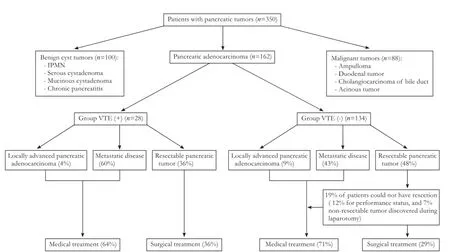
Fig. 1. Flow chart of the study population between May 2004 and January 2012. IPMN: intraductal papillary mucinous neoplasm.
The survival at three years was signifcantly different between the VTE (+) and VTE (-) groups (8% vs 40%, P=0.010) despite an equivalent number of progressionand recurrence in the two groups (Table 3 and Fig. 2). The median survival time was 12 months in the VTE (+) group (95% CI: 7.20-29.18) and 18 months (95% CI: 7.96-16.04) in the VTE (-) group (P=0.010). Predictive factors of overall survival were analyzed by univariate and multivariate statistical analysis. Variables that were included were clinical, VTE, operative treatment, patho-logical variables (tumor size, location and differentiation, number of harvested lymph node, nodal status, AJCC, microscopic R1 resection and retroperitoneal margin resection) and postoperative adjuvant therapy. Multivariate statistical analysis showed that VTE and treatment without surgery were independent prognostic factors of death (Table 4).
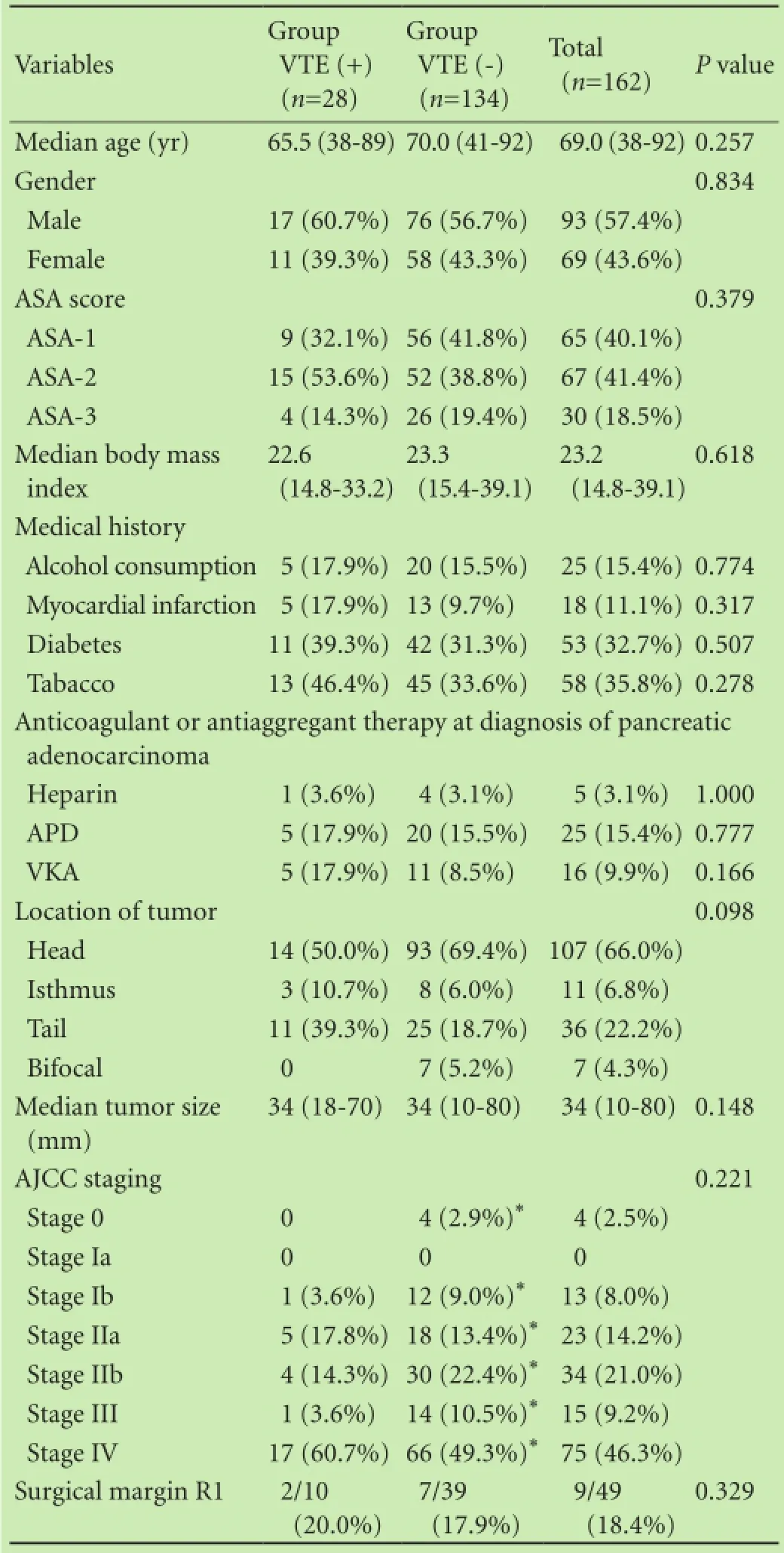
Table 1. Characteristics of patients and pathological fndings
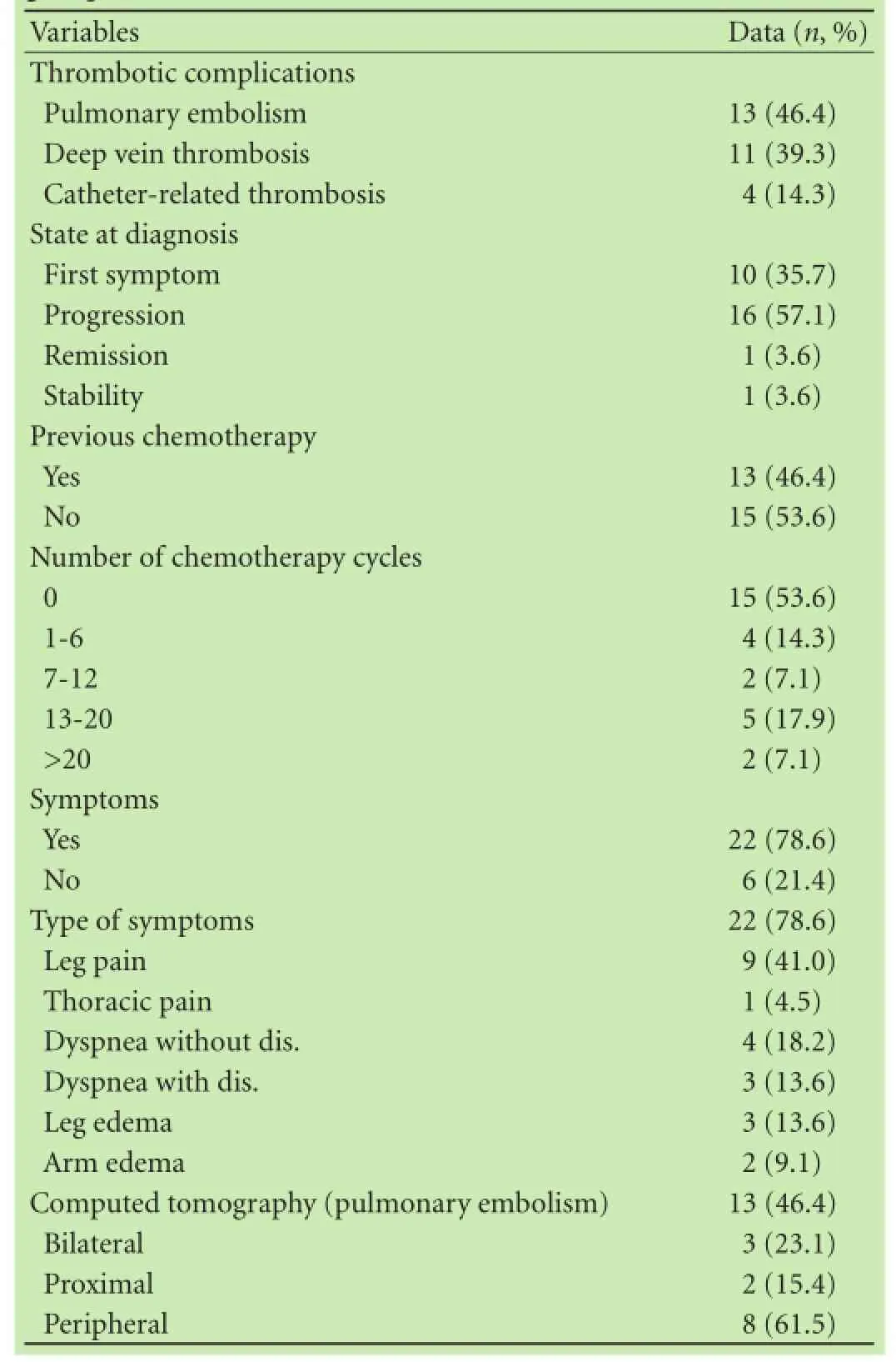
Table 2. Characteristics of patients and pathological fndings in group VTE(+)
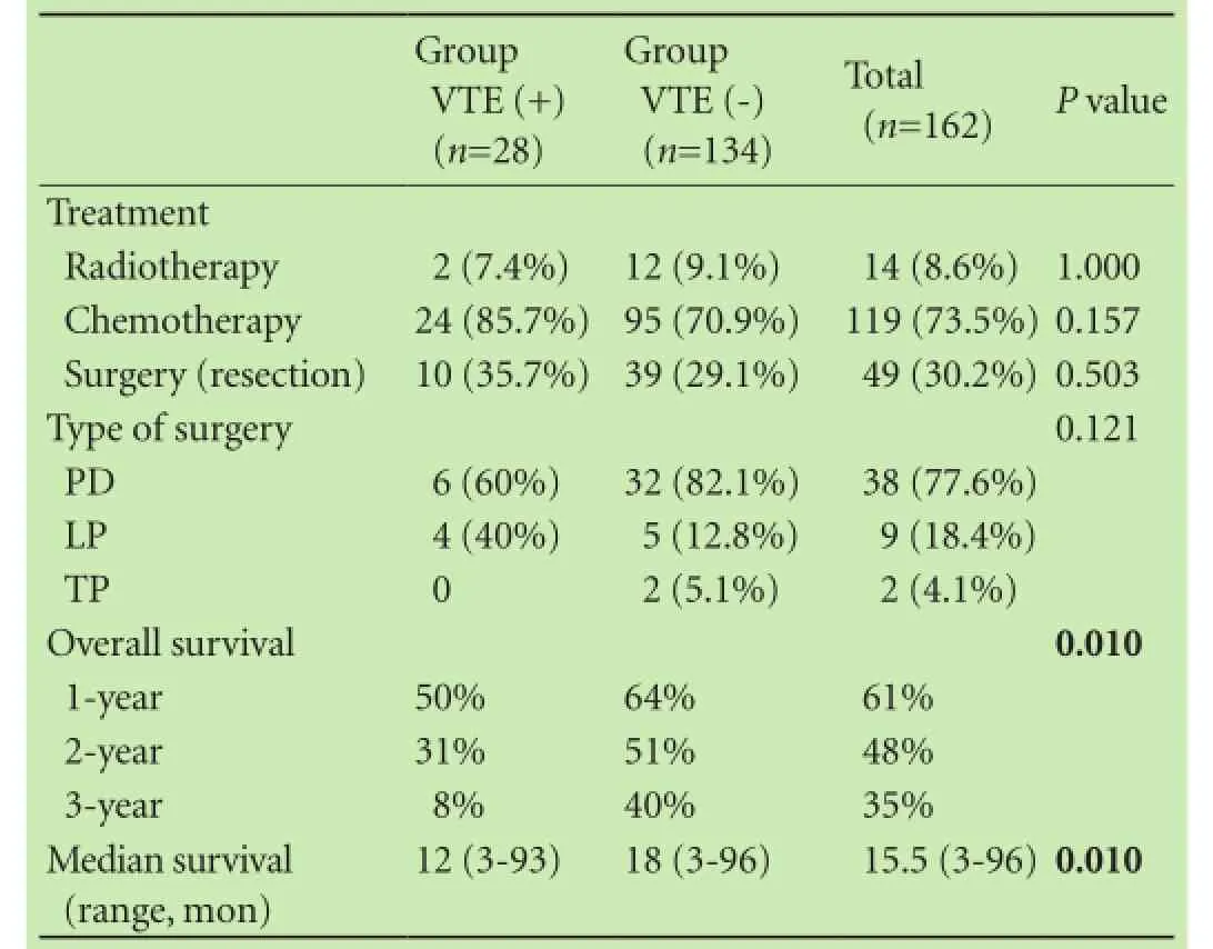
Table 3. Treatment and overall survival
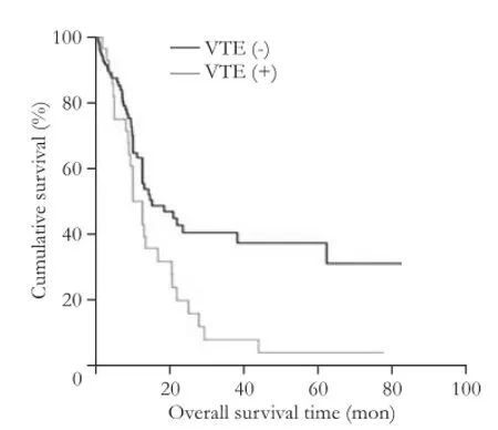
Fig. 2. Overall survival of the two groups.

Table 4. Predictive factors of overall survival in 162 patients treated for pancreatic adenocarcinoma
Discussion
In our study, the VTE incidence was 17.3%. This is consistent with the most recent studies of the literature in which VTE occurred in 5% to 36% of pancreatic cancers.[8,12,26]In contrast, Oh et al[12]reported a low incidence of 5%. The explanation given for the low risk was the Asian origin of the patients,[12]in whom the incidence of VTE was lower, even in the general population. Moreover, Epstein et al[8]found a much higher incidence of thrombosis (36%), but the present study also included arterial (myocardial and cerebral) thrombosis. Therefore, the incidence in our study seems to be in accordance with the incidence of venous thrombosis reported in the literature. VTE was equally distributed between PE and DVT in our population, and we noticed nearly 15% of catheter thrombosis, which was likely underestimated.
Furthermore, one-ffth of VTE cases was completely asymptomatic, and was found incidentally during routine investigations. This proportion would be much more important if systematic screening was implemented. In a prospective study, Font et al[26]found asymptomatic thrombosis in up to 28% of 340 consecutive cancer patients, with 50% demonstrating PE incidentally diagnosed and asymptomatic at clinical evaluation. Menapace et al[9]evaluated asymptomatic thrombosis in 135 pancreatic cancer patients; the proportions were similar, with 33.3% with asymptomatic PE and 21.4% with asymptomatic DVT. This type of thrombosis has the same impact as symptomatic thrombosis on cancer outcome and patient prognosis.[27]Thus, it would be interesting to determine whether there is a beneft in screening each cancer patient for VTE, mainly in cancers largely associated with thrombosis like pancreatic cancer and in case of metastasis.
Although no statistical difference has been found in comparing our two groups in terms of clinical, therapeutic (tumor resection rate and chemotherapy) and tumor characteristics, the overall survival was statistically different.
The present study focused on the effect of VTE on prognosis of patients with pancreatic cancer. The results showed a statistically signifcant worse effect of thrombosis on overall survival. Regardless of stage, the 3-year survival was lower in patients who developed thrombosis than in those who didn't.
In the literature, the effects of VTE on prognosis are conficting. Indeed, some series highlight a poor prognosis for patients with VTE. Epstein et al[8]found thrombosis (venous and arterial) in 36% of 1915 patients treated with chemotherapy for invasive pancreatic cancer. They found that thrombosis signifcantly increased the risk of death in multivariate analysis (HR=2.6, 95% CI 2.3-2.8, P<0.01).[8]In the study of Menapace et al[9]reported that nearly one-third of patients experienced at least one VTE. One-ffth to one-third of these events were incidentally diagnosed on routine restaging or surveillance scans. Both incidental and symptomatic VTE were associated with increased mortality.[9]Similar results were reported in 2007 by Mandalà et al[10]in 227 patients with unresectable pancreatic cancer, wherein both overall VTE and thrombosis during chemotherapy were associated with a decrease in overall survival (HR=1.76, 95% CI 1.30-2.40, P=0.0003; HR=1.95, 95% CI 1.32-2.87, P=0.0008). In our study, all deaths were attributed to pancreatic cancer. In accordance with other studies, thrombosis could have an impact on the oncologic evolution of pancreatic cancer.
Some studies[11,13,28]didn't fnd any impact of VTE on prognosis in pancreatic cancer. However, it is noteworthy that these studies included a heterogeneous population (PA, ampulloma). Shaib et al[11]compared only patients with stage IV cancer, which has the poorest prognosis. Median survival at this stage was too short forthromboembolic events to have any impact on the prognosis.
There are certain limitations to this report. First, the study was a retrospective analysis. Second, these results are based on the experience of a single institution and may therefore not be wholly generalizable to other institutions, given differences in treatment, diagnosis and practice.
Although clinical trials didn't fnd gemcitabine associated with dalteparin to have a signifcant thromboprophylactic effect in advanced pancreatic cancer on survival,[26,27]recent guidelines suggest that primary prophylaxis with low molecular weight heparin in patients treated with chemotherapy decreased the rate of VTE without an excess of bleeding in patients with locally advanced or metastatic pancreatic cancer (at subtherapeutic dosages).[25]In this population, we found that thrombosis predicts a poorer prognosis for patients with pancreatic cancer. This result could suggest the use systematic low molecular weight heparin in patients with locally advanced or metastatic PA.
Acknowledgement:The authors are deeply grateful to Anderson Loundou for the statistical analysis and ARCDO-PACA association for his help and advice.
Contributors:OM, FC and MD proposed the study concept and design, OM, FC and DL performed the acquisition of data. OM and FC contributed equally to this study. OM, FC and BL contributed to the draft of manuscript. PD and DF contributed to the writing of manuscript and data interpretation. FD is the guarantor.
Funding:This study was supported by institutional funding from INSERM (Paris, France) and the Aix-Marseille University (Marseille, France) and by a grant INCAa-DGSO-INSERM 6038 from sites de recherche intégré sur le cancer (SIRIC).
Ethical approval:Not needed.
Competing interest:No benefts in any form have been received or will be received from a commercial party related directly or indirectly to the subject of this article.
1 Trousseau A. Phlegmasia Alba Dolens. In Clinique Medicale de l'Hotel Dieu de Paris. Edited by Trousseau A. Paris: Ballier; 1865:654-712.
2 Blom JW, Doggen CJ, Osanto S, Rosendaal FR. Malignancies, prothrombotic mutations, and the risk of venous thrombosis. JAMA 2005;293:715-722.
3 Heit JA, Silverstein MD, Mohr DN, Petterson TM, O'Fallon WM, Melton LJ 3rd. Risk factors for deep vein thrombosis and pulmonary embolism: a population-based case-control study. Arch Intern Med 2000;160:809-815.
4 Haas S, Wolf H, Kakkar AK, Fareed J, Encke A. Prevention of fatal pulmonary embolism and mortality in surgical patients: a randomized double-blind comparison of LMWH with unfractionated heparin. Thromb Haemost 2005;94:814-819.
5 Kosuri K, Muscarella P, Bekaii-Saab TS. Updates and controversies in the treatment of pancreatic cancer. Clin Adv Hematol Oncol 2006;4:47-54.
6 Shah MM, Saif MW. Pancreatic cancer and thrombosis. Highlights from the "2010 ASCO Annual Meeting". Chicago, IL, USA. June 4-8, 2010. JOP 2010;11:331-333.
7 Oua?ssi M, Giger U, Louis G, Sielezneff I, Farges O, Sastre B. Ductal adenocarcinoma of the pancreatic head: a focus on current diagnostic and surgical concepts. World J Gastroenterol 2012;18:3058-3069.
8 Epstein AS, Soff GA, Capanu M, Crosbie C, Shah MA, Kelsen DP, et al. Analysis of incidence and clinical outcomes in patients with thromboembolic events and invasive exocrine pancreatic cancer. Cancer 2012;118:3053-3061.
9 Menapace LA, Peterson DR, Berry A, Sousou T, Khorana AA. Symptomatic and incidental thromboembolism are both associated with mortality in pancreatic cancer. Thromb Haemost 2011;106:371-378.
10 Mandalà M, Reni M, Cascinu S, Barni S, Floriani I, Cereda S, et al. Venous thromboembolism predicts poor prognosis in irresectable pancreatic cancer patients. Ann Oncol 2007;18:1660-1665.
11 Shaib W, Deng Y, Zilterman D, Lundberg B, Saif MW. Assessing risk and mortality of venous thromboembolism in pancreatic cancer patients. Anticancer Res 2010;30:4261-4264.
12 Oh SY, Kim JH, Lee KW, Bang SM, Hwang JH, Oh D, et al. Venous thromboembolism in patients with pancreatic adenocarcinoma: lower incidence in Asian ethnicity. Thromb Res 2008;122:485-490.
13 Mitry E, Taleb-Fayad R, Deschamps A, Mansencal N, Lepère C, Declety G, et al. Risk of venous thrombosis in patients with pancreatic adenocarcinoma. Gastroenterol Clin Biol 2007;31:1139-1142.
14 Aw A, Carrier M, Koczerginski J, McDiarmid S, Tay J. Incidence and predictive factors of symptomatic thrombosis related to peripherally inserted central catheters in chemotherapy patients. Thromb Res 2012;130:323-326.
15 Carrier M, Khorana AA, Zwicker JI, Lyman GH, Le Gal G, Lee AY, et al. Venous thromboembolism in cancer clinical trials: recommendation for standardized reporting and analysis. J Thromb Haemost 2012;10:2599-2601.
16 Owens WD, Felts JA, Spitznagel EL Jr. ASA physical status classifcations: a study of consistency of ratings. Anesthesiology 1978;49:239-243.
17 Tempero MA, Arnoletti JP, Behrman SW, Ben-Josef E, Benson AB 3rd, Casper ES, et al. Pancreatic Adenocarcinoma, version 2.2012: featured updates to the NCCN Guidelines. J Natl Compr Canc Netw 2012;10:703-713.
18 Yoshida T, Matsumoto T, Sasaki A, Shibata K, Aramaki M, Kitano S. Outcome of paraaortic node-positive pancreatic head and bile duct adenocarcinoma. Am J Surg 2004;187:736-740.
19 Doi R, Kami K, Ito D, Fujimoto K, Kawaguchi Y, Wada M, et al. Prognostic implication of para-aortic lymph node metastasis in resectable pancreatic cancer. World J Surg 2007;31:147-154.
20 Callery MP, Chang KJ, Fishman EK, Talamonti MS, William Traverso L, et al. Pretreatment assessment of resectable and borderline resectable pancreatic cancer: expert consensus statement. Ann Surg Oncol 2009;16:1727-1733.
21 Edge SB. Exocrine and endocrine pancreas. AJCC Cancer staging manual. 7th ed. New York, NY: Springer; 2010:241-249.
22 Lüttges J, Vogel I, Menke M, Henne-Bruns D, Kremer B, Kl?ppel G. The retroperitoneal resection margin and vessel involve-ment are important factors determining survival after pancreaticoduodenectomy for ductal adenocarcinoma of the head of the pancreas. Virchows Arch 1998;433:237-242.
23 Verbeke CS. Resection margins and R1 rates in pancreatic cancer--are we there yet? Histopathology 2008;52:787-796.
24 Wittekind C, Compton CC, Greene FL, Sobin LH. TNM residual tumor classifcation revisited. Cancer 2002;94:2511-2516.
25 Farge D, Debourdeau P, Beckers M, Baglin C, Bauersachs RM, Brenner B, et al. International clinical practice guidelines for the treatment and prophylaxis of venous thromboembolism in patients with cancer. J Thromb Haemost 2013;11:56-70.
26 Font C, Farrús B, Vidal L, Caralt TM, Visa L, Mellado B, et al. Incidental versus symptomatic venous thrombosis in cancer: a prospective observational study of 340 consecutive patients. Ann Oncol 2011;22:2101-2106.
27 Di Nisio M, Ferrante N, De Tursi M, Iacobelli S, Cuccurullo F, Büller HR, et al. Incidental venous thromboembolism in ambulatory cancer patients receiving chemotherapy. Thromb Haemost 2010;104:1049-1054.
28 Maraveyas A, Waters J, Roy R, Fyfe D, Propper D, Lofts F, et al. Gemcitabine versus gemcitabine plus dalteparin thromboprophylaxis in pancreatic cancer. Eur J Cancer 2012;48:1283-1292.
Received June 30, 2014
Accepted after revision March 4, 2015
I want to bring out the secrets of nature and apply them for the happiness of man. I don't know of any better service to offer for the short time we are in the world.
—Thomas Edison
Author Affliations: Departments of Digestive Surgery (Oua?ssi M, Frasconi C, Mege D, Boiron L and Sielezneff I) and Oncology (Dahan L), Timone Hospital, Aix-Marseille University, Marseille, France; Atelier Proven?ale d'Ecriture Médicale, Faculté de Médecine de Marseille (Oua?ssi M, Frasconi C and Mege D); Groupe Francophone Thrombose et Cancer, H?pital ST Louis, 1 avenue Claude Vellefaux, Paris, France (Oua?ssi M, Mege D, Panicot-dubois L, Debourdeau P, Dubois C and Farge D); INSERM UMR-1076, Faculty of Pharmacy, Aix-Marseille University, Marseille, France (Mege D, Panicot-dubois L and Dubois C) and INSERM UMR-1160, Internal Medicine and Vascular Unit, Assistance Publique-H?pitaux de Paris, Paris, France (Farge D); Department of Hematology, Hospital Sainte Catherine, Avignon, France (Debourdeau P)
Mehdi Oua?ssi, MD, PhD, Department of Digestive Surgery, Timone Hospital, 264 rue saint Pierre, 13385 Marseille, France (Tel: +33-4913885852; Fax: +33-4913885552; Email: mehdi.ouaissi@ mail.ap-hm.fr)
? 2015, Hepatobiliary Pancreat Dis Int. All rights reserved.
10.1016/S1499-3872(15)60397-6
Published online July 6, 2015.
 Hepatobiliary & Pancreatic Diseases International2015年4期
- Hepatobiliary & Pancreatic Diseases International的其它文章
- Influence of comorbidity on outcomes of older patients with acute pancreatitis based on a national administrative database
- Clinical characteristics and outcome of hepatocellular carcinoma in alcohol related and cryptogenic cirrhosis: a prospective study
- The response of Golgi protein 73 to transcatheter arterial chemoembolization in patients with hepatocellular carcinoma may relate to the influence of certain chemotherapeutics
- A serum metabolomic analysis for diagnosis and biomarker discovery of primary biliary cirrhosis and autoimmune hepatitis
- Development and validation of a predictive score for perioperative transfusion in patients with hepatocellular carcinoma undergoing liver resection
- Pancreaticoduodenectomy with portal vein/superior mesenteric vein resection for patients with pancreatic cancer with venous invasion
