Prevalence of coronavirus from diarrheic calves in the Republic of Korea
Jinho Park, Du-Gyeong Han, SuHee Kim, Jeong-Byoung Chae, Joon-Seok Chae, Do-Hyeon Yu,Kyoung-Seong Choi
1College of Veterinary Medicine, Chonbuk National University, Iksan 54596, Republic of Korea
2Department of Animal Science and Biotechnology, College of Ecology and Environmental Science, Kyungpook National University, Sangju 37224,Republic of Korea
3Animal Disease & Biosecurity Team, National Institute of Animal Science, Rural Development Administration, Wanju-Gun 55365, Republic of Korea
4Laboratory of Veterinary Internal Medicine, BK21 PLUS Program for Creative Veterinary Science Research, Research Institute for Veterinary Science and College of Veterinary Medicine, Seoul National University, Seoul 08826, Republic of Korea
5College of Veterinary Medicine, Gyeongsang National University, Jinju 52828, Republic of Korea
1. Introduction
Calf diarrhea leads to severe economic losses worldwide in the livestock industry as a result of morbidity, poor growth rates,treatment costs, and mortality among affected young calves[1,2].Approximately half of all mortality among calves up to the first month after birth has been attributed to diarrhea[3]. Calf diarrhea has a complex and multifactorial etiology, because infectious diseases,environmental, nutritional, and herd management factors such as housing type, colostrum intake, and hygienic conditions may be associated with field outbreaks, indicating that these factors may influence the severity and outcome of the disease[2,4]. Calves are at the greatest risk of developing diarrhea during the first month of life and the incidence of diarrhea decreases with age[4,5].
Mostly, outbreaks of calf diarrhea are associated with various infectious agents that present either individually or as part of a coinfection. Among the numerous infectious agents that can cause calf diarrhea, bovine coronavirus (BCoV) and bovine rotavirus (BRV)are recognized as the most important viral pathogens and these viruses can concomitantly infect calves[6-8]. In addition, bovine viral diarrhea virus (BVDV) is frequently associated with acute diarrhea in calves[9], although further studies are needed to evaluate its role in calf diarrhea. In the Republic of Korea (ROK), BVDV1 and BVDV2 are predominant and widespread among cattle[10,11].
To date, there have been few reports on the detection of viral pathogens associated with calf diarrhea in the ROK. There is little information available for Korean native calves owing to the small numbers of animals studied[12,13]. The main objective of the present study was therefore to investigate the prevalence of BCoV, BRV,and BVDV in diarrheic calves aged 1-81 days with feces of various consistencies, and to report any associations between these viral pathogens and diarrhea in calves.
2. Materials and methods
2.1. Ethical statement
This manuscript does not report on any studies performed with animals.
2.2. Fecal samples
Between April and October of 2016, a total of 143 fecal samples were collected from farms in six different regions in northern and southern ROK, namely Gimje (n=20), Gochang (n=20), Hoengseong(n=20), Iksan (n=7), Sancheong (n=14), and Wanju (n=62). Fecal samples were collected from Korean native calves aged 1-84 days.Among 143 calves, 51, 54, and 38 calves were 1-21, 22-42, and 43-84 days old, respectively. Each animal’s identification number, age,gender, and the number of animals in its herd were recorded. Fecal samples were scored for consistency based on accepted criteria(https://www.vetmed.wisc.edu/dms/fapm/fapmtools/calves.htm) and classified as normal, semi-formed (pasty), loose, or watery. All fecal samples were directly collected from the rectum, stored in plastic containers, photographed, and then categorized for consistency.A questionnaire was administered to farm managers on housing conditions, colostrum intake, and the vaccination of dams with the combined vaccine against coronavirus and rotavirus. The Korean native calves examined in this study were born to cows that were not vaccinated against BVDV. The collected samples were stored at -80℃ until analysis.
2.3. Extraction of RNA and reverse transcription-polymerase chain reaction (RT-PCR) for pathogen detection
Total RNA was extracted from fecal suspensions using RNAiso Plus Reagent (Takara Bio, Shiga, Japan) according to the manufacturer’s instructions. RT-PCR was performed to amplify BVDV, BCoV,and BRV RNA using DiaStar? 2× One-Step RT-PCR Smart Mix(Solgent, Daejeon, Korea). Briefly, reverse transcription was carried out at 50 ℃ for 30 min, followed by 94 ℃ for 5 min, and then 35 cycles at 94 ℃ for 1 min and 72 ℃ for 1 min, with a final extension step at 72 ℃ for 10 min. Details regarding each primer and the PCR conditions used to detect BCoV, BRV, and BVDV have been described previously[14-16]. The amplicons were analyzed in a 1.5%agarose gel and visual?ized after ethidium bromide staining.
2.4. Data analysis
The PCR results for each fecal sample were recorded as either positive or negative for each pathogen and categorized according to the age (1-21, 22-42, or 43-84 days) and fecal consistency(normal, semi-formed, loose, or watery feces) of each calf. The data were analyzed using the SPSS 23.0 software package (SPSS,Chicago, IL, USA). Pathogens that were associated with liquid fecal samples (semi-solid, loose, and watery feces) more frequently than normal fecal samples were investigated using a multinomial logistic regression model. The associations between diarrhea and each pathogen were calculated for all ages together and for each age group separately using Pearson’schi-square test or Fisher’s exact test, as appropriate. In addition, independent variables (infection,ages, colostrum intake, vaccination, and growth type) associated with diarrhea were predicted using binary univariable LogReg followed by binary multivariable LogReg only for variable withP<0.1 in univariable LogReg. Variables withP<0.05 were considered significant.
3. Results
3.1. Survey of enteric viral pathogens in 6 different regions
A total of 143 (50 normal and 93 diarrheal) fecal samples from Korean native calves aged 1-84 days (up to 12 weeks) were collected from six regions of the ROK, and these samples were tested for BCoV, BRV, and BVDV by RT-PCR. Among 143 fecal samples,the most prevalent pathogen was BVDV, which was detected in 86 animals (60.1%, 86/143) from five regions (Gimje, Gochang,Heongseong, Sancheong, and Wanju), while BVDV was not detected in fecal samples from the Iksan region (Table 1). As shown in Table 1, BCoV was identified in 51 samples (35.7%, 51/143) from all six regions surveyed. BRV was detected in only three calves (2.1%,3/143) from two regions (Gimje and Gochang), and these calves were co-infected with BCoV. In addition, co-infections with two pathogens (BCoV+BVDV, 17.5%; BCoV+BRV, 0.7%) and threepathogens (BCoV+BRV+BVDV, 1.4%) were detected in 26 and two calves, respectively (Table 1). Co-infections with BCoV and BVDV were found in four regions (Table 1). In 33 fecal samples (23.1%),none of the tested viral pathogens were found.

Table 1 Summary of viral pathogens detected in fecal samples from 143 Korean native calves in six regions of the Republic of Korea.
3.2. Prevalence of viral pathogens in feces from normal and diarrheic calves
As summarized in Table 2, the prevalence of each viral pathogen was determined in normal and diarrheal fecal samples. BVDV(62.4%) and BCoV (39.8%) were commonly detected in feces from diarrheic calves, whereas BRV was found at a lower frequency(3.2%). BVDV (56.0%) and BCoV (28.0%) were also detected in normal feces at lower frequencies than in diarrheic feces.BRV was not found in normal feces. Co-infections of BCoV and BVDV were detected more than twice as often diarrheal feces as compared to normal feces. Multiple infections with three pathogens(BCoV+BRV+BVDV) were found only in diarrheal samples. Among the pathogens identified, co-infection of BCoV and BVDV increased the odds ratio (OR) for diarrhea by 2.5-fold [95% confidence interval(CI): 0.9-7.0;P=0.08], but there were no significant associations between the individual pathogens and diarrhea.
3.3. Relationship between the presence of viral pathogens and fecal consistency
The associations between the viral pathogens and fecal consistency(semi-formed, loose, and watery feces) were investigated. Based on the multinomial regression analysis, BCoV detection was significantly correlated with the development of loose feces (Table 3). BCoV was detected in loose feces 2.9-fold more frequently than in normal feces (95%CI:1.1-7.6;P=0.03). Furthermore, coinfection with BCoV and BVDV was significantly associated with a loose fecal consistency (OR=3.6; 95%CI:1.0-12.4;P=0.04) (Table 3). There was no statistically significant association between the pathogens detected and semi-formed or watery feces.

Table 2 Detection frequency of viral pathogens in feces from normal and diarrheic calves among 143 Korean native calves.
3.4. Prevalence of viral pathogens according to the age of the calves
To analyze which pathogens are associated with diarrhea by age group among the calves, infections with BCoV, BRV, and BVDV in fecal samples from diarrheic calves aged 1-81 days were examined.In calves below 21 days of age, diarrhea was significantly associated with BCoV infection (Table 4). Among the three pathogens detected,the prevalence of BCoV was detected 9.3-fold more frequently in diarrheic feces than in normal feces among calves aged 1-21 days(OR=9.3; 95%CI:1.1-78.9;P=0.02), while BCoV infection showedno association with diarrhea in the other age groups (Table 4).BRV was detected only in the diarrheic feces of calves aged 1-21 days (Table 4). However, no statistically significant association between BRV and diarrhea was observed and the OR for BRV was not calculated because very few BRV infections were detected.These results revealed that BCoV infection was strongly associated with diarrhea in calves aged 1-21 days, whereas BRV and BVDV infections were not significantly associated with diarrhea in any age group.

Table 3 Multinomial logistic regression model to assess relationship between viral pathogens and three categories of fecal consistency among 143 Korean native calves.

Table 4 Relationship between viral pathogens and diarrhea among 143 Korean native calves (1-84 days).

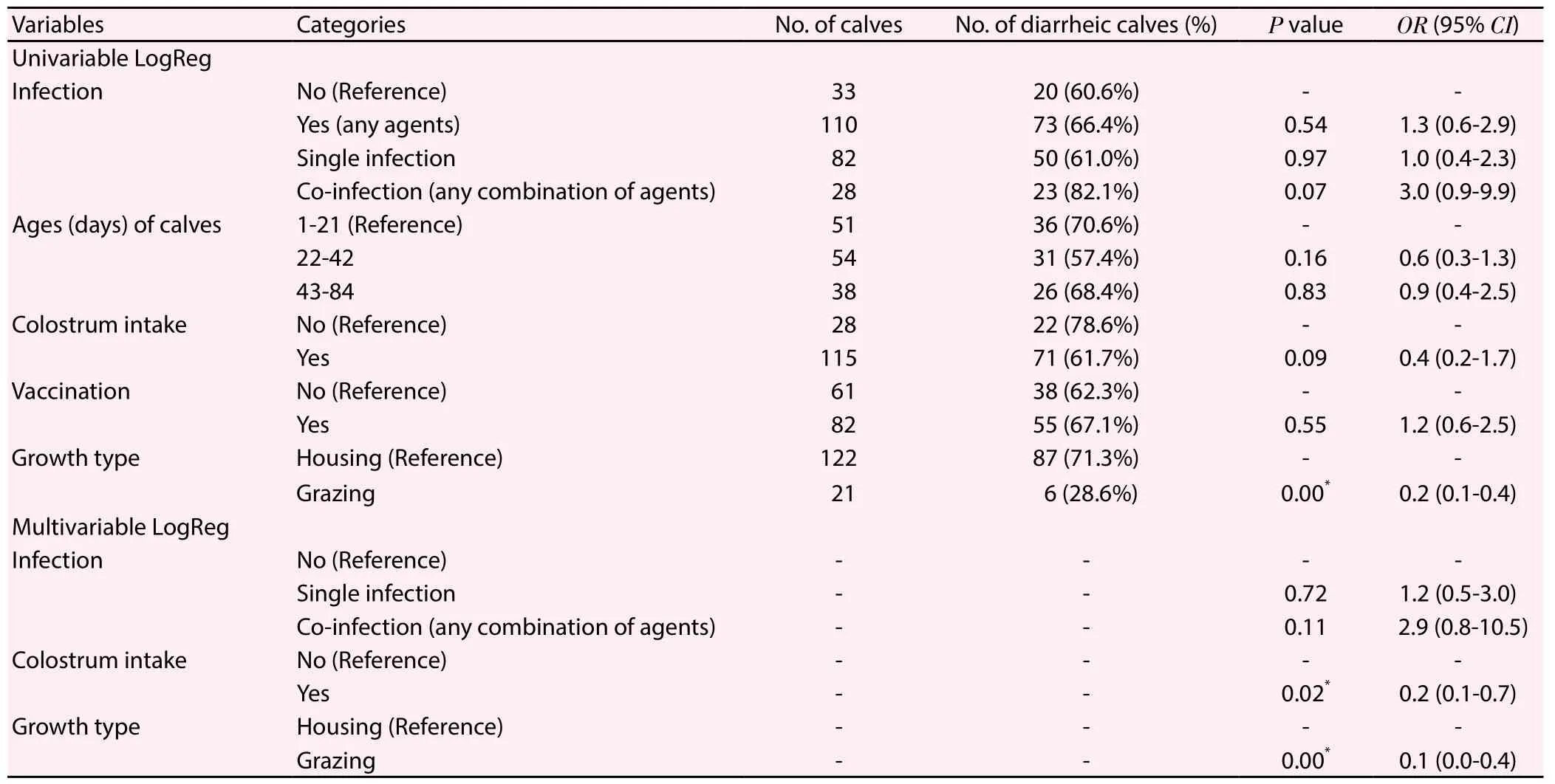
Table 5 Variables associated with diarrhea in fecal samples from 143 Korean native calves on 6 different regions of the Republic of Korea.
3.5. Variables associated with diarrhea
Information such as colostrum intake, vaccination, and herd management (grazing or housing) was obtained from the farms in the regions where fecal samples were collected. Initially, univariable binary LogReg identified 3 variables associated atP<0.01 with diarrhea (Table 5). Interestingly, grazing calves had a 5-fold lower OR for the incidence of diarrhea as compared to housed calves(OR=0.2; 95%CI:0.1-0.4;P=0.00). As shown in Table 5, vaccination of pregnant dams against BCoV and BRV did not prevent diarrhea among the calves. Although there were no significant associations,diarrhea may be slightly influenced by co-infection and colostrum intake. Co-infection increased the OR for the incidence of diarrhea by 3-fold (P=0.07) as compared to mono-infection. Next, 3 variables closely associated with diarrhea were analyzed with the multivariable LogReg model. The final model showed that colostrum intake decreased the OR for the incidence of diarrhea by 5-fold as compared to calves that had not been fed colostrum (P=0.02; Table 5). Grazing had a strong effect on the decrease in diarrhea (OR=0.1;95%CI:0.0-0.4;P=0.00). However, there was no statistical significance between diarrhea and co-infection.
4. Discussion
We investigated the prevalence of BCoV, BRV, and BVDV in normal and diarrheal (semi-formed, loose, or watery) feces of Korean native calves from six different regions in the ROK. In the present study, BVDV was the most frequently detected pathogen, followed by BCoV and then BRV; however, no association between diarrhea and the presence of BVDV was found. Co-infections with two or three pathogens were detected in 28 Korean native calves. BCoV was found in fecal samples from all six regions surveyed, reflecting the broad circulation of BCoV among Korean native calves. Our results revealed that the prevalence of BCoV was significantly higher in diarrheal calves aged 1-21 days than in non-diarrheal calves of the same age group. Infection with BCoV either alone or together with BVDV was associated with a significantly higher incidence of loose feces. Among the various factors evaluated, grazing and colostrum intake were associated with a significant reduction in the incidence of diarrhea. Interestingly, no viral pathogens were found in 33 diarrheal feces. It is possible that these cases were associated with other pathogens, including bacteria (e.g.,Escherichia coliandSalmonella), parasites (e.g.,CryptosporidiumandGiardia), or other viruses (e.g., torovirus and norovirus). Further studies are necessary to identify additional infectious agents in the feces of diarrheic calves.
In this study, BCoV was the second most prevalent pathogen and was detected in 35.7% (51 out of 143) of all fecal samples. This pathogen was present alone or as part of a co-infection with other viral pathogens (BVDV and/or BRV). According to our results,the prevalence of BCoV was higher than that of BRV in the ROK.Several studies have reported the lack of an association between BCoV infection and calf diarrhea[17,18]. However, our results showed that BCoV infection was more commonly detected in loose feces(OR=2.9;P=0.03) and the incidence of loose feces was significantly higher among calves that were co-infected with BCoV and BVDV(OR=3.6;P=0.04) as compared with those that were infected with BCoV alone. Consequently, we demonstrated a statistically significant relationship between the detection of BCoV and the occurrence of loose feces. In addition, cases of diarrhea caused by BCoV were most prevalent among diarrheic calves aged 1-21 days in the ROK (OR=9.3;P=0.02). Consequently, it seems that BCoV infection may contribute to calf diarrhea either alone or as a coinfection with other viral pathogens, or in conjunction with other factors.
BRV is a primary cause of acute diarrhea in neonatal calves[19,20].In this study, the prevalence of BRV among diarrheic calves was very low and no correlation between the BRV infection and diarrhea was found. However, we cannot rule out the possibility that other BRV infections, such as group B and group C rotaviruses, might be circulating in Korean native calves. A previous study showed that group C rotavirus caused diarrhea in Korean native calves[12].Although the present results do not show the importance of BRV as a major enteric pathogen, BRV should be continuously surveyed to monitor its circulation among Korean native calves.
BVDV was widespread among both normal and diarrheal fecal samples in the ROK. In the present study, we could not demonstrate any significant relationship between BVDV infection and diarrhea, despite a high positive rate of BVDV among all age groups. We cannot rule out the possibility of BVDV transmission to calves through colostrum feeding or the presence of persistently infected animals on these farms. Although there was no statistical significance, it is estimated that co-infection with BVDV and BCoV may increase the occurrence of diarrhea (P=0.08). The current study did not demonstrate the role of BVDV in calf diarrhea;however, the clinical significance of BVDV infections, especially persistently infected, should not be overlooked. Future studies are therefore necessary to clarify the association between BVDV and the occurrence of diarrhea.
To date, no studies have estimated the relationship between viral pathogens and fecal consistency (semi-formed, loose, or watery feces). According to our findings, BRV, which has previously been shown to be highly associated with acute watery diarrhea, was not detected in watery feces, whereas BCoV and BVDV were detected in watery feces. BCoV and BVDV were also detected in normal feces.Our results indicate that the consistency of feces did not necessarily correlate with the viral pathogens detected. The present study demonstrated that only the incidence of loose feces was significantly related with BCoV infection. Such a difference in fecal consistency may be a useful criterion for determining the clinical significance of BCoV detection during diagnostic investigations. Larger epidemiological surveys are needed to identify any associations between fecal consistency and the presence of infectious pathogens.Given such large scale data, it should be possible to estimate which pathogens cause diarrhea according to the consistency of the fecal samples.
We investigated the associations between viral pathogens and diarrhea among calves by age group. Interestingly, the detection rate of the three viral pathogens examined in this study gradually decreased as the age of calves increased (data not shown). Our results showed that BCoV was strongly associated with diarrhea among calves aged 1-21 days. This observation was quite different from the results of previous studies[18,21,22]. Bartelset al. mentioned that because BCoV is an opportunistic infection, its prevalence may be affected by calf immunity and environmental factors such as overcrowding, housing of calves in age-mixed rather than agespecific groups, and poor hygiene[17]. Moreover, it is speculated that the prevalence of BCoV at this age may be due to the presence of infected animals that were shedding virus. The results highlight the importance of farm management strategy,i.e., good hygienic procedures, as a protective factor in calf-rearing.
Diarrhea in claves is closely associated with infectious agents,nutritional quality, environmental stress, and other management factors. The present study indicated that vaccination was not associated with the occurrence of diarrhea, whereas grazing(P=0.00) and colostrum intake (P=0.02) may significantly reduce the incidence of diarrhea. Several studies have shown that delayed colostrum intake, feeding with infected or purchased colostrum,calving season, and parity (i.e., primiparous dams) are related to the incidence of calf diarrhea because of the low levels of maternal antibodies in calves[23,24]. The results of the present study emphasized the importance of the first colostrum intake as soon as possible after birth. Grazing may not only reduce the incidence of diarrhea, but also should be encouraged in terms of animal welfare in the ROK. Taken together, the occurrence of diarrhea may be more commonly associated with other factors such as passive immunity,cattle management systems, herd size, and access to grazing. Further studies are necessary to determine the effects of these variables in preventing calf diarrhea.
In conclusions, this study showed that BCoV was strongly associated with diarrhea in Korean native calves aged 1-21 days.However, further investigations to identify the presence of other enteropathogens such asEscherichia coliandCryptosporidiumare needed. The prevalence of BCoV described in this paper highlights the need for in-depth epidemiological studies. Therefore, grazing and colostrum intake is recommended for preventing and controlling calf diarrhea caused by BCoV.
Conflict of interest statement
The authors declare no conflict of interests.
Acknowledgments
This work was supported by the National Research Foundation of Korea (NRF), funded by the Korea government (No.2015R1C1A2A01053080). This work was also carried out with the support of the “Cooperative Research Program for Agriculture Science&Technology Development (Project No. PJ01194503)” from the Rural Development Administration, the Republic of Korea.
[1] Al Mawly J, Grinberg A, Prattley D, Moffat J, Marshall J, French N. Risk factors for neonatal calf diarrhoea and enteropathogen shedding in New Zealand dairy farms.Vet J2015; 203(2): 155-160.
[2] Meganck V, Hoflack G, Piepers S, Opsomer G. Evaluation of a protocol to reduce the incidence of neonatal calf diarrhoea on dairy herds.Prev Vet Med2015; 118(1): 64-70.
[3] Brickell JS, McGowan MM, Pfeiffer DU, Wathes DC. Mortality in Holstein-Friesian calves and replacement heifers, in relation to body weight and IGF-I concentration, on 19 farms in England.Animal2009;3(8): 1175-1182.
[4] Ammar SS, Mokhtaria K, Tahar BB, Amar AA, Redha BA, Yuva B, et al.Prevalence of rotavirus (GARV) and coronavirus (BCoV) associated with neonatal diarrhea in calves in western Algeria.Asian Pac J Trop Biomed2014; 4(Suppl 1): S318-322.
[5] Lorenz I, Fagan J, More SJ. Calf health from birth to weaning. II.Management of diarrhoea in pre-weaned calves.Ir Vet J2011; 64(1): 9.
[6] Bok M, Mi?o S, Rodriguez D, Badaracco A, Nu?es I, Souza SP, et al. Molecular and antigenic characterization of bovine Coronavirus circulating in Argentinean cattle during 1994-2010.Vet Microbiol2015;181(3-4): 221-229.
[7] Collins PJ, Mulherin E, Cashman O, Lennon G, Gunn L, O’Shea H, et al.Detection and characterisation of bovine rotavirus in Ireland from 2006-2008.Ir Vet J2014; 67(1): 13.
[8] Cho YI, Yoon KJ. An overview of calf diarrhea-infectious etiology,diagnosis, and intervention.J Vet Sci2014; 15(1): 1-17.
[9] Mohamed FF, Mansour SM, El-Araby IE, Mor SK, Goyal SM. Molecular detection of enteric viruses from diarrheic calves in Egypt.Arch Virol2017; 162(1): 129-137.
[10] Choi KS, Song MC. Epidemiological observations of bovine viral diarrhea virus in Korean indigenous calves.Virus Genes2011; 42(1): 64-70.
[11] Oem JK, Hyun BH, Cha SH, Lee KK, Kim SH, Kim HR, et al.Phylogenetic analysis and characterization of Korean bovine viral diarrhea viruses.Vet Microbiol2009; 139(3-4): 356-360.
[12] Park SI, Jeong YJ, Kim HJ, Park JG, Kang SY, Woo SK, et al. Genetically diverse group C rotaviruses cause sporadic infection in Korean calves.J Vet Med Sci2011; 73(4): 479-482.
[13] Park SJ, Lim GK, Park SI, Kim HH, Koh HB, Cho KO. Detection and molecular characterization of calf diarrhoea bovine coronaviruses circulating in South Korea during 2004-2005. Zoonoses Public Health 2007; 54(6-7): 223-230.
[14] Isegawa Y, Nakagomi O, Nakagomi T, Ishida S, Uesugi S, Ueda S.Determination of bovine rotavirus G and P serotypes by polymerase chain reaction.Mol Cell Probes1993; 7(4): 277-284.
[15] Tsunemitsu H, Smith DR, Saif LJ. Experimental inoculation of adult dairy cows with bovine coronavirus and detection of coronavirus in feces by RT-PCR.Arch Virol1999; 144(1): 167-175.
[16] Vilcek S, Paton DJ, Durkovic B, Strojny L, Ibata G, Moussa A, et al.Bovine viral diarrhoea virus genotype 1 can be separated into at least eleven genetic groups.Arch Virol2001; 146(1): 99-115.
[17] Bartels CJ, Holzhauer M, Jorritsma R, Swart WA, Lam TJ. Prevalence,prediction and risk factors of enteropathogens in normal and non-normal faeces of young Dutch dairy calves.Prev Vet Med2010; 93(2-3): 162-169.
[18] Uhde FL, Kaufmann T, Sager H, Albini S, Zanoni R, Schelling E, et al.Prevalence of four enteropathogens in the faeces of young diarrhoeic dairy calves in Switzerland.Vet Rec2008; 163(12): 362-366.
[19] Izzo MM, Kirkland PD, Mohler VL, Perkins NR, Gunn AA, House JK.Prevalence of major enteric pathogens in Australian dairy calves with diarrhoea.Aust Vet J2011; 89(5): 167-173.
[20] Gulliksen SM, Lie KI, L?ken T, Oster?s O. Calf mortality in Norwegian dairy herds.J Dairy Sci2009; 92(6): 2782-2795.
[21] Delafosse A, Chartier C, Dupuy MC, Dumoulin M, Pors I, Paraud C.Cryptosporidiumparvum infection and associated risk factors in dairy calves in western France.Prev Vet Med2015; 118(4): 406-412.
[22] de la Fuente R, Garcia A, Ruiz-Santa-Quiteria JA, Luzon M,Cid D, Garcia S, Orden JA, et al. Proportional morbidity rates of enteropathogens among diarrheic dairy calves in central Spain.Prev Vet Med1998; 36(2): 145-152.
[23] Beam AL, Lombard JE, Kopral CA, Garber LP, Winter AL, Hicks JA, et al. Prevalence of failure of passive transfer of immunity in newborn heifer calves and associated management practices on US dairy operations.J Dairy Sci2009; 92(8): 3973-8390.
[24] Raboisson D, Trillat P, Cahuzac C. Failure of passive immune transfer in calves: a meta-analysis on the consequences and assessment of the economic impact.PLoS One2016; 11(3): e0150452.
 Asian Pacific Journal of Tropical Biomedicine2018年1期
- Asian Pacific Journal of Tropical Biomedicine的其它文章
- A comprehensive review on anti-diabetic property of rice bran
- Ethnobotanical survey of antimalarial plants in Awash-Fentale District of Afar Region of Ethiopia and in vivo evaluation of selected ones against Plasmodium berghei
- Larvicidal activity of Neem oil and three plant essential oils from Senegal against Chrysodeixis chalcites (Esper, 1789)
- Protective effect of ashwagandha (Withania somnifera) against neurotoxicity induced by aluminum chloride in rats
- Oxidative stress mitigation, kinetics of carbohydrate-enzymes inhibition and cytotoxic effects of flavonoids-rich leaf extract of Gazania krebsiana (Less.): An in vitro evaluation
- Identification of commonly regulated genes in HPV18- and HPV16-infected cervical cancer cells treated with the curcumin analogue 1,5-bis(2-hydroxyphenyl)-1,4-pentadiene-3-one
