Kidney injury in COVID-19
Adeel Rafi Ahmed, Chaudhry Adeel Ebad, Sinead Stoneman, Muniza Manshad Satti, Peter J Conlon
Adeel Rafi Ahmed, Chaudhry Adeel Ebad, Sinead Stoneman, Department of Nephrology, Beaumont Hospital, Dublin D09 V2N0, Ireland
Muniza Manshad Satti, Department of Medicine, Connolly Hospital, Dublin D15X40D, Ireland
Peter J Conlon, Department of Nephrology, Beaumont Hospital and Royal College of Surgeons in Ireland, Dublin D09 V2N0, Ireland
Abstract
Coronavirus disease 2019 (COVID-19) continues to affect millions of people around the globe. As data emerge, it is becoming more evident that extrapulmonary organ involvement, particularly the kidneys, highly influence mortality. The incidence of acute kidney injury has been estimated to be 30% in COVID-19 non-survivors. Current evidence suggests four broad mechanisms of renal injury: Hypovolaemia, acute respiratory distress syndrome related, cytokine storm and direct viral invasion as seen on renal autopsy findings. We look to critically assess the epidemiology, pathophysiology and management of kidney injury in COVID-19.
Key Words: COVID-19; SARS-CoV-2; Acute kidney injury; Cytokine storm; Acute respiratory distress syndrome; Renal replacement therapy
INTRODUCTION
The severe acute respiratory syndrome coronavirus 2 (SARS-CoV-2) infection leading to the coronavirus disease 2019 (COVID-19) is affecting millions of people worldwide, carrying a case fatality rate between 0.9% to 7.2% depending on the demographics, implementation of preventative measures, testing strategies and availability of health care resources[1-3]. Severe disease is seen in approximately 20% of cases, of which around 6% represents the critically ill COVID-19 patients[4,5]. Amongst the critically ill, 65% to 95% have acute respiratory distress syndrome (ARDS), followed by acute kidney injury (AKI) and acute cardiac injury/cardiomyopathy[6-8]. AKI is common among critically ill patients with COVID-19 and is an independent marker of mortality[9,10]. Prompt recognition and management of AKI in COVID-19 can limit its progression and contribute to reducing morbidity and mortality[9]. Multiple mechanisms of kidney injury have emerged as we learn more about SARS-CoV-2[11]. In this review, we look to answer the many pertinent questions regarding the epidemiology, pathophysiology and management of AKI in COVID-19 patients.
MAIN BODY
Epidemiology
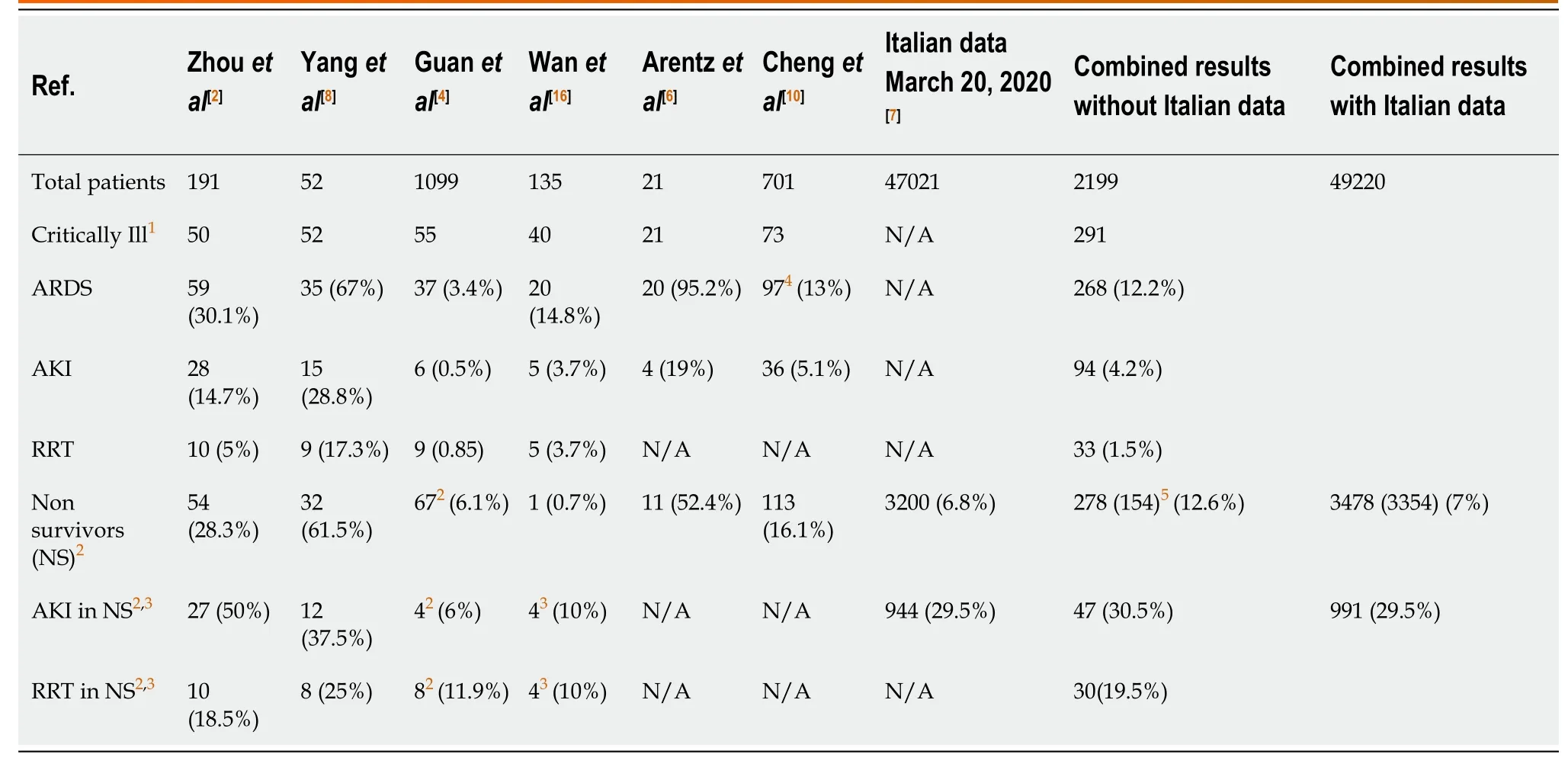
AKI, in general, has an incidence of around 3%-18% in hospitalised patients and is associated with 10%-20% mortality in the non-intensive care hospital setting, with up to 50% mortality in the intensive care setting[12-14]. There is a paucity of evidence identifying the role AKI plays in COVID-19. Majority of studies use Kidney Disease: Improving Global Outcomes (KDIGO) criteria to define AKI in COVID-19[15]. Assessment of data from major published cohorts on COVID-19, combining results from intensive care unit (ICU) admissions with non-ICU admission, reveals an overall AKI incidence of around 4.2% (Table 1)[2,4,6-8,10,16]. Amongst the non-survivors (NS), the incidence of AKI is approximately 30% and renal replacement therapy (RRT) is required in 19.5% (Table 1). Comparatively in the severe acute respiratory syndrome (SARS) outbreak in 2003, the incidence of AKI was around 6.7%, and multivariate analysis showed AKI as a significant independent risk factor for predicting mortality (relative risk: 4.057; 99% confidence interval: 1.461-11.27;P< 0.001)[17].
What is the mechanism of AKI in COVID-19? Four possible key mechanisms are becoming evident in the COVID-19 pandemic (Table 2): Hypovolaemia[9,18], ARDS related AKI[19,20], cytokine storm syndrome (CSS) associated AKI[21-23], and direct viral tropism for proximal tubular cells and podocytesviathe angiotensin-converting enzyme 2 (ACE2) carboxypeptidase[24,25].
Hypovolaemia
A majority of patients have significant insensible water losses due to high-grade pyrexia and tachypnoea on presentation[26]. A subgroup of patients has substantial gastrointestinal symptoms leading to extrarenal volume loss[18]. These patients are particularly prone to developing pre-renal AKI.
ARDS related AKI
AKI is seen in around 35%-50% of patients who develop ARDS and substantially increases mortality by nearly two-fold in the ICU[27-30]. ARDS and its associated mechanical ventilation strategies can cause or aggravate renal injuryviamultiple pathways[31]. There are broadly five categories; haemodynamic effects, gas exchange impairment (hypoxemia/hypercapnia), acid-base dysregulation, hyper inflammation and neurohormonal effects[32]. In COVID-19, significant AKI generally develops after the onset of ARDS, suggesting lung- kidney crosstalk as the dominant mechanism of kidney injury[2,33].

Table 1 Summary of acute kidney injury incidence in coronavirus disease 2019 patients

Table 2 Summary of the mechanism of kidney injury in coronavirus disease 2019
The haemodynamic effects of acute pulmonary disease result in increased pulmonary artery pressures, right ventricular failure, venous congestion and increased intra-abdominal/intrathoracic pressures[34-38].
Impaired gaseous exchange with hypercapnia leads to a reduction of renal vasodilatory response and renal blood flow with altered diuresis and increased oxygen utilisation in the proximal tubule[39-41]. Severe hypoxemia also causes a reduction in renal blood flow with possible activation of the hypoxia-inducible factor system, influencing lung and kidney outcomes[42]. There is the activation of reninangiotensin-aldosterone system, with increased aldosterone secretion with resultant activation of the sympathetic nervous system and release of non-osmotic vasopressin[31,43]. An immune-mediated/inflammatory response is noted in ARDS with the release of interleukin (IL)-6, tumour necrosis factor (TNF alpha), IL-1, transforming growth factor and substance P[44-47].
Mechanical ventilation can worsen the haemodynamic effects and cause ventilatorinduced lung injury leading to further cytokine release and multi-organ dysfunction syndrome[48]. The effects of excessive positive end-expiratory pressure (and high tidal volumes) on kidney function include a further increase in intrathoracic pressures, which causes increased right ventricular dysfunction, reduced venous return and reduced cardiac output[34-36].
AKI independently worsens ARDS. AKI leads to increased production, decreased clearance of inflammatory cytokines and down-regulation of lung aquaporin and ion channels[49,50]. The rise in circulatory cytokines, particularly IL-6, leads to increased infiltration of lungs with neutrophils and macrophages, and increased pulmonary vasculature permeability worsens ARDS[51,52]. In the later phase of inflammation, IL-6 promotes IL-10 production, which has anti-inflammatory and organ protective effects[53]. Limited data suggest AKI promotes neutrophil dysfunction, causing reduced clearance of infection and increasing lung permeability[54,55]. Haemodynamically, the inflammatory state and increased alveolar-capillary permeability combined with decreased urine output in AKI worsens pulmonary oedema[56,57]. Most immunological studies are based on animal models, however, observational data support the negative impact of AKI on pulmonary outcomes in critically ill patients, with two times more requiring invasive mechanical ventilation[58,59].
The incidence of shock is variable in COVID-19 based on the reported cohort studies; in the ICU setting it may be as high as 35%[2,8]. This vasopressor dependent state causes renal blood flow dysregulation, including ischaemia-reperfusion injury, metabolic reprogramming and inflammation resulting in AKI[60]. Preliminary reports suggest rhabdomyolysis is not a major component of COVID-19, but data vary in each centre with some case reports showing a significant rise in creatine kinase and other viral infections (H1N1 and SARS) have reported this complication[4,61-63].
Cardio-renal syndrome can play a significant role in critically ill COVID-19 patients[64,65]. In cardio-renal syndrome, excessive inflammation and rise in cytokines seem central to the pathophysiological process[64,66]. The high levels of IL-6, TNF and IL-1 have a direct cardio-depressant effect and may promote myocardial cell injury[67,68]. Acidaemia promotes pulmonary vasoconstriction, increases right ventricular afterload and exacerbates negative inotropic effect[69,70]. Myocarditis may also occur in COVID-19[71].
The overall combined effect of this entire process is an inflammatory, cardiodepressant, acidotic, volume retaining state with high intrathoracic and intraabdominal pressures resulting in high renal back pressures, decreased and dysregulated renal blood flow and severe renal tubular injury.
Cytokine storm syndrome associated AKI
Observational data from a subgroup of patients with COVID-19 suggest the development of features consistent with CSS triggered by SARS-CoV-2 virus characterised by high serum ferritin, D-dimer, lactate dehydrogenase, cytopenia, ARDS, acute cardiac injury, abnormal liver function test, raised IL-6 and coagulation abnormalities[72-75]. Viral infections have been reported as one of the most common triggers for cytokine storms[76]. One study demonstrated similar or lower levels of cytokines in COVID-19 pneumonia when compared to other critically ill patients, questioning the hypothesis of CSS[77]. However, the use of dexamethasone, a potent anti-inflammatory steroid, has demonstrated a significant reduction in mortality amongst critically ill COVID-19 patients, highlighting the major role of hyperinflammation[78].
Can this hyperinflammatory state cause AKI? Various case series have indicated significant renal involvement, particularly in CSS associated with secondary haemophagocytic lymphohistiocytosis (sHLH)[22,79-82]. The majority present with AKI with or without nephrotic range proteinuria[79]. Histological and observational findings indicate polymorphic renal lesions with acute tubular necrosis (ATN) being the most common, followed by tubulointerstitial nephritis (TIN), collapsing glomerulopathy (with podocytopathies) and thrombotic microangiopathy (TMA)[22,79,82]. ATN and TIN are most likely due to sepsis-related haemodynamic changes, coagulopathy (disseminated intravascular coagulopathy) and perhaps the direct toxic effect of raised cytokines (IL-6 and TNF) on renal epithelial cells[83]. Nephrotic syndrome with collapsing glomerulopathy and podocytopathies are generally seen in severe cases of sHLH with African ethnic predisposition[80]. It is hypothesised a circulating cytokine during CSS phase of sHLH may cause podocytopathy[82]. Hyperinflammation, as seen in COVID-19, also leads to a hypercoagulable state that can cause fibrin thrombi occlusions in renal capillaries (TMA pattern of renal injury)[84-86].
Renal biopsy histology of patients of black ethnicity who had AKI and were subsequently SARS-CoV-2 positive showed collapsing glomerulopathy, severe podocyte effacement with acute tubular injury (ATI)[87,88]. The APOL1 genotyping on the biopsy material was performed, and the patients were found to be homozygous for the G1 risk allele. Genetic predisposition with CSS may lead to collapsing glomerulopathy in COVID-19[89].
The hyperinflammatory state can cause renal injuryviamultiple mechanisms as highlighted, however, the discussion is incomplete without further assessing the role of direct viral tropism for renal parenchyma and renal autopsy findings.
Direct viral invasion
Viruses must gain entry into a cell and use the host cell machinery to replicate. The ACE2 is the coreceptor used by SARS-CoV-2 to gain entry to the cells[90]. ACE2 forms part of the renin-angiotensin-aldosterone system, a cascading peptide-pathway that regulates vascular tone and salt and water balance. The ACE2 degrades angiotensin II to angiotensin, resulting in vasodilation and countering the effects of ACE[91-94].
The ACE2 is expressed in the kidney, staining abundantly in the brush border of tubular epithelial cells, moderately in parietal epithelial cells and absent in glomerular or mesangial endothelial cells[92]. Although hypertension may be a risk factor for poor prognosis with SARS-CoV-2 infection, inferences that this is due to effects on ACE2 expression as a consequence of ACE inhibitor or angiotensin receptor blocker (ARB) use are not supported by data[91,95]. Previous studies have not shown that there is upregulation of plasma ACE2 activity in patients taking ACE inhibitors or ARBs compared to patients not on these agents[94,96,97].
There is currently no data to suggest that even if ACE inhibitors or ARBs did upregulate ACE2 expression that this would facilitate faster or greater viral entry of SARS-CoV-2 into cells[91].
SARS-CoV-2 shares 79.6% sequence identity to SARS-CoV; therefore, the mechanism of COVID-19 associated AKI may share some similarities with SARS[93].
The data on whether SARS-CoV-2 caused direct kidney injury through viral entry are conflicting. In an autopsy series of 18 patients who died of SARS infection, viral sequences were located in the epithelial cells of the renal distal tubules[98]. Similarly, using a murine monoclonal antibody specific for SARS-CoV-2 nucleoprotein in four patients who died of SARS, SARS-CoV-2 antigen and RNA was found in the epithelial cells of distal convoluted renal tubules[99]. However, in a smaller case series in which autopsy findings from kidney specimens of seven SARS patients were presented, there was no virus or viral-like particles in the tubular epithelial or glomerular cells. Similarly, SARS-CoV-2 was not detected in these seven kidney samples usingin situhybridization[17]. Data from the Middle East respiratory syndrome (MERS) suggested the presence of the virus in the proximal tubular epithelial cells[100].
Observational and histopathological studies on COVID-19 have suggested renal parenchymal involvement[10,25,87,101-104]. A retrospective study from Tongji Hospital in Wuhan, China showed the prevalence of haematuria and proteinuria at presentation among NS was significantly more compared to recovered patients (86% and 82%vs50% and 38%)[101]. This coincided with significantly higher levels of inflammatory markers on presentation. A prospective analysis of 701 patients with COVID-19 from the same hospital showed a prevalence of 43.9% with proteinuria and 26.7% with haematuria[10]. This study further demonstrated haematuria and proteinuria were independent markers of in-hospital mortality in COVID-19, suggesting more aggressive disease and early features of possible direct viral invasion and hyperinflammation.
Early histopathological analysis from autopsies conducted on COVID-19 patients demonstrated on light microscopy primarily proximal ATI and ATN with vacuolar degeneration, TIN, endothelial injury, diffuse red blood cell aggregation in peritubular capillaries and glomerular capillary loops, rarely with focal fibrin thrombi[25,102]. Electron microscopy showed SARS-CoV-2 viral particles in the cytoplasm of the proximal tubule, distal tubule and podocytes. The ACE2 expression was prominent in proximal tubular cells, particularly in areas with severe ATI. Furthermore, focal strong parietal epithelial cells staining was present as well as occasional weaker podocyte staining of ACE2. Six autopsy cases showed the presence of CD68+ macrophages and membrane attack complex, C5b-C9, in the tubulointerstitium[25].
Based on limited evidence, it is plausible that during severe infection and high viral loads, SARS-CoV-2 infection and replication in renal tubular cells and podocytes causes ATI and ATN with subsequent TIN, which is further exacerbated by CSS. Fibrin thrombi and a TMA pattern of renal injury may be present due to hypercoagulable state. This entire process of kidney injury with the presence of SARSCoV-2 in the renal parenchyma can be described as COVID-19 nephropathy. Patients with dysregulation or a genetic variant of ACE2, allowing rapid SARS-CoV-2 infiltration, may show early signs of intrinsic renal injury by new-onset proteinuria and haematuria[103-105]. Larger studies looking into renal histology in COVID-19 are required to elucidate the detailed mechanism of renal injury.
Renal management of COVID-19
The COVID-19 can be divided into three phases with the first phase being mild symptoms, characterised by fever and cough, continuing for approximately 5 d, progressing to the second phase with new-onset or worsening of dyspnoea and or hypoxia (silent hypoxia), which lasts 2 to 5 d, and the final phase demonstrating severe viral pneumonitis and ARDS requiring ICU management[2,8,106]. Majority of the patients (81%) remain in the first phase and do not require significant hospitalisation[1]. As mentioned previously, AKI significantly increases in-hospital mortality, particularly in the ICU setting, which also holds in case of COVID-19[10,27,29,103].
Risk factors
A majority of the patients that present to the hospital with COVID-19 are 60 years or older with a high proportion having diabetes, hypertension and ischaemic heart disease[1,2,4]. These co-morbidities are associated with micro and macrovascular complications, all affecting renal blood flow. Any minor haemodynamic or nephrotoxic insult can lead to a substantial AKI in these patients[66,107].
All patients presenting with symptoms of COVID-19 should have urinalysis (urine dipstick, midstream urine and spot urine protein to creatinine ratio) and should be possibly repeated at each phase of the disease[108,109]. Identification of haematuria and proteinuria may allow early recognition of patients with a high risk of disease progression to ARDS, AKI and increased mortality[10,103,104,109-111]. Active urinary sediments are seen in a much larger proportion of COVID-19 patients than those with only diabetes and hypertension[10,25,102-104]. Urinalysis should be considered in conjunction with other baseline investigations such as FBC, renal profile, liver function tests, D-dimer, fibrinogen, ferritin, procalcitonin, lactate dehydrogenase, IL-6, Creactive protein, troponins, creatine kinase and Sequential Organ Failure Assessment (SOFA) score[72].
Data extrapolated from research looking at risk factors for AKI in ARDS highlights age, presence of diabetes and heart failure, worsening acidosis on day 1 of ARDS, higher severity of illness score (SOFA and APACHE III) and obesity as strongly associated with the development of AKI[20]. Similar risk factors for AKI, with the inclusion of black race, have been identified in data specific to COVID-19[33].
Drug dosing needs to be adjusted as per creatinine clearance and potential nephrotoxic treatment options need to be assessed for risk-benefit[111]. All drugs can cause acute interstitial nephritis, and a high diagnostic suspicion is of paramount importance. Remdesivir, an antiviral drug, has shown some evidence of quicker recovery and trend towards lower mortality amongst patient with severe COVID-19[112]. However, the drug is primarily renally excreted and is currently not recommended in patients with an estimated glomerular filtration rate below 30 mL/min/1.73 m2[113]. Animal models at high doses showed it can potentially cause AKI[113].
Volume management
The primary management of severe COVID-19 revolves around oxygenation and achieving an appropriate volume status. From a volume perspective, patients that present early during the disease can be hypovolaemic with gastrointestinal symptoms, fever and/or have an exacerbation of heart failure; therefore, volume management should aim to achieve euvolemia and stabilisation of blood pressure, which may be achieved through diuretics or intravenous fluids[4,114,115]. The minimum required volume should be used to achieve effective arterial volume.
Choice of fluids remains a matter of literature debate, however, current data suggest large volume resuscitation should be through balanced crystalloids rather than isotonic saline due to lower incidence of AKI[116,117]. Isotonic saline can lead to the development of hyperchloremic acidosis, which harms organ perfusion[118,119]. Acidosis is also an independent risk factor for developing AKI in ARDS[20]. Isotonic bicarbonate can be considered in hypovolemic patients with significant metabolic acidosis (particularly in pH < 7.20) and AKI[120].
Once initial volume resuscitation is accomplished, the next aim should be to achieve and maintain cumulative net even balance[121-124]. The most well-established data comes from the comparison of Two Fluid-Management Strategies in the Acute Lung Injury (FACTT) trial, consisting of 1000 patients with ARDS, where the conservative fluid strategy (cumulative 7-d fluid balance -136 ± 491 mL) compared liberal fluid strategy (cumulative 7-d fluid balance 6992 ± 502 mL) had significantly more ventilator-free days and ICU-free days. A post-hoc analysis showed a non-statistically significant higher incidence of AKI in the liberal fluid strategy group[124]. A large retrospective study comparing conservative fluid strategy (FACTT) with semi-conservative fluid strategy (FACTT lite: Cumulative 7-d fluid balance 1918 ± 323 mL) in ARDS showed similar ventilator-free days and incidence of AKI and lower incidence of new-onset shock[122]. Both FACTT and FACTT lite protocols contained instructions to withhold furosemide until patients achieved a mean arterial pressure of greater than 60 mmHg for 12 h. However, specific fluid management in new-onset shock was not defined in both protocols. Despite its inaccuracies, targeting a central venous pressure of around 8 mmHg and pulmonary artery occlusion pressure of around 12 mmHg with monitoring of urine output provided the best outcomes in both protocols[121,122]. Volumes assessment is based on many other factors including passive leg raise response, inferior vena cava diameter, lung ultrasound, ejection fraction, capillary refill time and blood pressure (vasopressor requirements). It is important to note volume management strategies need to be individualised and various other factors such as ethnicity may impact decision making[125].
Until more robust evidence is available in volume management of COVID-19 induced ARDS, we continue to support a relatively conservative fluid management strategy.
Role of continuous renal replacement therapy in COVID-19
Around 20% of NS in COVID-19 required RRT, which was primarily continuous renal replacement therapy (CRRT) (Table 1)[2,4]. Many of these patients required it due to AKI with severe electrolyte derangements and/or volume overload intending to achieve net even or negative fluid balance. The timing of initiating CRRT varies amongst centres, however, two major randomised control trials over the last decade showed a delayed strategy of either absolute indications developing or AKI KDIGO stage 3 for more than 48 h compared to an early strategy of RRT within 6-12 h of AKI KDIGO stage 3 that had no difference in mortality, ICU-free days, ventilator-free days and vasopressor-free days[126-129]. Many patients did not require CRRT in the delayed group due to recovery of native renal function. However, a large proportion of COVID-19 patients are in ARDS at the time of AKI and some small randomised control trials have suggested early initiation of CRRT in ARDS improved oxygenation and mechanical ventilation-free days[130-132]. A post-hoc analysis of Artificial Kidney Initiation in Kidney Injury trial assessing subgroup of patients with ARDS (n= 207) showed no difference in ventilator-free days between the two RRT strategies and quicker renal function recovery with delayed strategy once AKI KDIGO 3 had occurred[126]. Some observational data are suggesting a higher incidence of circuit clotting in COVID-19, thus regional citrate anticoagulation should be first-line based on the availability of trained staff and centre experience[9,133].
CRRT timing should be based on an individual patient’s physiological reserve. This depends on age, cardiovascular risk factors, pulmonary comorbidities, baseline renal function and the trend of inflammatory and renal injury markers[129]. A delayed strategy of waiting for 48–72 h after progressing to AKI KDIGO 3 or until an absolute indication that arises may apply to most COVID-19 patients with septic shock[129]. CRRT can be applied earlier in ARDS patients, who despite optimum volume management with diuretics are not able to attain an early cumulative net even or negative fluid balance.
Some authors have suggested using extracorporeal blood purification technologies particularly in the context of CSS seen in COVID-19 patients[134,135]. These technologies are primarily direct haemoperfusion, plasma adsorption on a resin, CRRT with hollow fibre filters with adsorptive properties and high-dose CRRT with medium cut-off or high cut-off membranes[134]. Extracorporeal cytokine adsorption and removal can be potentially beneficial in patients with CSS[136-138]. Yet, no conclusive data exist regarding its benefits, particularly when managing COVID-19. Previous studies involving these technologies have been either too small to reach a conclusion or showed no benefit[139-143]. Some data are emerging from Italy and Germany where cytokine adsorption technology was applied in managing COVID-19. We cannot currently recommend the use of extracorporeal blood purification outside the standard use of CRRT in COVID-19 due to inconclusive evidence but look forward to future studies.
Management of COVID-19 in renal transplant recipients
Renal transplant recipients with COVID-19 have a higher incidence of AKI and mortality compared to the general population[144-150]. Data from case series show AKI in 30%-57% of presentations with a mortality of up to 28%[144-146]. A relatively high percentage present with gastrointestinal symptoms, particularly diarrhoea, causing hypoperfusion of renal parenchyma and loss of bicarbonate ion[144,145,149]. Appropriate volume resuscitation in the early phase, stabilisation of blood pressure and withholding nephrotoxic medication including ACE-inhibitors and ARBs remain the principles of treatment. Diarrhoea can also cause supra-therapeutic calcineurin inhibitor (CNI) levels causing AKI.
Viral infection can be severe in patients on immunosuppression as immune system response, particularly T cell-mediated, is diminished. Transplant recipients with suspected or confirmed COVID-19 should have immunosuppression adjusted immediately based on case to case and severity of disease[151].
In general, antimetabolites (mycophenolate mofetil, azathioprine) should be stopped completely. CNI (tacrolimus, cyclosporin) dose should be reduced by up to 50% or stopped completely in severe cases[148,150]. The aim is target trough tacrolimus 3-5 ng/mL and cyclosporine 25-50 ng/mL. The mammalian target of rapamycin (mTOR) inhibitors (sirolimus, everolimus) should be stopped or switched to CNI. The mTOR inhibitors have a well-known side effect of pneumonitis, which may worsen pneumonia associated with COVID-19 disease. Steroids dose should be increased to stress dose strengths during this initial phase[151].
The time frame to restart immunosuppression is not clear and can be considered based on improvement in clinical parameters and negative results of SARS-CoV-2 swab polymerase chain reaction. Each case needs to be evaluated in-depth merited on risks and benefit to recommence immunosuppressive therapy with a multidisciplinary team approach, especially infectious disease and transplant physicians.
A suggested approach is to restart CNI at a half dose of usual maintenance dose with aim of trough level at a lower threshold in cases where CNI was stopped, with aim of up-titration of dose in further 2 wk. Steroids dose should be continued at a stress dose level during this titration period. Anti-metabolites and mTOR inhibitor recommencement can be considered after 2-4 wk based on case outcome[151].
Drug interactions need to be considered in transplant recipients with new potential therapies in the management of the disease. Risk of rejection will persist when off immunosuppression, which may need to be balanced on an individual case severity basis.
CONCLUSION
AKI leads to worse outcomes in COVID-19. Multiple mechanisms of renal injury are involved but can broadly be categorised into hypovolaemic, ARDS related, CSS associated and direct viral invasion of the renal parenchyma. Haematuria and proteinuria are associated with higher mortality and may signify aggressive disease early, thus all patients should have a baseline urinalysis. SARS-CoV-2 has an affinity towards the renal parenchyma and is seen in renal autopsies with associated intrinsic renal damage, collectively termed as COVID-19 nephropathy. Volume assessment is key in managing COVID-19; patients can present hypovolaemic during the early phase, particularly transplant recipients due to a high incidence of gastrointestinal symptoms, and aim should be to achieve euvolemia. Current evidence supports a conservative fluid management strategy during ARDS. Standard indications for CRRT apply, however, early initiation can be considered in ARDS if diuretics fail to support a conservative fluid management strategy. Renal transplant recipients have a higher case fatality rate, and immunosuppression needs to be reduced in COVID-19.
 World Journal of Nephrology2020年2期
- World Journal of Nephrology的其它文章
- Findings on intraprocedural non-contrast computed tomographic imaging following hepatic artery embolization are associated with development of contrast-induced nephropathy
- Restructuring nephrology services to combat COVID-19 pandemic: Report from a Middle Eastern country
