Double cystic duct without being aware of the anatomic anomaly during laparoscopic cholecystectomy:The first case of reverse Y type anomaly
Suhyun Lim,Chang Hwan Kim,Kwang Yeol Paik ,Wook Kim
Department of Surgery, Yeouido St. Mary’s Hospital, College of Medicine, The Catholic University of Korea, #10, 63-ro, Yeongdeungpo-gu, Seoul 07345, Korea
TotheEditor:
Variations in cystic duct anatomy are quite common,but double cystic ducts arising from one gallbladder is extremely rare,which have only been reported in less than 20 cases in literature [1–4].It is difficult to diagnose the anatomic anomaly preoperatively,and the rate of open conversion and post-operation complications were high due to ductal injury during surgery [2].Therefore,it is important for surgeons to recognize the anatomic variations preoperatively to prevent possible complications.In this case report,we presented an extremely rare case of a patient who had double cystic ducts and underwent cholecystectomy without being aware of the anatomic anomaly.
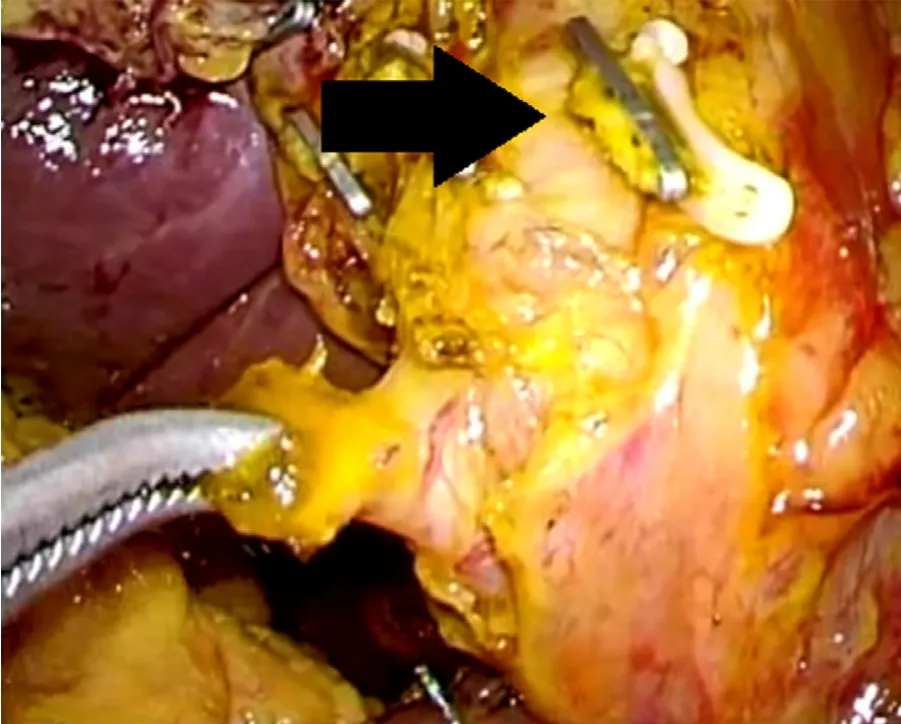
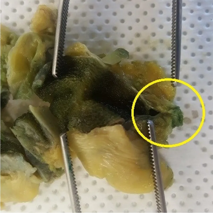
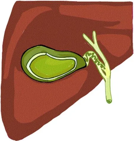
An 82-year-old woman planned for gastrectomy for early gastric cancer also had 1.6 cm-sized gallstone.Although the patient was asymptomatic,laparoscopic cholecystectomy was performed together with laparoscopic distal gastrectomy,considering the possibility of inflammation which can be vulnerable after gastrectomy in the future.The surgery was carried out under laparoscopy,distal gastrectomy was performed and then cholecystectomy.Intraoperatively we identified only one cystic duct and it was safely ligated with clips.Operation was finished without any complication.The day after the operation,it was evident that Jackson-Pratt drain filled with bilous fluids.The patient had no clinical symptoms,physical examination revealed no remarkable findings,and laboratory studies showed normal-ranged white blood cell (WBC)count (5720/μL) and elevated aspartate aminotransferase (147 U/L)and alanine aminotransferase (121 U/L).Re-operation was planned immediately although the patient did not have any symptoms.The second surgery was also done laparoscopically,and we found that the bile leakage was from another cystic duct that was not identified on previous surgery (Fig.1).After identifying the second cystic duct,we ligated it with clip.After the second operation,the drain changed to serous and patient started diet on 4th postoperative day from second surgery and was discharged on 6th day without any complication.The pathology result showed chronic cholecystitis and we found only one opening of cystic duct in the gross description of gallbladder side (Fig.2).Overall,the patient had double cystic ducts joining to form one duct,which then goes into gallbladder,shaped like a reverse-Y (Fig.3).

Fig.1.Bile leakage from a cystic duct (grabbed by instrument),taken during reoperation.Black arrow points to cystic duct ligated on the 1st surgery.
There had only been about 20 cases of double cystic duct in English literatures [1–4].It was found that most of the cases were identified intraoperatively and 3 cases were converted to laparotomy due to bile duct injury [1–4].In this case the surgery was completed laparoscopically but second operation was needed because we could not identify the accessory cystic duct in the first operation.During cholecystectomy,the surgeon should be aware of the possibility of double cystic duct,to prevent possible bile duct injury.The types of double cystic ducts are named and classified according to their configurations into H,Y,and trabecular type,as described in Table 1 .In this case where the double cystic ducts diverge from one cystic duct coming out of gall bladder had not beenreported previously.It figured like the reverse–Y,and we named it“reverse Y-type”.

Table 1 Different type of double cystic ducts.

Fig.2.Gallbladder shows one cystic duct opening (Yellow circle).

Fig.3.Schematic view of“reverse Y type”.
In conclusion,we presented a very first case of a patient with“reverse Y-type”double cystic duct,which had not been introduced yet in literature.Although sometimes it is not easy to get an exact information of anatomic variations of biliary tree,surgeons should bear in mind the possibility of these variations.The whole process of surgery should be carried out with cautions because unintentional injury to these structures can lead to serious complications.Complete mapping and confirmation of these variations before hepatobiliary surgeries are key to successful surgery and somewhat essential to prevent possible complications.
Acknowledgments
None.
CRediT authorship contribution statement
Suhyun Lim:Writing -original draft.Chang Hwan Kim:Data curation,Writing -original draft.Kwang Yeol Paik:Conceptualization,Supervision,Validation,Writing -review &editing.WookKim:Conceptualization,Validation.
Funding
None.
Ethical approval
Not needed.
Competing interest
No benefits in any form have been received or will be received from a commercial party related directly or indirectly to the subject of this article.
 Hepatobiliary & Pancreatic Diseases International2021年2期
- Hepatobiliary & Pancreatic Diseases International的其它文章
- Meetings and Courses
- Novel 3D preclinical model systems with primary human liver cells:Recent progresses,applications and future prospects
- A new nomogram for predicting lymph node positivity in pancreatic cancer
- Comparison of the effects of retrieval balloons and basket catheters for bile duct stone removal on the rate of post-ERCP pancreatitis
- Novel mutation of the TJP2 gene in a Chinese child with progressive cholestatic liver disease coexistent with hearing impairment
- Acute cardiogenic liver injury caused by heart failure in an adolescent
