The characteristic and dynamic electrocardiogram changes on hyperkalemia in a hemodialysis patient with heart failure:a case report
Rui ZHAO, Xin HAO, Fei WANG, Jing FENG, Jing PAN, Ce JING, Ming-Ren MA,Yan LIU, Ling MA,?, Xiao-Qing CAI,?
1. Department of Cardiology, the 940th Hospital of Joint Logistics Support Force of Chinese PLA, Lanzhou, China; 2. VIP Outpatient Department of HMI, the Second Medical Center of Chinese PLA General Hospital, Beijing, China; 3. Department of Nephrology, the 942th Hospital of Joint Logistics Support Force of Chinese PLA, Lanzhou, China; 4. Department of Medical Service, the 940th Hospital of Joint Logistics Support Force of Chinese PLA, Lanzhou, China
Hyperkalemia, usually associated with predisposing chronic diseases as heart failure (HF), severe kidney disease or diabetes mellitus (DM),[1] is defined as an elevation of potassium concentration more than 5.5 mmol/L,[2]at which the potassium homeostasis is disrupted and the following neuromuscular manifestations appeared. Notably, its consequences on cardiac conduction system are potentially lethal, but prone to be overlooked in clinical scenario. The electrocardiogram (ECG) change in hyperkalemia is characteristic and helpful for diagnosis and prognosis, whereas merely appears in less than 50% of patients in studies.[3–5]Herein, we report an interesting case of lethal hyperkalemia presenting dynamic and characteristic ECG change during emergent dialysis.
A 65-year-old male with end-stage renal disease(ESRD) suffered gradually aggravated chest distress and debilitation for one day. He was taken to emergency department, where transient loss of consciousness occurred, accompanied by nausea and vomiting. The patient’s medical history included hypertension, type 2 DM, chronic renal anemia, congestive HF and ESRD undergoing routine hemodialysis for the past two years. However, he ceased hemodialysis over one week due to personal reasons.His medications at the time of admission included metoprolol, amlodipine, isosorbide, acarbose, erythropoietin and sacubitril/valsartan for managing the aforementioned chronic diseases. The physical examination in emergency department showed his blood pressure was merely 70/40 mmHg with a pulse of 70 beats/min under the support of intravenous isoprenaline, and meanwhile the respiration rate was 28 times/min with moderate distress. The cardiac auscultation reflected weak heart sounds. The rapid blood gas analysis showed the pH value of blood as merely 7.012, serum potassium soared to 8.9 mmol/L and lactic acid as high as 8.0 mmol/L,which all indicated disturbed homeostasis and critical condition.
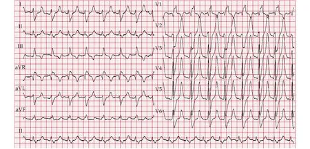
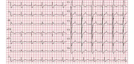
Characteristically, the 12-lead ECG revealed a wide QRS complex rhythm blended with T wave, presented in sine-wave shape, with a rate of 68 beats/min and QRS duration more than 300 ms (Figure 1), which harbingered impending ventricular fibrillation and asystole. In such emergency circumstance, calcium,insulin and glucose were administered intravenously to antagonize the effects of hyperkalemia and promote influx of potassium into cells, as well as sodium bicarbonate for rectifying acidosis. Meanwhile,dopamine and isoprenaline were maintained to facilitate hemodynamic support. Then bedside dialysis in urgency were performed. We monitored the ECG and potassium concentrations during hemodialysis (Figures 2 & 3). Through the dynamic ECG changes from Figure 1 to Figure 3, we could observe the transition from sine-wave shape to narrowbased, peaked T waves in short duration of 250 ms,and finally to the baseline ECG presented as complete right bundle branch block. These characteristics at different time points are listed in Table 1. After dialysis, the brain natriuretic peptide level was 4,551 pg/mL indicating the concurrent congestive HF, while echocardiography showed significant dilation of the whole heart and widened pulmonary trunk, with enlarged left ventricular diastolic diameter as 57 mm and the left ventricular ejection fraction preserved as 59%. Through reviewing the medication list, we reduced the dose of sacubitril/valsartan and metoprolol to minimize influence of serum potassium. After comprehensive management of anemia, volume overload and hyperglycemia, as well as routine hemodialysis, the patient was finally discharged.

Figure 1 Admission electrocardiogram at emergency department showed sinoventricular rhythm in sine-wave shape cause by severe hyperkalemia at 8.9 mmol/L before dialysis.

Figure 2 Electrocardiogram during dialysis delineated peaked T wave, widen QRS complex and minor P wave cause by moderate hyperkalemia at 7.1 mmol/L.

Figure 3 Post-dialysis electrocardiogram showed complete right bundle branch block back to baseline with serum potassium at 4.9 mmol/L in physiologic range.

Table 1 Serum potassium levels, pH value and electrocardiogram characteristics at different time points.
Potassium, as the most important ion for the resting membrane potential, only 2% is extracellular,whereas the other 98% is intracellular.[6]The physiological concentration of serum potassium between 3.5 mmol/L and 5.5 mmol/L is achieved by complex interplay of potassium intake, excretion and cellular shift. Potassium levels are mainly regulated by kidney, except 5%?10% excreted through intestinal canal.[7]Once renal function is impaired, the homeostasis of potassium would be disrupted, thus hyperkalemia occurs. Then not only neuromuscular manifestations as muscle weakness, paresthesia, or paralysis appeared, but also the impaired cardiac conduction may result in lethal arrhythmias or cardiac arrest. Notably, there are two types of hyperkalemia:(1) inherent hyperkalemia caused by chronic kidney diseases, hormonal disorders, DM and diseases with cell membrane instability;[8]and (2) treatment related hyperkalemia induced by medications as herbs (insidious potassium source), renin-angiotensin-aldosterone system inhibitors, mineralocorticoid receptor antagonists, heparin and nonsteroidal antiinflammatory drugs. In this case, the patient had inherent hyperkalemia due to ESRD and DM. Moreover,the treatment related hyperkalemia as sudden cease of hemodialysis and continuity medication of valsartan, surely added insult on injury.
The orderly progression of ECG changes induced by hyperkalemia has been proven in experimental models.[7]Interestingly, the ECG progression has been reversely revealed in this patient from severe to moderate hyperkalemia during urgent dialysis.When the patient firstly in the emergency department with severe potassium at 8.9 mmol/L, the sinoatrial node activity stimulated ventricles directly yet slowly accompanied by repolarization abnormality, thus presented as sinusoidal waves without P waves.[4,9]Due to efficient medication and timely dialysis, the potassium levels regressed to 7.1 mmol/L after two hours. At that period, moderate hyperkalemia may increase myocyte excitability,shorten action potential and promote potassium efflux at plateau phase, resulted in relative narrow QRS and peaked T wave. Then after ten hours dialysis, as serum potassium level got back to physiologic range, the ECG return to the baseline.
Notably, though acute hyperkalemia is a lethal scenario, it can be effectively treated in three ways.Firstly, liquid calcium should be administered to antagonize the cellular effects of hyperkalemia via membrane stabilization effect. Secondly, we can promote cellular influx of potassium by administering insulin and beta-2 adrenoreceptor agonists. Last but not the least, we could remove potassium from the body through dialysis. In this patient, we implemented all three methods to abate hyperkalemia and finally solve the tough scenario. In addition, patiromer (RLY5016) and sodium zirconium cyclosilicate(ZS-9) are new treatments for hyperkalemia, in advantage of normalizing and maintaining potassium levels in HF state.[10,11]
In conclusion, after receiving urgent dialysis and comprehensive management, the patient survived from lethal hyperkalemia and rehabilitated well. It is worth paying special attention that the strength of our case is that characteristic ECG plays a center role in deciding on the urgency of hyperkalemia and type of intervention. Furthermore, the successful treatment experience provides a certain reference value for dynamic ECG monitoring in judgment of appropriateness and promptness of hyperkalemia management.
ACKNOWLEDGMENTS
This study was supported by the Gansu Provincial Health Research Project (GSWSKY2020-01). All authors had no conflicts of interest to disclose.
 Journal of Geriatric Cardiology2022年2期
- Journal of Geriatric Cardiology的其它文章
- Lactobacillus levels and prognosis of patients with acute myocardial infarction
- Usefulness of Impella support in different clinical settings in cardiogenic shock
- Translational proteomics in cardiogenic shock: from benchmark to bedside
- Challenges in the conduct of randomised controlled trials in cardiogenic shock complicating acute myocardial infarction
- Mechanical circulatory support in cardiogenic shock and postmyocardial infarction mechanical complications
- Cardiogenic shock due to left main related myocardial infarction:is revascularization enough?
