Association of Blastocystis hominis with colorectal cancer:A systematic review of in vitro and in vivo evidences
lNTRODUCTlON
(
)is one of the most commonly recovered microorganisms in faecal specimens,and its widespread presence is found in colorectal cancer(CRC)patients[1].Its distribution is known to be prevalent in both rural and urban areas[2].This microorganism has been in discussion since early 1900s[3,4];however,the taxonomic position of
remains unanswered.
treatment is often difficult due to its drug resistance and the failure of the host defenses to counter the infection[5].Previous studies suggest that
is a common and diverse element of microbiota in human host,as it has been highly prevalent in healthy individuals[6-8].
is commonly found in both patients with gastrointestinal symptoms and in healthy people widely across the world.More recently,researchers consider
as an emerging zoonotic disease,and its pathogenic potential in human is somewhat controversial[9].Although accumulating data suggest that
is a pathogen,the pathogenic role in humans is still a matter of debate.
In order to test the truth of his statement about the dogs, he said at once, Salt, I am hungry, and before the words were out of his mouth the dog had disappeared, and returned in a few minutes with a large basket full of the most delicious food
It is suggested that around 20% of cancer reported worldwide could have been due to infectious agents[10,11].Viruses such as hepatitis B virus,human papilloma virus and Epstein-Barr virus have been associated with carcinogenesis.Various other bacteria also have been described previously to exacerbate cancer[12].There are numerous epidemiological evidences that strongly support the fact that parasites can be a factor of various malignant tumours[13],but it is challenging to validate this relationship.Previously,a review article highlighted the correlation of various protozoan parasites including
with carcinogenesis[13].In addition,there was a case report in India that demonstrated a possible association of subtype 3
in the worsening of CRC[14].
There are a few other systematic reviews on the interventional studies done on
,but we did not find any systematic reviews on the association between
and CRC.Therefore,this systematic review aimed to(1)identify prevalence of
in CRC patients;(2)review
colorectal carcinoma cell line study models on the cytopathic and immunological effects of
antigens;and(3)review an
experimental animal model study to investigate the effect of infection with
on exacerbation of colorectal carcinogenesis.
MATERlALS AND METHODS
PRISMA guidelines were utilised in conducting this review[15].
Eligibility criteria
For
studies,the quality of the studies was assessed using Quasi experimental appraisal tool[16],which contained nine questions.For
studies,checklist of Systematic Review Center for Laboratory Animal Experimentation’s Risk of Bias tool for assessing risk of bias as quality assessment was used[17].The quality of all included studies was acceptable.
Exclusion
Articles that reported the association of
with cancer in general without specific findings on its association with CRC,reviews papers,conference proceedings and case reports were excluded from this review.
Search
PubMed,Science Direct,Scopus and Google scholar databases were searched.
If the newspapers, or even the whole world, said of a certain subject, It is so-and-so; and a little schoolboy declared he had learned quite differently, they would take his assertion as the only true one, because he belonged to the family
The various keywords used were:
infection and CRC and their MESH terms and synonyms.Manual searching through reference lists of included journal articles was done to find the missed studies during online search.
Study selection
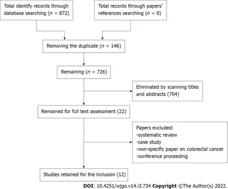
We identified 872 papers in the initial screening(Figure 1).
The storms threwvessel after vessel on the fatal reefs; there were snow-storm andsand-storms; the sand flew up to the houses, blocking the entrances,so that people had to creep up through the chimneys; that wasnothing at all remarkable here
Identification
First,two authors developed a search strategy with different key words and their synonyms.All articles were moved to the Endnote X7 software(Clarivate Analytics,Philadelphia,PA,United States),and 146 duplicate papers were removed.Two authors independently went through the titles and abstracts of the remaining 726 papers.Subsequently,a total of 22 papers were retained for full text review.The eligibility of retained papers was evaluated by two other authors.Authors checked the reference lists of all included articles for any relevant studies that met this systematic review inclusion criteria and had not been found during the database searches.
After looking at the portraits and reading the card, her entire demeanor12 changed. She bounced out of her chair, handed the card to me and commissioned my brothers to hang the paintings facing each other over the fireplace. She stepped back and looked for a long while. With sparkling, tear-filled eyes and a wide smile, she quickly turned and said, I knew Daddy would be with us on Christmas Day!
After full review of the 22 papers,12 were selected.All authors have completed data extraction and agreed eligibility of included papers(Figure 1).

Quality assessment
Original articles that reported the prevalence and association of
subtypes with CRC patients,
studies using
antigen and an
study using an animal model published before February 2,2020 were included.
Data extraction
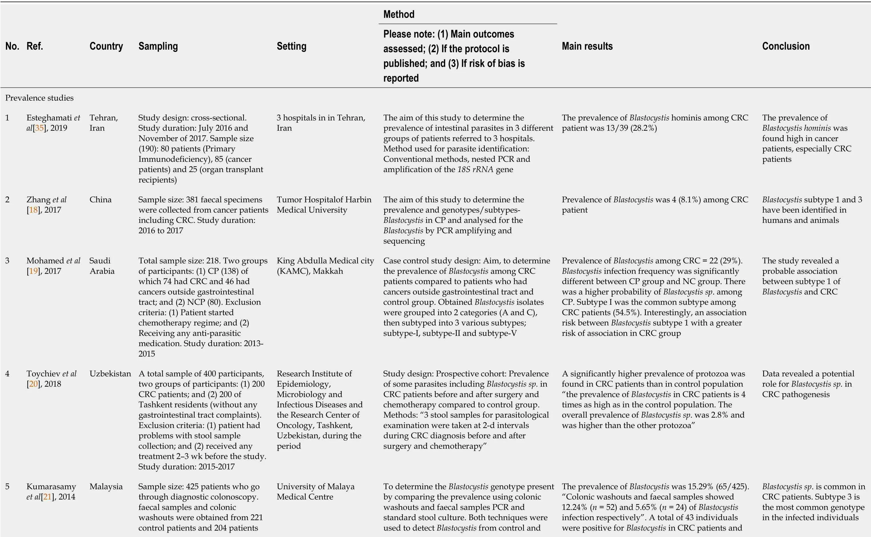
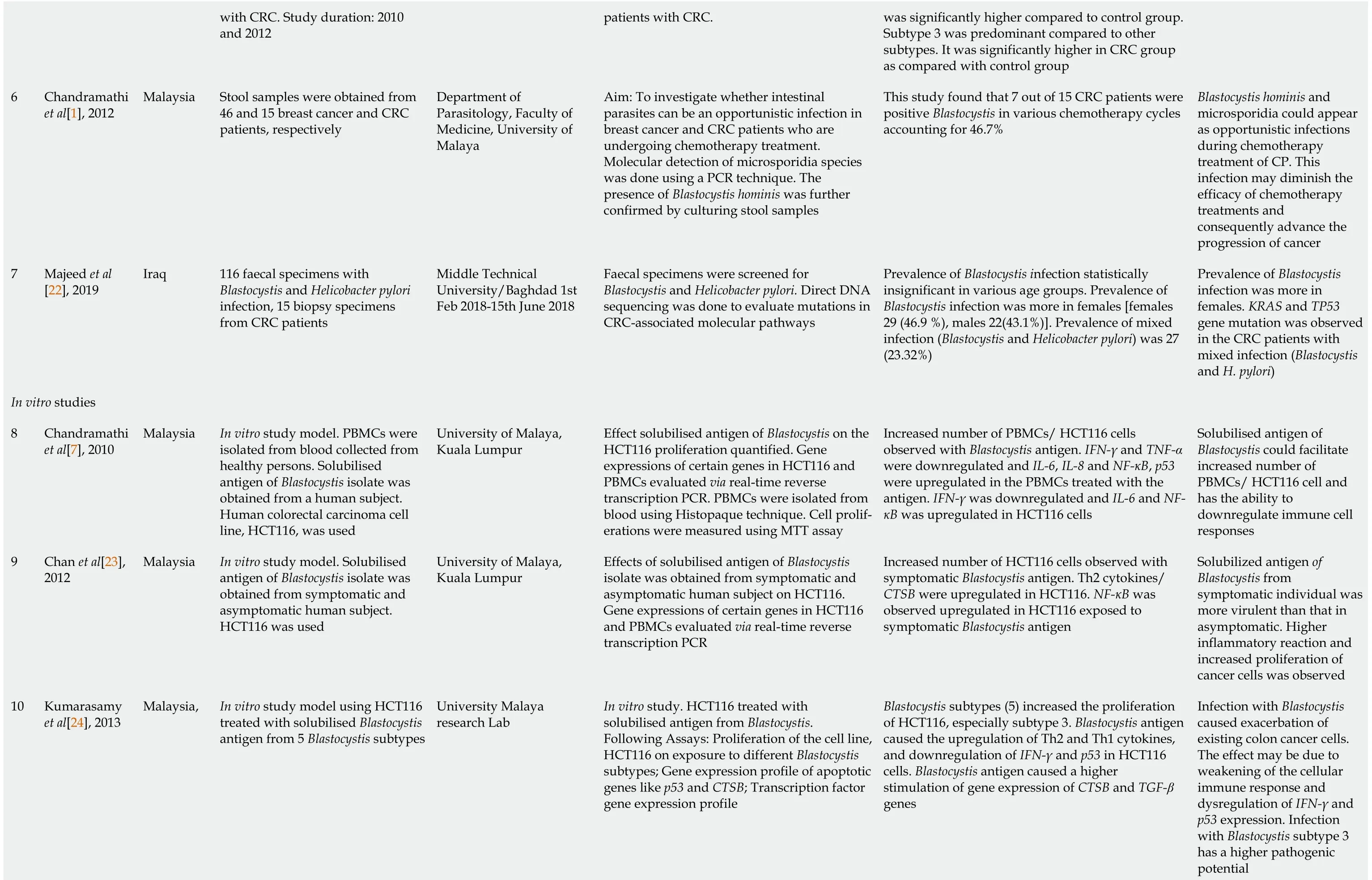
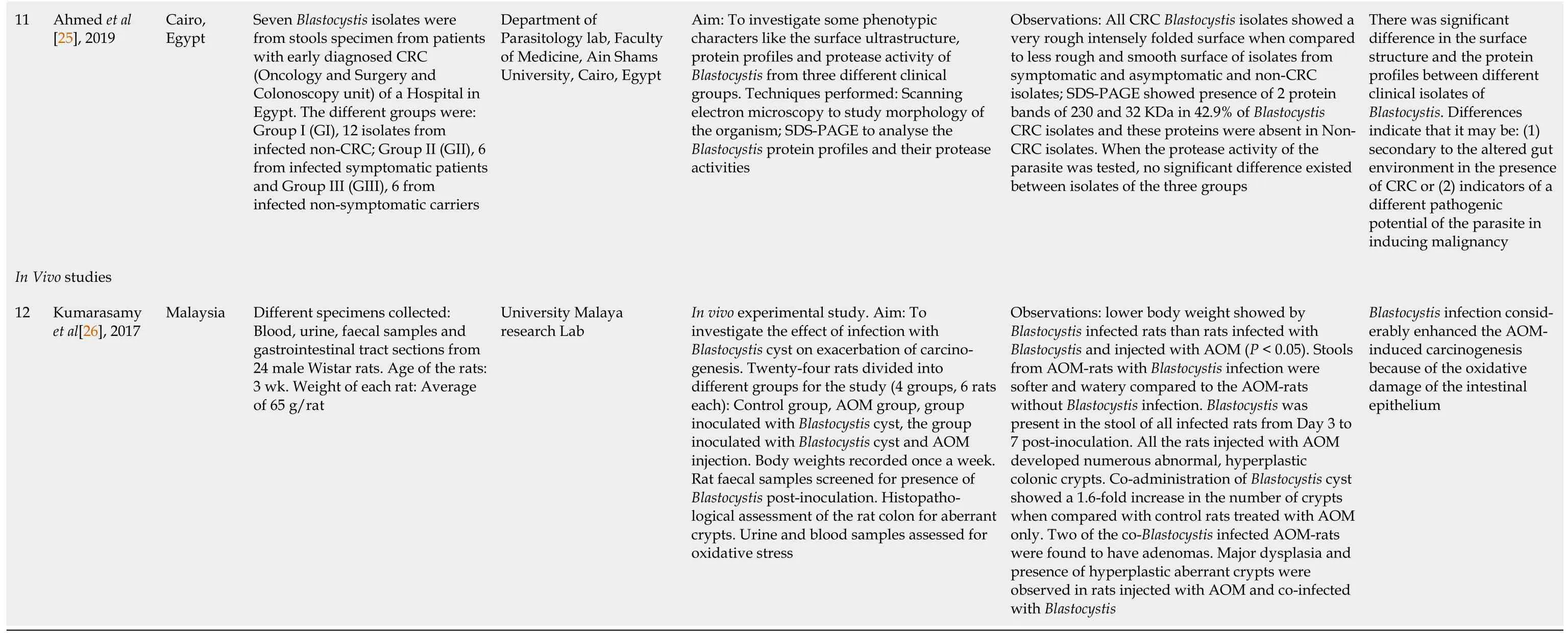
The data extracted included author-year,country,sampling,setting,methods,results and study conclusion(Table 1).


RESULTS
Prevalence of Blastocystis in CRC patients
Based on the seven reviewed articles,the prevalence of
in CRC patients were found to be between 2.8%-46.7%,whereby subtype 1 and subtype 3 were predominantly isolated.
A study by Esteghamati
[35]evaluated the prevalence of
in 85 cancer patients,including 39 CRC and 46 cancers outside gastrointestinal tract(COGT).In this study,
was identified in 11/39(28.2%)among CRC group.Another study in China by Zhang
[18]showed the prevalence of
in 4 among 49(8.1%)patients with CRC.In another study conducted in Saudi Arabia,the prevalence of
among CRC patients was 29.7%[19].Subtype I was thepredominant(54.5%)among CRC patients,while subtype II was predominant(43.7%)among COGT patients[19].Higher prevalence of intestinal helminths and protozoa was observed in CRC patients than in the control population in a study conducted in Uzbekistan.The prevalence of
in CRC patients was four times higher than that in the control population.The overall prevalence of
(2.8%)was significantly higher than the other protozoa[20].In a study by Kumarasamy
[21]in Malaysia,among 221 control patients and 204 CRC patients with colorectal malignancies,the overall prevalence of
infection was 15.29%(65/425).A total of 43(21.08%)samples were positive for
infection in CRC patients and was significantly higher compared to normal individuals(
=22,9.95%,
< 0.01).Subtype 3 was present at higher levels compared to other subtypes detected in both groups and was significantly higher in CRC patients as compared with control patients[21].
Another study sought to investigate some phenotypic characteristics such as the surface ultrastructure,protein profiles and protease activity of
isolated from three different clinical groups:CRC patients,non-CRC symptomatic and asymptomatic infected persons.This study showed the presence of two protein bands of 230 and 32 KDa in 42.9% of
CRC isolates with their complete absence from non-CRC isolates.There was no significant difference in the protease activity of the protein among isolates of the three groups,CRC and Non-CRC
isolates[25].
In vitro colorectal carcinoma cell line studies on the cytopathic and immunological effects ofBlastocystis antigen
Intestinal colonisation of
(
)as a risk factor to the worsening of colorectal cancer(CRC).
The three
model studies used the human colorectal carcinoma cell line HCT116 and the
isolated from a human subject[7,23,24].One of the studies demonstrated the cytopathic effect of
antigen on peripheral blood mononuclear cells.This study findings showed increased cell proliferations in
antigen-stimulated HCT116 cell-lines,which suggested that the infection by
may facilitate the growth of colon cancer cells[7].Another
study showed that the five subtypes of
significantly increased the proliferation of HCT116,especially subtype 3.
antigen caused the upregulation of T helper(Th)2 and Th1 cytokine gene expressions,and downregulation of interferon gamma and
gene expressions in HCT116 cells.In addition,
antigen caused a significantly higher stimulation of Cathepsin B(CTSB)and Transforming growth factor beta(TGF-β)gene expression,which indicates the pathogenic potential of this protozoan[24].Another study showed an increase in cell proliferation in HCT116 cells inoculated with the symptomatic
antigen.Gene expression studies carried out in this research also showed a significant upregulation of Th2 cytokines,which indicates the parasites’ potential in weakening the cellular immune response[23].HCT116 cells exposed to symptomatic and asymptomatic
antigen caused a significant upregulation of
,which led to the postulation that the
antigen may enhance the invasive and metastasis properties of CRC[23].
Another study was designed to investigate the emergence of
and Microsporidia infections in breast and CRC patients undergoing chemotherapy treatment.This study found that 7 out of 15 CRC patients were positive
in various chemotherapy cycles,accounting for 46.7%.However,the researchers did not mention whether the isolate was from the same patients in different cycles[1].In a study carried out in Iraq,stool samples from 116 patients with
and
infections were investigated.Fifteen tissue samples of CRC were taken from 15 suspected patients out of 116 infected cases,and it was shown that the infection with
and
was associated with pathological gene mutation in the CRC patients[22].
In vivo experimental animal model to investigate the effects of infection with Blastocystis on exacerbation of colorectal carcinogenesis
is a commonly identified microorganisms in CRC patients.These studies have provided supportive data that
could exacerbate existing CRC
alteration in host immune response and increased oxidative damage.
DlSCUSSlON
is one of the most common gut microorganisms found in healthy individuals[27].Besides being associated with a healthy gut microbiota[27,28],
infection is also known to be opportunistic in immunocompromised patients[29].CRC is the third most common cancer diagnosed worldwide and one of the major causes of cancer-associated fatality[30].The reason for high mortality is due to the asymptomatic progression of the disease that usually results in late diagnosis[31,32].Certain gut microorganisms are known to be one of the important factors that had been associated with initiation and development of CRC[33].However,data on the roles of parasites are vague and restricted.Various findings have been reported regarding the association between
among CRC patients,whereby positive association was shown in all the studies[1,23,24].Therefore,the aim of this systematic review was to quantify the studies published so far that revealed the direct association of
and CRC.This paper outlines the results of a systematic review to evaluate the prevalence of
in CRC patients,
studies using
antigen and an
study using animal models.
Out of the data extracted from 12 studies relevant to this topic,all the studies showed positive association between
and CRC.Prevalence studies,
investigations and
studies were used to evaluate the pathogenicity of
with CRC.
The global prevalence of
infection ranged from 1.5%-20% in developed countries,which was much less than that in developing countries,which was 30%-50%[34].Based on our review,the prevalence of
infection in CRC patients ranged between 2.8%-46.7%.It has been widely reported in the world,in developing countries such as Iran,China,Saudi Arabia,Uzbekistan and Malaysia[18-20,24,35].The first demonstration of
infection in Iran was reported in 11 CRC patients in Tehran province[35].In Malaysia,a total of 43 samples were positive for
from 204 CRC patients[21].This study utilised colonic washout in addition to stool sample to recover the parasites.Subtype 3
was detected predominantly as compared to other subtypes[21].Some of these findings highlighted the high prevalence of certain subtypes of
among these patients[19,21].A previous study showed that subtype 1 was the most common genotype identified(54.5%)among CRC patients[19].DNA was extracted from
cultures
conventional method and subtyped using multiplex polymerase chain reaction(PCR)with restriction fragment length polymorphism and sequence-tagged site primers-based PCR.In another study,subtype 3 was predominant compared to other subtypes found in both CRC patients and healthy individuals[21].Subtype 3 is speculated as the most pathogenic subtype in symptomatic individuals[36].Some researchers have attributed subtype 3 to be more pathogenic compared to other subtypes[37,38].
was initially screened
culture and conventional PCR using stool samples and colonic washouts.The presence of
infection in CRC patients could be contributed by various reasons including health status.For instance,positive cases were more likely in patients with gastrointestinal symptoms compared to healthy individuals[18].Besides,
was also identified in higher frequency in immunosuppressed CRC patients who were undergoing chemotherapy treatment[1,18].
A total of three
studies were carried out using colorectal carcinoma cell line models to study cytopathic and immunological effects of
antigen[7,23,24].Some of the research studies suggested that solubilised antigen of
could facilitate the exacerbation of CRC cells,HCT116[7,23].In another study by Kumarasamy
[24],
subtype 3 stimulated significantly higher
and
gene expression in HCT116,which indicates the pathogenic potential of this protozoan.Result of
studies that were performed in Malaysia were similar[7,23,24].
is commonly found in both patients with gastrointestinal symptoms and in healthy people widely across the world.More recently,researchers consider
as an emerging zoonotic disease,and its pathogenic potential in human is unclear[9].The pathogenic potential of
was widely debated,as they are found in both symptomatic and asymptomatic patients.The significant expression of nuclear factor kappa light chain enhancer of activated B cells was observed in HCT116 exposed to
antigen isolated from individuals with gastrointestinal symptoms,but such observations were not found when the colon cells treated with
antigen isolated from asymptomatic individuals.This finding shows the potential pathogenicity of symptomatic
in CRC patients[23].Similarly,HCT116 cells exposed to symptomatic and asymptomatic
antigen caused a significant upregulation of
.These gene expression findings lead to a postulation that the
antigen may enhance the invasive and metastasis properties of CRC[23].Besides,proliferation of HCT116 when exposed with
antigen could be a result of higher levels of interleukin(IL)-6 and IL-8 expression[23].
Another study revealed that solubilised antigen isolated from subtype 2 and 3 isolates introduced to colon cancer cells showed significant IL-8 and IL-6 expression[24].The production of inflammatory cytokines such as IL-8 together with reactive oxygen species could contribute to the pathogenesis of cancer[38].In a few studies conducted previously,IL-6 expression was associated with proliferation of colon carcinoma[39,40].Besides that,subtype 3
also triggered positive expression of
in cancer cells.A previous study showed that
expression is significant in CRC patients[41].
Only one study was conducted to investigate the
effect of
in Wistar rats.In parallel with
studies,an
study showed similar findings on the possible role of
to exacerbate CRC.The results demonstrated that
may cause damage to the intestinal mucosal layer and result in increased crypts formation.Furthermore,an increased oxidative stress was also observed in these rats.There have been numerous animal models of human CRC and animal model of tumour carried out
quantification of aberrant crypt foci[42,43].This allows the study of gut microbiome and its role in pathogenesis.In humans,there are many potential pathogenic and nonpathogenic gut microbial infections,and various animal models have been used for such studies.Aberrant crypt foci are known as putative precancerous lesions of the colon in both animal models and humans[44,45].Even though various studies associating
and CRC were carried out
model using colon cancer cells,this study utilised animal model to bridge between
findings in the laboratory and studies in humans.As such,this extensive
study showed that
had a major impact on normal intestinal function in Wistar rats resulting in damage to the intestinal mucosal layer and inducing oxidative stress,which caused increase in crypts formation in AOM-treated rat models.The study establishes that
is a pathogen,and there is a need to screen cancer patients for harbouring this parasite.
This systematic review has some limitations.According to these investigations,a greater prevalence of
was found in CRC patients,but the question whether increased prevalence of
could be linked with increased high risk to CRC is unclear.The studies discussed in this review did not highlight the association of
according to cancer stages,and it was unclear if
itself could result in the initiation of malignancy as
acquisition alone is insufficient for cancer development.Even though strong association between
and CRC is apparent,some questions remain unanswered.Therefore,we propose future studies should focus on the pathogenicity of
in various stages of CRC by concentrating on the molecular pathways involved in tumorigenesis.
CONCLUSlON
In conclusion,according to various recent studies,
is one of the most commonly identified microorganisms in CRC patients,whereby subtype 1 and subtype 3 were predominantly isolated.It is apparent in most cases that the prevalence is higher in developing countries compared to developed countries.These studies have provided supportive data that
could exacerbate existing CRC
alteration in host immune response and increased oxidative damage.An
study wellestablished that
infections resulted in tissue damage from host inflammatory responses that may predispose the host towards neoplasm exacerbation.Upregulation of gene expression responsible for proinflammatory cytokines and downregulation of apoptotic genes was observed in
studies.Through continued research in
and CRC,we may discover new findings as well as develop new effective means of prevention.Future studies of CRC and
should attempt to determine the various stages of CRC that are most likely to be associated with
and its association with other intestinal bacteria.In addition,future
studies should evaluate exposure to various subtypes of
.
ARTlCLE HlGHLlGHTS
Research background
Three of the reviewed articles used
study models and observed considerable cytopathic and immunological effects induced by the solubilised antigen of
to the human colorectal carcinoma cell line[7,23,24].These findings speculated that
infection may enhance the proliferation,invasiveness and metastatic properties of CRC cells.One study investigated some phenotypic characteristics of
isolated from CRC patients[25].
Research motivation
There has been an increase in the prevalence of
in CRC patients.Besides,various
and in vivo studies have highlighted
as an important risk factor for the worsening of CRC.
Over the decades moviegoers have heard the straight version of “Lilli” in dozens of feature films and documentaries. And she is still heard today whenever old soldiers gather to sing of loneliness and loves gone by.
She laid her hands together across her bosom, and then she darted forward as a fish shoots through the water, between the supple79 arms and fingers of the ugly polypi, which were stretched out on each side of her
Research objectives
To perform a systematic review on all evidence on the association between CRC and
Good people, you who are reaping, if you do not tell the King that all this corn belongs to the Marquis of Carabas, you shall be chopped as small as herbs for the pot.
The King45 himself, old as he was, could not help watching her, and telling the Queen softly that it was a long time since he had seen so beautiful and lovely a creature.
Research methods
Future studies of CRC and
should attempt to determine the various stages of CRC that are most likely to be associated with
and its association with other intestinal diseases.
Research results
Out of 12 studies selected for this systematic review,seven studies have confirmed the prevalence of
.A total of four studies employing
human colorectal carcinoma cell line study models showed significant cytopathic and immunological effects of
.One
experimental animal model study showed that there was a significant effect of infection with
on exacerbation of colorectal carcinogenesis.
Research conclusions
An animal model study compared the effects of
infected rats and rats infected with
co-administered with Azoxymethane(AOM),a potent carcinogen.This finding showed that the co-administration of
cyst resulted in a 1.6-fold increase in the number of colonic crypts when compared with control rats treated with AOM only.Two of the co-
infected AOM rats were found to have adenomas.Major dysplasia and the presence of hyperplastic aberrant crypts were also observed in rats injected with AOM and co-infected with
[26].
Research perspectives
A systematic review of the literature was performed by searching PubMed,Science Direct,Scopus and Google scholar databases up to February 2020.
FOOTNOTES
Azzani M designed the research,performed the literature search and extracted the data;Kumarasamy V wrote the discussion and extracted the data;Atroosh WM wrote the methodology and extracted the data;Anbazhagan D wrote the results and extracted the data;Abdalla M wrote the introduction and extracted the data;All authors read and approved the final manuscript.
It doesn t matter when. I m sure the Addisons are nice people, but I m not going to waste an evening socializing with people who don t have any eligible3 daughters.
Dr.Azzani has nothing to disclose.
The authors have read the PRISMA 2009 Checklist,and the manuscript was prepared and revised according to the PRISMA 2009 Checklist.
This article is an open-access article that was selected by an in-house editor and fully peer-reviewed by external reviewers.It is distributed in accordance with the Creative Commons Attribution NonCommercial(CC BYNC 4.0)license,which permits others to distribute,remix,adapt,build upon this work non-commercially,and license their derivative works on different terms,provided the original work is properly cited and the use is noncommercial.See:https://creativecommons.org/Licenses/by-nc/4.0/
With arms outstretched and hands clutching the broomstick inserted through the sleeves of an old plaid shirt, I wore a felt hat and faded overalls stuffed with straw
Malaysia
Vinoth Kumarasamy 0000-0002-8895-1796;Wahib Mohammed Atroosh 0000-0003-3809-3460;Deepa Anbazhagan 0000-0002-2527-4126;Mona Mohamed Ibrahim Abdalla 0000-0002-4987-9517;Meram Azzani 0000-0002-7939-1539.
Chang KL
Filipodia
So early one morning with his pole in his hand, he crept past the red barn on McFeeglebee s land out to the edge of the grove to the pond, where he baited his hook, sinking it deep. Then Georgie P. Johnson fell asleep.
You can go, said the Lieutenant, but I don t think it will be worth it. Your friend is probably dead and you may throw your own life away. The Lieutenant s words didn t matter, and the soldier went anyway.
Chang KL
1 Chandramathi S,Suresh K,Anita ZB,Kuppusamy UR.Infections of Blastocystis hominis and microsporidia in cancer patients:are they opportunistic?
2012;106:267-269[PMID:22340948 DOI:10.1016/j.trstmh.2011.12.008]
2 Aksoy U,Akisü C,Bayram-Deliba? S,Ozko? S,Sahin S,Usluca S.Demographic status and prevalence of intestinal parasitic infections in schoolchildren in Izmir,Turkey.
2007;49:278-282[PMID:17990581]
3 Brumpt E.
n.sp.et formes voisines.
1912;5:725-730
4 Low GC.Two Chronic Amoebic Dysentery Carriers treated by Emetine,with some Remarks on the Treatment of Lamblia,Blastocystis and E.
Infections.
1916;19:29-34
5 Roberts T,Ellis J,Harkness J,Marriott D,Stark D.Treatment failure in patients with chronic Blastocystis infection.
2014;63:252-257[PMID:24243286 DOI:10.1099/jmm.0.065508-0]
6 Borralho PM,Kren BT,Castro RE,da Silva IB,Steer CJ,Rodrigues CM.MicroRNA-143 reduces viability and increases sensitivity to 5-fluorouracil in HCT116 human colorectal cancer cells.
2009;276:6689-6700[PMID:19843160 DOI:10.1111/j.1742-4658.2009.07383.x]
7 Chandramathi S,Suresh K,Kuppusamy UR.Solubilized antigen of Blastocystis hominis facilitates the growth of human colorectal cancer cells,HCT116.
2010;106:941-945[PMID:20165878 DOI:10.1007/s00436-010-1764-7]
8 Cirioni O,Giacometti A,Drenaggi D,Ancarani F,Scalise G.Prevalence and clinical relevance of Blastocystis hominis in diverse patient cohorts.
1999;15:389-393[PMID:10414382 DOI:10.1023/a:1007551218671]
9 Basak S,Rajurkar MN,Mallick SK.Detection of Blastocystis hominis:a controversial human pathogen.
2014;113:261-265[PMID:24169810 DOI:10.1007/s00436-013-3652-4]
10 Bouvard V,Baan R,Straif K,Grosse Y,Secretan B,El Ghissassi F,Benbrahim-Tallaa L,Guha N,Freeman C,Galichet L,Cogliano V;WHO International Agency for Research on Cancer Monograph Working Group.A review of human carcinogens--Part B:biological agents.
2009;10:321-322[PMID:19350698 DOI:10.1016/s1470-2045(09)70096-8]
11 zur Hausen H.Streptococcus bovis:causal or incidental involvement in cancer of the colon?
2006;119:xi-xii[PMID:16947772 DOI:10.1002/ijc.22314]
12 Lax AJ.Opinion:Bacterial toxins and cancer--a case to answer?
2005;3:343-349[PMID:15806096 DOI:10.1038/nrmicro1130]
13 El-Gayar EK,Mahmoud MM.Do protozoa play a role in carcinogenesis?
2014;7:80[DOI:10.4103/1687-7942.149553]
14 Padukone S,Mandal J,Parija SC.Severe
subtype 3 infection in a patient with colorectal cancer.
2017;7:122-124[PMID:29114493 DOI:10.4103/tp.TP_87_15]
15 Moher D,Liberati A,Tetzlaff J,Altman DG;PRISMA Group.Preferred reporting items for systematic reviews and metaanalyses:the PRISMA statement.
2009;6:e1000097[PMID:19621072 DOI:10.1371/journal.pmed.1000097]
16 (JBI)TJBI.Critical Appraisal Tools.[cited 1 January 2021].Available from:http://joannabriggs.org/research/criticalappraisal-tools.html
17 Hooijmans CR,Rovers MM,de Vries RB,Leenaars M,Ritskes-Hoitinga M,Langendam MW.SYRCLE's risk of bias tool for animal studies.
2014;14:43[PMID:24667063 DOI:10.1186/1471-2288-14-43]
18 Zhang W,Ren G,Zhao W,Yang Z,Shen Y,Sun Y,Liu A,Cao J.Genotyping of
and Subtyping of
in Cancer Patients:Relationship to Diarrhea and Assessment of Zoonotic Transmission.
2017;8:1835[PMID:28983297 DOI:10.3389/fmicb.2017.01835]
19 Mohamed AM,Ahmed MA,Ahmed SA,Al-Semany SA,Alghamdi SS,Zaglool DA.Predominance and association risk of
subtype I in colorectal cancer:a case control study.
2017;12:21[PMID:28413436 DOI:10.1186/s13027-017-0131-z]
20 Toychiev A,Abdujapparov S,Imamov A,Navruzov B,Davis N,Badalova N,Osipova S.Intestinal helminths and protozoan infections in patients with colorectal cancer:prevalence and possible association with cancer pathogenesis.
2018;117:3715-3723[PMID:30220046 DOI:10.1007/s00436-018-6070-9]
21 Kumarasamy V,Roslani AC,Rani KU,Kumar Govind S.Advantage of using colonic washouts for Blastocystis detection in colorectal cancer patients.
2014;7:162[PMID:24708637 DOI:10.1186/1756-3305-7-162]
22 Majeed GH,Mohammed NS,Muhsen SS.The synergistic effect of Blastocystis hominis and H.pylori in Iraqi colorectal cancer patients.
2019;11:523-526
23 Chan KH,Chandramathi S,Suresh K,Chua KH,Kuppusamy UR.Effects of symptomatic and asymptomatic isolates of Blastocystis hominis on colorectal cancer cell line,HCT116.
2012;110:2475-2480[PMID:22278727 DOI:10.1007/s00436-011-2788-3]
24 Kumarasamy V,Kuppusamy UR,Samudi C,Kumar S.Blastocystis sp.subtype 3 triggers higher proliferation of human colorectal cancer cells,HCT116.
2013;112:3551-3555[PMID:23933809 DOI:10.1007/s00436-013-3538-5]
25 Ahmed MM,Habib FSM,Saad GA,El Naggar HM.Surface ultrastructure,protein profile and zymography of
species isolated from patients with colorectal carcinoma.
2019;43:294-303[PMID:31263336 DOI:10.1007/s12639-019-01092-9]
26 Kumarasamy V,Kuppusamy UR,Jayalakshmi P,Samudi C,Ragavan ND,Kumar S.Exacerbation of colon carcinogenesis by Blastocystis sp.
2017;12:e0183097[PMID:28859095 DOI:10.1371/journal.pone.0183097]
27 Scanlan PD,Stensvold CR,Rajili?-Stojanovi? M,Heilig HG,De Vos WM,O'Toole PW,Cotter PD.The microbial eukaryote Blastocystis is a prevalent and diverse member of the healthy human gut microbiota.
2014;90:326-330[PMID:25077936 DOI:10.1111/1574-6941.12396]
28 Audebert C,Even G,Cian A;Blastocystis Investigation Group,Loywick A,Merlin S,Viscogliosi E,Chabé M.Colonization with the enteric protozoa Blastocystis is associated with increased diversity of human gut bacterial microbiota.
2016;6:25255[PMID:27147260 DOI:10.1038/srep25255]
29 Bednarska M,Jankowska I,Pawelas A,Piwczyńska K,Bajer A,Wolska-Ku?nierz B,Wielopolska M,Welc-Fal?ciak R.Prevalence of Cryptosporidium,Blastocystis,and other opportunistic infections in patients with primary and acquired immunodeficiency.
2018;117:2869-2879[PMID:29946765 DOI:10.1007/s00436-018-5976-6]
30 Favoriti P,Carbone G,Greco M,Pirozzi F,Pirozzi RE,Corcione F.Worldwide burden of colorectal cancer:a review.
2016;68:7-11[PMID:27067591 DOI:10.1007/s13304-016-0359-y]
31 O'Connell JB,Maggard MA,Liu JH,Etzioni DA,Ko CY.Are survival rates different for young and older patients with rectal cancer?
2004;47:2064-2069[PMID:15657655 DOI:10.1007/s10350-004-0738-1]
32 Siegel RL,Ward EM,Jemal A.Trends in colorectal cancer incidence rates in the United States by tumor location and stage,1992-2008.
2012;21:411-416[PMID:22219318 DOI:10.1158/1055-9965.EPI-11-1020]
33 Zhu Q,Gao R,Wu W,Qin H.The role of gut microbiota in the pathogenesis of colorectal cancer.
2013;34:1285-1300[PMID:23397545 DOI:10.1007/s13277-013-0684-4]
34 Su FH,Chu FY,Li CY,Tang HF,Lin YS,Peng YJ,Su YM,Lee SD.Blastocystis hominis infection in long-term care facilities in Taiwan:prevalence and associated clinical factors.
2009;105:1007-1013[PMID:19488784 DOI:10.1007/s00436-009-1509-7]
35 Esteghamati A,Khanaliha K,Bokharaei-Salim F,Sayyahfar S,Ghaderipour M.Prevalence of Intestinal Parasitic Infection in Cancer,Organ Transplant and Primary Immunodeficiency Patients in Tehran,Iran.
2019;20:495-501[PMID:30803212 DOI:10.31557/APJCP.2019.20.2.495]
36 Souppart L,Sanciu G,Cian A,Wawrzyniak I,Delbac F,Capron M,Dei-Cas E,Boorom K,Delhaes L,Viscogliosi E.Molecular epidemiology of human Blastocystis isolates in France.
2009;105:413-421[PMID:19290540 DOI:10.1007/s00436-009-1398-9]
37 Ragavan ND,Govind SK,Chye TT,Mahadeva S.Phenotypic variation in Blastocystis sp.ST3.
2014;7:404[PMID:25174569 DOI:10.1186/1756-3305-7-404]
38 Rajamanikam A,Hooi HS,Kudva M,Samudi C,Kumar S.Resistance towards metronidazole in Blastocystis sp.:A pathogenic consequence.
2019;14:e0212542[PMID:30794628 DOI:10.1371/journal.pone.0212542]
39 Chung YC,Chang YF.Serum interleukin-6 Levels reflect the disease status of colorectal cancer.
2003;83:222-226[PMID:12884234 DOI:10.1002/jso.10269]
40 Galizia G,Orditura M,Romano C,Lieto E,Castellano P,Pelosio L,Imperatore V,Catalano G,Pignatelli C,De Vita F.Prognostic significance of circulating IL-10 and IL-6 serum levels in colon cancer patients undergoing surgery.
2002;102:169-178[PMID:11846459 DOI:10.1006/clim.2001.5163]
41 Herszényi L,Farinati F,Cardin R,István G,Molnár LD,Hritz I,De Paoli M,Plebani M,Tulassay Z.Tumor marker utility and prognostic relevance of cathepsin B,cathepsin L,urokinase-type plasminogen activator,plasminogen activator inhibitor type-1,CEA and CA 19-9 in colorectal cancer.
2008;8:194[PMID:18616803 DOI:10.1186/1471-2407-8-194]
42 Kim J,Ng J,Arozulllah A,Ewing R,Llor X,Carroll RE,Benya RV.Aberrant crypt focus size predicts distal polyp histopathology.
2008;17:1155-1162[PMID:18483337 DOI:10.1158/1055-9965.EPI-07-2731]
43 Reddy BS.Studies with the azoxymethane-rat preclinical model for assessing colon tumor development and chemoprevention.
2004;44:26-35[PMID:15199544 DOI:10.1002/em.20026]
44 Bird RP.Observation and quantification of aberrant crypts in the murine colon treated with a colon carcinogen:preliminary findings.
1987;37:147-151[PMID:3677050 DOI:10.1016/0304-3835(87)90157-1]
45 Roncucci L,Stamp D,Medline A,Cullen JB,Bruce WR.Identification and quantification of aberrant crypt foci and microadenomas in the human colon.
1991;22:287-294[PMID:1706308 DOI:10.1016/0046-8177(91)90163-j]
 World Journal of Gastrointestinal Oncology2022年3期
- World Journal of Gastrointestinal Oncology的其它文章
- Re:Association between intestinal neoplasms and celiac disease -beyond celiac disease and more
- Clinical efficacy and prognostic risk factors of endoscopic radiofrequency ablation for gastric low-grade intraepithelial neoplasia
- Pancreatic head vs pancreatic body/tail cancer:Are they different?
- Computed tomography-based radiomic to predict resectability in locally advanced pancreatic cancer treated with chemotherapy and radiotherapy
- Cost-effective low-coverage whole-genome sequencing assay for the risk stratification of gastric cancer
- RNA-Seq profiling of circular RNAs in human colorectal cancer 5-fluorouracil resistance and potential biomarkers
