Malignant biliary obstruction due to metastatic non-hepato-pancreato-biliary cancer
Takeshi Okamoto
Abstract Malignant biliary obstruction generally results from primary malignancies of the pancreatic head, bile duct, gallbladder, liver, and ampulla of Vater. Metastatic lesions from other primaries to these organs or nearby lymph nodes are rarer causes of biliary obstruction. The most common primaries include renal cancer,lung cancer, gastric cancer, colorectal cancer, breast cancer, lymphoma, and melanoma. They may be difficult to differentiate from primary hepato-pancreatobiliary cancer based on imaging studies, or even on biopsy. There is also no consensus on the optimal method of treatment, including the feasibility and effectiveness of endoscopic intervention or surgery. A thorough review of the literature on pancreato-biliary metastases and malignant biliary obstruction due to metastatic non-hepato-pancreato-biliary cancer is presented. The diagnostic modality and clinical characteristics may differ significantly depending on the type of primary cancer. Different primaries also cause malignant biliary obstruction in different ways, including direct invasion, pancreatic or biliary metastasis, hilar lymph node metastasis, liver metastasis, and peritoneal carcinomatosis. Metastasectomy may hold promise for some types of pancreato-biliary metastases. This review aims to elucidate the current knowledge in this area,which has received sparse attention in the past. The aging population, advances in diagnostic imaging, and improved treatment options may lead to an increase in these rare occurrences going forward.
Key Words: Bile duct obstruction; Obstructive jaundice; Pancreas; Metastasis; Endoscopic retrograde cholangiopancreatography
lNTRODUCTlON
Metastatic lesions from other primaries to the pancreatic head, biliary tree, liver, ampulla of Vater, and hilar or peripancreatic lymph nodes have the potential to cause biliary obstruction. The pancreas and biliary tree are rare destinations for metastases. However, such metastases can result from almost every primary, from brain tumor to melanoma of the toe.
Of all the precious wives in the whole wide world, how did Mark get so lucky to marry the best one? He married you, Michelle, and he is so lucky! I am so proud of you, my little girl.
In a multicenter analysis of 159 patients with pancreatic metastases, the most common primaries were renal cell carcinoma (38 %), lung cancer (2 %), colorectal cancer (11 %), and sarcoma (6 %)[1 ]. About 41 %were isolated metastases, 78 % were single masses, and were more or less uniformly distributed throughout the pancreas. About 24 % were symptomatic and only ten cases presented with jaundice.Two-thirds were diagnosed by endoscopic ultrasound-fine-needle aspiration (EUS-FNA) and most of the remainder were diagnosed by surgery. Median overall survival was 43 mo, with extrapancreatic metastasis, tumor-related symptoms at diagnosis, and pathology of primary tumors independently predicting survival. In a literature review of 234 subjects, synchronous presentation with the primary tumor and surgical resection were important prognostic factors in addition to tumor-related symptoms at diagnosis and pathology of primary tumors[2 ]. There were marked differences in the studied subjects:68 % had renal cell carcinoma, 86 % were isolated metastases, 79 % involved the pancreatic head, and 62 %were symptomatic at diagnosis, but extrapancreatic metastases were only observed in 14 % of cases. As may be expected, metastases from lung cancer and melanoma had worse prognoses than those from renal cell carcinoma.
Autopsy studies report extremely high rates of pancreatic metastases. An autopsy database study found 81 pancreatic metastases among only 190 pancreatic tumors (43 %). Major primaries included the lung (42 %), gastrointestinal tract (25 %), kidney (5 %), and breast (4 %)[3 ]. A Japanese autopsy study found 103 cases of pancreatic metastases among 690 cadavers with malignant tumors, implying that 15 % of all metastatic malignancies involve the pancreas[4 ]. Specifically, pancreatic metastases were found in 35 % of gastric cancers, 25 % of bladder cancers, 21 % of ovarian cancers, 15 % of lung cancers,and 9 % of renal cell carcinomas and breast cancers. One-third of pancreatic metastases were not visible macroscopically. About 17 % involved the pancreasviadirect invasion rather than distant metastasis.
Metastases to the bile duct are extremely rare. There are isolated case reports of from primaries including ovarian cancer, colon cancer, rectal cancer, esophageal cancer, gallbladder cancer, hepatocellular carcinoma, breast cancer, and malignant melanoma[5 -13 ].
Herein, hepato-pancreato-biliary metastases from non-hepato-pancreato-biliary malignancies and resulting malignant biliary obstruction (MBO) are discussed. Characteristics of each primary are discussed after a brief overview of diagnosis and treatment of obstructive metastases.
DlAGONSlS
Diagnostic imaging
Computed tomography (CT) is the most common imaging modality used for cancer follow-up. One study found pancreatic metastases in 0 .3 % of 6623 patients followed up for various malignancies[14 ].An analysis of 192 cases in seven imaging studies of pancreatic metastases from various primaries reveals that the most common primaries were renal cell carcinoma (30 %), lung (26 %), stomach/colon(13 %), and breast (10 %) (Table 1 )[14 -20 ]. Of these, 66 % were solitary masses, 27 % had multiple nodules,and 7 % had diffuse involvement. Isolated pancreatic metastases were observed in 5 %-33 % of cases.Pancreatic metastases were found synchronously with the primary malignancy in 22 %-44 % of cases. A mass in the pancreatic head was found in 11 %-29 % of cases, but only 5 %-6 % had biliary dilation.Pancreatic duct dilatation was observed in 11 %-22 % of cases, with pancreatitis rarely observed. CA19 -9 was elevated in 8 %-28 % of cases, but most primaries of such cases were gastrointestinal and may havebeen unrelated to pancreatic involvement.

Table 1 Computed tomography findings of pancreatic metastases
CT characteristics of metastatic lesions were generally concordant with the primary lesions, except some cases of gastric cancer[19 ]. Metastases from renal cell carcinoma are generally hypervascular with well-defined margins, while most other metastases are hypoattenuating with unclear borders, similar to primary adenocarcinoma[14 -19 ]. In a comparison with primary pancreatic adenocarcinoma, metastases had significantly less pancreatic duct dilatation, vascular involvement, parenchymal atrophy, or peripancreatic fluid[20 ]. Biliary dilatation was also less frequently observed, possibly due to the small number of metastases located in the pancreatic head in the study.
In summary, most metastases present as solitary masses or multiple nodules on CT and generally do not cause symptoms or biliary obstruction. Renal cell carcinoma metastases may be identified by hypervascular, well-defined lesions, while the absence of pancreatic duct dilatation or parenchymal atrophy may be clues for metastatic disease in general.
When the fish were all taken out, he fetched the net which he had laid out to dry, folded it up very small, and ran down to the river, hoping that he might find a place narrow enough for him to jump over; but he soon saw that it was too wide for even the best jumper in the world
Studies on magnetic resonance imaging (MRI) and fluorodeoxyglucose-positron emission tomography/CT (FDG-PET/CT) for diagnosing pancreatic metastases are limited. MRI of metastases of renal cell carcinoma may also be distinguished from primary adenocarcinoma due to their vascularity[21 ]. One FDG-PET/CT study of 26 lesions in 19 patients found no difference in maximum standardized uptake values between pancreatic metastases and primary pancreatic adenocarcinoma[22 ]. However,several isodense intrapancreatic nodules missed on contrast CT were discovered by FDG-PET/CT.FDG-PET/CT may also be useful in detecting other unsuspected distant metastases.
Endoscopic ultrasound
Endoscopic ultrasound (EUS) may also provide clues to identify metastatic disease. In a study comparing 28 pancreatic metastases (23 solitary, four multiple, and one diffuse lesion) to 60 cases of pancreatic adenocarcinoma, Hjiiokaet al[23 ] reported that main pancreatic duct dilatation was also observed significantly more frequently in pancreatic adenocarcinoma than in metastatic lesions. Regular borders and absence of retention cysts predicted pancreatic metastases.
In a small study of contrast-enhanced harmonic EUS, pancreatic metastases from renal cell carcinoma and lymphoma were hyperenhancing, while most other metastases were hypoenhancing[24 ]. While both contrast-enhanced harmonic EUS and elastography may assist in differentiating malignant lesions from benign lesions, there is insufficient evidence in their ability to differentiate metastases from primary adenocarcinoma[25 ,26 ].
Fine-needle aspiration
Pancreatic metastatic accounted for 0 .9 %-2 .5 % of EUS-FNA samples of the pancreas and 4 .7 %-7 .2 % of pancreatic malignancies[27 -29 ]. These figures may be higher than those of other modalities because lesions with typical findings of pancreatic adenocarcinoma may be resected without EUS-FNA. In the largest review of 582 pancreatic metastases diagnosed by EUS-FNA, renal cell carcinoma (34 %), lung cancer (15 %), colon cancer (10 %), melanoma (7 %), and breast cancer (6 %) were the most common primaries[28 ].
EUS-FNA is a safe and well-accepted method of diagnosing such metastatic lesions. EUS-FNA for pancreatic metastases and primary adenocarcinoma have reported sensitivities of 75 %-94 % and 75 %-84 %, respectively, and specificities of 60 %-100 % and 97 %-100 %, respectively[30 -33 ]. No significant complications including tumor seeding of the needle tract have been reported in EUS-FNA of pancreatic metastases[34 ]. Immunohistochemistry andKRASmutation analysis can be crucial in reaching the final diagnosis[23 ].
TREATMENT
Endoscopic biliary drainage
In studies of endoscopic biliary drainage which include metastases from non-hepato-pancreato-biliary primaries, metastases are not necessarily a rare cause of MBO. Metastases were the cause of biliary obstruction in 14 % of 1346 patients in 14 such studies[35 -49 ]. The pooled average across six studies limited to distal MBO (n= 395 ) was 9 %, while that of three studies limited to hilar MBO (n = 256 ) was 22 %[40 -48 ]. Gastric cancer (including lymph node metastases) was the cause of 80 % of MBO across four studies focused on surgically altered anatomies[49 -52 ]. MBO due to metastases from primaries with better prognoses than primary pancreato-biliary cancer may be able to better generally tolerate and benefit from endoscopic retrograde cholangiopancreatography (ERCP), although they may not be candidates for surgical resection. These figures may be overstated, as some reports which include no metastatic cases did not intentionally exclude them. Selection bias may also be a factor; most reports were from university-affiliated tertiary care centers or cancer institutes.
Studies focused exclusively on biliary drainage for metastatic cancer are mainly limited to those on percutaneous drainage[53 ,54 ]. One study on 93 patients achieved clinical success in 73 % of patients and found that survival differed significantly depending on the primary tumor[54 ]. Subsequent chemotherapy prolonged median survival from 1 to 5 mo, with the greatest benefit observed in the 28 cases with colorectal cancer. Another study with 42 cases each of gastric and colorectal cancer found that performance status and absence of peritoneal metastases were associated with longer survival[55 ].
There is only one study on prognostic factors after ERCP stenting for biliary drainage in metastatic cancers of various origins[56 ]. Colorectal cancer was the most common primary (25 %), and there was only one case of renal cell carcinoma. Technical success was achieved in 91 .7 % of patients and 67 % of successfully cases received subsequent treatment. Performance status and treatment after drainage were independent predictors of overall survival.
One study on metallic stent placement in hilar MBO reported that metastatic disease from other primaries was an independent risk factor for technical failure[57 ]. The study only included five patients,of which one had pancreatic cancer. The authors reported that the extrinsic nature of the biliary stricture necessitated multiple procedures, including percutaneous drainage. Larger studies have failed to reproduce this result. EUS-guided intervention has also been reported with success in hilar lymph node or hepatic metastases from colorectal, breast, gastric, urogenital, and anal cancer[57 ]. With respect to endoscopic biliary drainage, it appears acceptable to approach MBO due to metastases in a similar manner as MBO from primary hepato-pancreato-biliary cancer.
Surgery
Metastasectomy accounted for 1 .4 % of 5745 pancreatic resections across six studies which provided total figures (range: 0 .7 %-3 .1 %)[58 -63 ]. A review of 399 metastasectomies found that renal cell carcinoma accounted for 62 .6 % of cases[64 ]. Forty percent had symptoms at presentation and, in a separate review,22 % had jaundice[64 ,65 ]. About 10 % were found synchronously with the primary tumor[64 ]. Median survival was 50 .2 mo after surgery overall, but 71 .7 mo for renal cell carcinoma. On the other hand,perioperative mortality was observed in 2 .2 % of all metastasectomies. Patients with isolated pancreatic metastases had better prognoses than those with other metastases (45 mo vs 26 mo). In the separate review[65 ], renal cell carcinoma patients had a long post-surgical median survival of 105 mo, compared to 54 mo for colon cancer, 40 mo for sarcoma, 34 mo for ovarian cancer, 26 mo for breast cancer, 14 mo for melanoma, and 6 mo for lung cancer. On the other hand, pooled analysis showed long 5 -year postsurgical survival of 61 .1 % for isolated pancreatic metastases (of which 74 % were renal cell carcinomas)and 58 .9 % for local invasion from colon or gastric cancer[66 ].
A study of 98 metastasectomies found that old age, non-renal cell carcinomas, vascular invasion, and positive margins were independently associated with increased mortality risk[67 ]. Resection can generally be considered for isolated metastases. Many reports suggest long-term survival in renal cell carcinoma. Symptomatic relief may also be achieved; all colorectal cancer patients experienced symptomatic relief after metastasectomy in a surgical review[68 ]. While data is lacking for other primaries, metastasectomy should only be attempted when margin-negative resection can be expected.As melanoma and lung cancer patients have poor prognosis even after surgery, indication for metastasectomy should be considered with caution.
PRlMARY MALlGNANClES
Renal cell carcinoma
Renal cell carcinoma is the most common primary for pancreatic metastases in CT, EUS-FNA, and surgical series. When such metastases occur, they are discovered synchronously with primary renal cell carcinoma in 7 % of cases[69 ]. There is generally a long time lag of up to 32 years between nephrectomy and pancreatic metastases. Most have multiple extrapancreatic metastases. About half of isolated metastases occur in the pancreatic head. One study suggested a predilection for fatty pancreas[70 ].
Counterintuitively, many reports note that renal cell carcinoma patients with pancreatic metastases have better prognoses than those without, even when there are concomitant extrapancreatic metastases[69 ,71 -73 ]. In one study, median overall survival with and without pancreatic metastases were 39 and 23 mo, respectively[73 ]. The size of isolated metastases, the number of metastatic pancreatic lesions, and time interval from nephrectomy to pancreatic metastasis had no impact on survival[69 ]. The laterality of the primary renal cancer has no impact on the portion of the pancreas affected. Affinity of indolent types of renal cell carcinomas to the pancreas has been suggested, with characteristic genetic mutations,high sensitivity to antiangiogenic treatment and resistance to immune check point inhibitors[69 ,74 ]. The extreme rarity of metastases to the biliary tree may also provide support for this affinity[75 ,76 ].
It should be noted that none of the studies reported pathological confirmation of lymph node metastases.
With that voice inside your head, tell your finger how much you love it and how much you need it. I could see Timmy s little face focused in deep concentration. Tell your finger that you need it to dial the phone. I paused, watching his little lips silently repeat my words. And to write your sentences in school. I paused again so he could say the words after me. And tell it how much you need it to point at things. I waited for another moment and then continued, Now just say, grow for me finger, grow. I love you and I need you.
Despite the large number of reports on pancreatic metastases, there is a surprisingly small number of reports of biliary obstruction. Only two of 307 ERCP cases in eight studies involved metastases from renal cell carcinoma. There are isolated reports of MBO from ampullary or bile duct involvement[79 ,80 ].Simultaneous gastric outlet obstruction can rarely occur[80 ].
About 80 % of pancreatic metastases are resectable[81 ]. While there are no direct comparative studies,surgery for both single and multiple metastases to the pancreas is generally considered safe and associated with long-term survival[82 -84 ]. A systematic review showed that extrapancreatic disease was a risk factor for recurrence after surgery but had no impact on survival[85 ]. However, it is not clear whether surgery is required. There was no significant difference in overall survival between tyrosine kinase inhibitors and surgery in one study[86 ]. Another study reported that the existence of pancreatic metastasis did not affect survival in patients treated with first-line tyrosine kinase inhibitors[87 ].Radiotherapy was also effective in a small case series[88 ].
Lung cancer
There are ten reports of squamous cell lung cancer metastasizing to the pancreas[105 ]. All were single lesions, 67 % involved the pancreatic head, 50 % had biliary or pancreatic duct dilation, and 30 % had obstructive jaundice. There are two reports of lung adenocarcinoma metastasizing to the common bile duct, both causing biliary obstruction[106 ,107 ].
Small cell lung cancer presents initially with increased serum bilirubin in about 10 % of cases, caused by hepatic (6 %) or pancreatic (4 %) metastases[94 ]. Obstructive jaundice due to hilar lymph node compression from metastatic small cell lung carcinoma has also been reported[95 ]. Most reports of metastasis-induced acute pancreatitis come from small cell lung cancer or gastric cancer metastasizing to the pancreatic head, with isolated reports of squamous cell lung cancer, adrenocortical carcinoma,and breast cancer[96 -99 ]. Twenty-six cases from small cell lung cancer have been reported to date[100 ].While initially estimated to occur in 0 .12 % of small cell lung cancer cases, the figure may be higher as 14 cases were reported from a single institution[96 ,101 ]. Chemotherapy may provide a survival benefit[96 ].
At least 23 reports of primary bile duct small cell carcinoma have been reported[102 ]. While there are no reports of small cell lung cancer metastasizing to the bile duct, there are more than ten reports of biliary obstruction as the initial presentation of small cell lung cancer[103 ]. In one study, 4 .0 % of small cell lung cancers presented initially with extrahepatic obstruction due to pancreatic metastases, while another 5 .6 % had jaundice due to hepatic metastases[104 ]. While there was good overall response to chemotherapy, the latter group had a worse prognosis.
Lung cancer is the leading cause of cancer death worldwide[89 ]. About 57 % have metastatic disease at diagnosis[90 ]. About 3 % of lung cancer patients develop pancreatic metastases[91 ]. The frequency of pancreatic metastases depends on the histological subtype, occurring most commonly in small cell carcinoma (10 %), followed by adenocarcinoma (2 .4 %), large cell carcinoma (1 .9 %), and squamous cell carcinoma (1 .1 %)[92 ]. Most cases were asymptomatic[93 ]. A majority of pancreatic metastases present as solitary lesions (73 %) but can also be multiple (12 %) or diffuse (15 %). Concomitant liver and adrenal metastases are observed in 73 % and 69 % of cases, respectively.
Seventeen cases of duodenal metastases from lung cancer have been reported, caused by adenocarcinoma (47 %), squamous cell (29 %), small cell (12 %) or large cell (12 %) cancers and arising in all four parts of the duodenum[108 ,109 ]. MBO occurred from two metastases occupying the second part(squamous cell and adenocarcinoma), both managed endoscopically[109 ,110 ].
CT and MRI may not be contributory in differentiating metastases from primary adenocarcinoma, or in hyperenhancing lesions, neuroendocrine tumors[105 ]. FDG-PET/CT found abnormal pancreatic accumulations in 1 .6 %-2 .3 % of lung cancer cases, discovering metastases from adenocarcinoma and small and large cell lung cancer as small as 6 mm[111 ]. All had metastases to at least one other organ.While one patient had acute pancreatitis, none developed obstructive jaundice.
As with metastases from other primaries, diagnosis can be made by EUS-FNA[112 ]. Thyroid transcription factor-1 can aid in differentiating pancreatic metastases from primary pancreatic adenocarcinoma and small cell lung cancer metastases from primary small cell biliary cancer[102 ,113 ,114 ].KRASG12 C mutations and napsin A are also associated with lung adenocarcinoma, while KRAS G12 R mutations, CK20 , and CDX2 support the diagnosis of pancreatic adenocarcinoma[114 ].
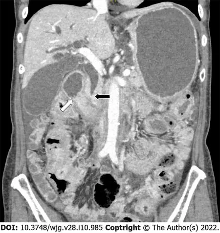
ERCP has acceptable technical success and adverse event rates, offering a chance for long-term survival if chemotherapy can be resumed[99 ,162 ]. Concomitant duodenal strictures may preclude ERCP in 18 % of cases and may be treated using EUS-guided interventions (Figure 2 )[99 ]. When resectable,metastatic breast cancer to the pancreas have a median survival of 26 mo[2 ]. While cases series have suggested potential for improved survival[163 ], there is insufficient data to determine whether solitary metastatic lesions to the pancreas should be resected. Survival in MBO with extensive metastases may average only 2 mo with palliative care alone[99 ].
Gastric cancer
Most gastric cancers occur in the non-cardia and are commonly associated withHelicobacter pyloriinfection, leading to geographic differences in incidence. Liver metastases are observed in almost half of metastatic gastric cancer patients[118 ]. There are only 11 reports of pancreatic metastases, mostly resulting from moderately or poorly differentiated adenocarcinoma[119 ].
All night long he tossed about, and awoke the next morning in a high fever.17 The queen, who had no other child, and lived in a state of perpetual anxiety about this one, at once gave him up for lost, and indeed his sudden illness puzzled the greatest doctors, who tried the usual remedies in vain. At last they told the queen that some secret sorrow must be at the bottom of all this, and she threw herself on her knees beside her son’s bed, and implored31 him to confide32 his trouble to her. If it was ambition to be king, his father would gladly resign the cares of the crown, and suffer him to reign33 in his stead; or, if it was love, everything should be sacrificed to get for him the wife he desired, even if she were daughter of a king with whom the country was at war at present!
Obstructive jaundice arises most commonly from extrinsic lymph node compression, followed by intraductal metastases which present as band-like wall thickening with enhancement on contrast CT,much like primary cancer of the bile duct[120 -122 ]. A minority arise from direct tumor invasion,extrinsic compression from liver metastases, or peritoneal carcinomatosis. Most result from Borrmann type 3 (63 %-72 %) or type 2 (21 %-24 %) adenocarcinomas with antral involvement (60 %-98 %), after total or partial gastrectomy (79 %-89 %)[121 ,123 ]. Histological composition of the primary tumors varies significantly across studies, with differentiated adenocarcinomas accounting for 9 %-90 % of total cases[121 ,123 ,124 ]. Obstructive jaundice occurs in 1 .4 %-2 .3 % of post-operative patients, with a median interval of 10 -15 mo after surgery[125 ,126 ].
The beauties no longer pleased him, their witty33 speeches had ceased to amuse; and indeed, for their parts, they found the Prince far less amiable34 than of yore, and were not sorry when he declared that, after all, a country life suited him best, and went back to the Leafy Palace
The blade was always bright and clear; each time she looked she had the happiness of knowing that all was well, till one evening, tired and anxious, as she frequently was at the end of the day, she took it from its drawer, and behold15! the blade was red with blood
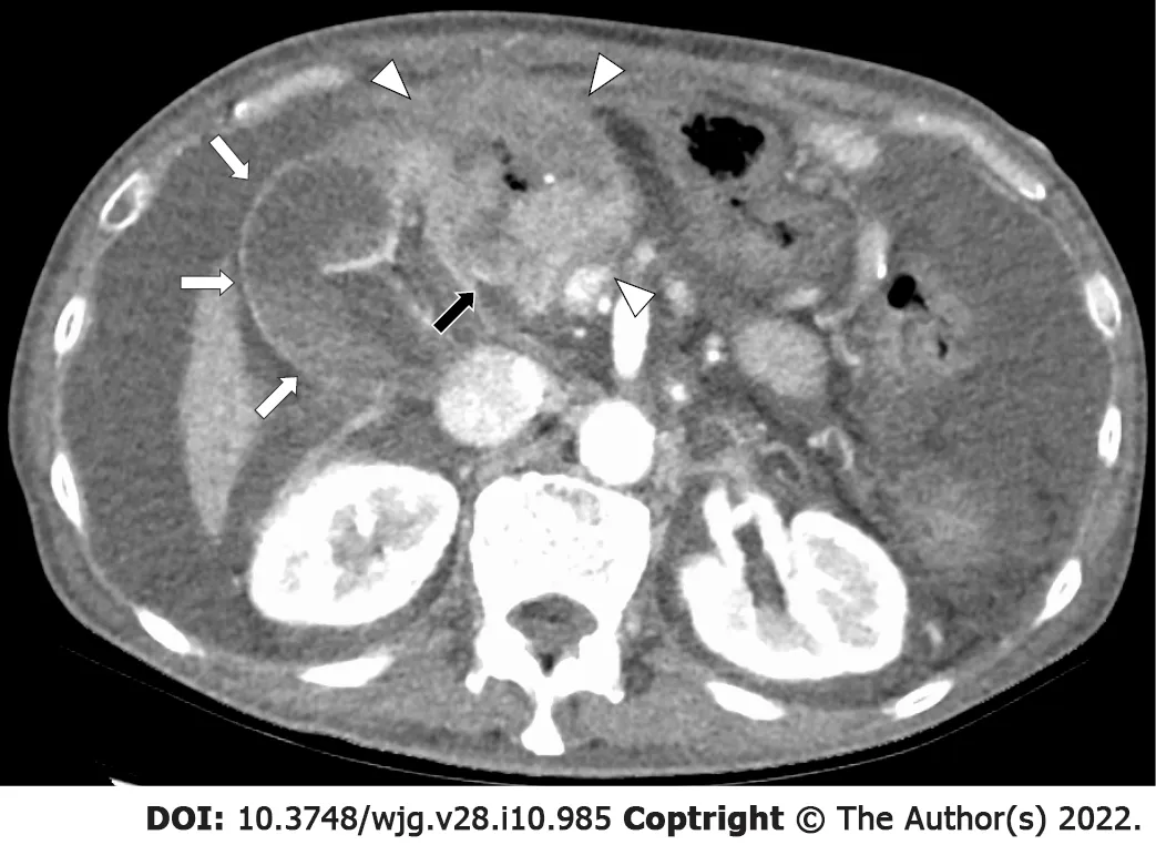
Drainage for MBO due to gastric cancer can involve two major issues: surgically altered anatomy and concomitant gastric outlet obstruction (GOO). As most affected patients have undergone total or partial gastrectomy, approaching the papilla can be extremely difficult (Figure 1 ). While percutaneous biliary drainage has largely been successful, poor prognostic factors after drainage include significant liver metastases, hilar strictures and high carbohydrate antigen (CA) 19 -9 [127 ,128 ]. The main cause of stent occlusion was sludge buildup (13 %) in one study, with no cases of tumor ingrowth[122 ]. When ERCP is contraindicated, EUS-guided biliary drainage was comparable to percutaneous drainage in terms of both technical success rate and stent patency period[129 ]. As in the case of post-operative pancreatic cancer patients, ERCP and stent placement using a double-balloon enteroscope has also been reported for this purpose[49 ,52 ,130 ]. Technical success was reported in all 26 gastric cancer cases across two studies[49 ,52 ]. Median time to recurrent biliary obstruction for seven cases in one study was 7 .4 mo,with two cases of tumor ingrowth and one case of mucosal hyperplasia[49 ].
The clerk disappeared and came back a moment later with three robes in sturdy terry cloth. He chose blindly, hardly glancing down, taking the one on top. Three sizes, the clerk was saying, and a better selection of colors next month, but he was already in the aisle24, a coral-colored robe draped over his arm, his shoes squeaking28 on the tiles as he moved impatiently between the other shoppers to where she stood.
After hepatobiliary cancers, gastric cancer is the most common cause of combined MBO and GOO,accounting for 4 %-8 % of such cases[131 -133 ]. Other rare cases include colon, breast, and renal cancer[131 ]. The extrahepatic bile duct and first or second parts of the duodenum are most commonly involved. Regardless of the primary site, MBO tends to precede GOO. Double stenting of the duodenum and bile duct has success rates approaching 100 %, with only rare reports of post-procedural pancreatitis[133 ,134 ]. Metallic stents tended to have longer patency than plastic stents, while more adverse events resulted from EUS-guided drainage when compared with ERCP[133 ].

Figure 1 A 69 -year-old man presented with obstructive jaundice due to recurrence 18 mo after distal gastrectomy and Roux-en-Y reconstruction for gastric cancer. A recurrent mass with central necrosis (white arrowheads) obstructed the extrahepatic bile duct (black arrow), causing dilatation of intrahepatic bile ducts and gallbladder (white arrows). While endoscopic ultrasound-guided hepaticogastrostomy led to symptomatic relief, the patient died 1 mo later.
Colorectal cancer
Almost half of colorectal cancer patients experience metastatic disease. Metastases to the liver ultimately occur in 25 %-30 % of affected patients, of which only about 25 % can be resected[135 -137 ]. Liver metastases tend to occur more commonly in left-sided cancers and in relatively young patients[137 ].Other reported sites of metastases which may cause biliary obstruction include lymph nodes, pancreas,peritoneum, and the extrahepatic bile duct[138 ,139 ].
Cytokeratin (CK) 7 negativity and CK20 positivity in EUS-FNA specimens aid in differentiating from primary pancreato-biliary adenocarcinoma, which is generally CK positive and CK20 negative[140 ].However, some types of primary pancreatic adenocarcinoma such as the colloid type may be CK7 negative/CK20 positive, complicating the differential diagnosis[30 ].
Biliary obstruction is associated with poor outcomes not only because it reflects widespread disease,but also because of chemotherapy cannot be performed at the desired dose. One study found jaundice in about 10 % of metastatic colorectal cancer patients due to liver metastases (53 %) or metastatic lymph nodes (47 %)[141 ]. Endoscopic or percutaneous biliary drainage was only successful in 42 % of cases with median overall survival of 1 .5 mo, which improved to 9 .6 mo when chemotherapy could be restarted.The study was unable to identify predictors of drainage failure, although drainage was attempted less often in cases with hilar involvement. In another study[142 ], biliary drainage mostly by ERCP was successful in about 65 % of cases, allowing 70 % of successful cases to restart chemotherapy and improving median survival from 33 to 262 d. A study on both colorectal (n = 32 ) and gastric cancers (n=60 ) found that multiple hepatic metastases and hilar strictures were associated with unsuccessful percutaneous drainage, while poor performance status, multiple liver metastases, ascites, history of treatment with multiple chemotherapy regimens, undifferentiated carcinoma, and high CA 19 -9 Levels were associated with poor prognosis[127 ].
A characteristic almost unique to colorectal cancer is the ability to spread along epithelial surfaces and grow intraductally, mimicking neoplasms of the lung, bladder, or intrahepatic bile ducts[143 ]. Liver metastases of colorectal cancer can exhibit intrabiliary extension in 3 .6 %-10 .6 % of cases, compared to 0 .7 %-1 .9 % of metastases from other primaries[144 ]. Such phenomena are most commonly observed in well-differentiated adenocarcinomas originating in the rectosigmoid[145 ,146 ]. Microscopic intrabiliary extension has been reported in up to 40 .6 % of liver metastases from colorectal cancer[145 ]. There are also reports of intrabiliary extension from hepatocellular carcinoma as well as liver metastases of neuroendocrine tumor, gastrointestinal tumor, and invasive lobular breast cancer[144 ,147 ]. Of the other reports on liver metastases, only the neuroendocrine tumor case presented signs of biliary obstruction[144 ].
Two patterns of intrabiliary growth were identified by Estreallaet al[144 ]: Bile duct colonization which replace the biliary epithelium and tumor plugs which may or may not affect the biliary epithelium. Such growth was limited to intrahepatic ducts in 72 % of cases, while the remainder involved the hilum. Laboratory, imaging, and histological abnormalities suggesting biliary obstruction were all positively associated with the degree of biliary growth. While intrabiliary extension may mimic intrahepatic cholangiocarcinoma on imaging studies, they can generally be differentiated on immunohistochemistry with CK7 and CK20 [140 ]. In contrast with liver metastases from other primaries, biliary biopsy may be useful in achieving a preoperative diagnosis[138 ]. Intrabiliary extension is paradoxically associated with a better prognosis, possibly because it occurs more frequently in well-differentiated adenocarcinomas which have longer survival periods[145 ,146 ].
As isolated liver and lung metastasectomy have been established in colorectal cancer, resection of isolated pancreatic metastases may also be justifiable in selected patients. A review of 24 studies revealed that out of 37 colorectal cancer cases undergoing pancreatic resection, 19 had disease recurrence, with median survival of 21 mo[68 ]. In addition, all studied patients experienced symptomatic relief after surgery, which lasted until recurrence of cancer.
Breast cancer
Approximately 6 % of breast cancer are metastatic at diagnosis in developed countries, and 20 %-30 %eventually develop metastases[148 ]. Most common sites of metastases are bone, lung, liver and brain[149 ,150 ]. Jaundice is found in 6 %-25 % of breast cancer patients with liver metastases, generally resulting from hepatic failure rather than MBO[151 ]. Some studies suggest worse prognosis when jaundice is present, most likely because chemotherapy must be reduced or discontinued as a result[152 ]. Liver metastases occur in about 5 % of all breast cancer patients and 32 % of metastatic cases[152 ,153 ].
While about 80 % of invasive breast cancers are invasive ductal carcinomas, lobular carcinoma is the most common histological subtype observed in gastrointestinal metastases[154 ,155 ]. Pancreatic metastases are found in 5 %-13 % of autopsies of breast cancer patients[156 ,157 ]. In a review of 24 cases of periampullary breast cancer metastases, MBO was observed at initial diagnosis in five cases, with metachronous MBO cases occurring 1 -23 years later[158 ]. Our institution reported eleven cases of obstructive jaundice due to metastatic breast carcinoma, resulting from metastases to hilar or peripancreatic lymph nodes (36 %), pancreas (27 %), liver (18 %), gallbladder (9 %), and peritoneal carcinomatosis(9 %)[99 ]. MBO due to metastases to the duodenum have also been reported[159 ].
Metastases can be difficult to differentiate from primary pancreatic cancer, both clinically and radiologically[28 ,147 ,155 ,160 ]. CA15 -3 elevation may suggest breast cancer metastasis in some cases. As breast cancers are adenocarcinomas, diagnosis by EUS-FNA may require immunohistochemical analysis with monoclonal antibodies such as gross cystic disease fluid protein-15 [161 ].KRASpoint mutation analysis has also been shown to be useful[23 ].
In addition to ERCP for MBO and standard treatment for metastatic lung cancer, one study found a survival benefit in resecting pancreatic metastases, with a median survival of 29 mo for curative intent resectionvs8 mo for palliative surgery or medical management[115 -117 ].
Malignant melanoma and Merkel cell carcinoma
Metastatic melanoma has a poor prognosis with median survival of about 8 mo[164 ]. Visceral metastases, particularly when multiple, are associated with poor survival[165 ]. Pancreato-biliary involvement and obstructive jaundice from malignant melanoma can take various forms: primary malignant melanoma of the biliary tract, melanoma of unknown primary arising in the pancreas,pancreato-biliary metastasis, and bile duct compression from hepatic or lymph node metastases.
Autopsy studies on malignant melanoma have shown metastases to the liver in 54 %-88 %, intestines in 26 %-58 %, and pancreas in 38 %-53 % of cases, although discovery rates in the clinical setting are much lower[166 ]. It should be noted that primaries as well as metastases may not be black, as amelanotic melanoma accounts for up to 27 .5 % of melanoma cases[167 ].
Primary malignant melanoma of the biliary tract is a rare type of mucosal melanoma which can only be diagnosed after excluding primaries in other locations including the skin, eye, and gastrointestinal tract. There are 13 reports in the literature, of which 12 presented with jaundice[168 ]. It tends to present in relatively young males as black, polypoid lesions exhibiting endoluminal growth. Most cases arise in the common bile duct, but can involve intrahepatic bile ducts or the gallbladder. Surgery is the treatment of choice, with a good prognosis if complete resection is achieved. One case was successfully treated with immunotherapy.
Isolated metastases to the pancreas are relatively common. While findings on imaging are generally non-specific, isolated reports describe multiple hypoechoic nodules with hyperchoic septa and central necrosis on EUS[169 ,170 ]. T2 -weighted MRI may help differentiate pancreatic melanoma, which tends to be hypointense, from pancreatic adenocarcinoma, which tends to be hyperintense[171 ]. If melanoma can be raised in the differential diagnosis, such lesions can be diagnosed by EUS-FNA with the help of immnohistochemical markers such as Human Melanoma Black 45 and Melan A[169 ,170 ]. Large analyses suggest a survival benefit for surgical resection[172 ,173 ]. Additional support comes from Woodet al[174 ], who resected eight cases of melanoma metastases to the pancreas and achieved curative resection in six, of which three survived for over five years.
Melanoma of unknown primary, characterized by metastases in lymph nodes or other areas where primary lesions are unlikely to arise without evidence of a separate primary lesion, accounts for 2 .2 %-3 .2 % of malignant melanomas[172 ,173 ,175 ]. There are ten reports of isolated pancreatic melanoma with no other lesions. There is no consensus on whether they can be considered primary pancreatic melanoma. They are often resected surgically, justified based on the reports of isolated pancreatic metastases described above[172 -175 ].
37.Plaited in a crown: Plaits, also known as braids, can be created in various styles. The most common interweaves three sections of hair into a thicker, stronger rope of hair. In many cultures, young girls would wear their braids down, while women would wear their hair pinned up as a sign of maturity.Return to place in story.

Figure 2 A 62 -year-old woman presented with jaundice, nausea, and vomiting 13 years after partial mastectomy for breast cancer. Biopsyproven duodenal metastases caused both bile duct (black arrow) and duodenal (white arrow) obstruction. Double stenting led to temporary symptomatic relief. The patient subsequently opted for palliative care.
There are at least 18 reports of metastatic melanoma to the common bile duct[176 ]. Painless obstructive jaundice is the usual finding, and prognosis is dismal unless curative resection can be achieved. Five cases of ampullary metastasis causing obstructive jaundice have also been reported, all requiring endoscopic drainage or surgery[177 -181 ].
Merkel cell carcinoma is another type of aggressive skin cancer which has a high recurrence rate after resection. About one-third of Merkel cell carcinoma patients eventually develop metastases, of which 8 % develop pancreatic metastases[182 ]. It can be diagnosed by EUS-FNA and may cause obstructive jaundice when located in the pancreatic head[183 ]. While a relatively rare disease, the reported incidence is increasing. A high index of suspicion is required for patients with a history of resection and a thorough skin examination should be considered.
Soft tissue sarcoma
Soft tissue sarcomas are mesenchymal tumors which account for less than 1 % of all cancers, but are extremely heterogeneous with over 75 subtypes[184 ]. Metastatic soft tissue sarcomas are generally refractory to chemotherapy and have median survival of less than one year. There are over 50 reports of pancreatic metastases from various types of soft tissue sarcoma, leiomyosarcoma being the most reported subtype[185 -188 ]. Diagnosis was achieved by fine-needle aspiration in 20 cases, of which 11 were conducted by EUS-FNA[189 ].
In a CT study of 13 leiomyosarcoma cases with pancreatic metastases, 85 % were women and primaries were mostly located in the uterus (39 %), retroperitoneum (31 %), or extremities (23 %).Pancreatic metastases developed after a median interval of 24 mo after diagnosis, 38 % were multiple,46 % contained necrosis, and 77 % were hypovascular in the arterial phase[185 ]. While 69 % had pancreatic head involvement, lesions were small (1 .0 -3 .5 cm) and biliary and pancreatic duct obstruction were only observed in one patient each. Among 27 unique cases contained in two literature reviews,only two of twelve cases with pancreatic head metastases had jaundice[187 ,188 ].
There are over 50 cases of primary pancreatic leiomyosarcoma, which is the most common type of pancreatic stromal tumor and occurs most frequently in middle-aged females[189 ]. Cystic features may lead to misdiagnoses as pseudocysts or cystic neoplasms and to false-negative EUS-FNA results. There are also 23 reports of primary pancreatic carcinosarcoma as well as reports of various subtypes of sarcoma originating in the bile duct as well as the ampulla of Vater[190 -194 ]. More than half of pancreatic carcinosarcomas occur in the pancreatic head, of which about half cause obstructive jaundice[190 ]. Thus, sarcoma should be included in the differential diagnosis of isolated pancreato-biliary tumors even in the absence of a coexisting primary tumor.
Surgery has been performed for both primary and metastatic sarcoma arising in the pancreas and duodenum[195 ,196 ]. The largest case series describes seven out of 17 existing reports of resection for pancreatic metastases, with one postoperative death and recurrence in all other six cases despite margin-free resection in four cases, with a median survival of 21 mo[195 ]. The benefits of resection therefore remain unclear.
Now listen towhat I tell you! You can snore; you are snoring the whole night, and Ihardly a quarter of an hour! And the blood rose to the head of theexcited criminal; he threw himself upon his comrade, and beat him with his clenced fist in the face
Lymphoma
35.The forest: Woods or forests frequently appear as settings in fairy tales. Forests symbolize the female principle, the unconscious, danger, mistakes, problems, fertility, and enchantment68. They often serve as the homes for outlaws69 such as in the Robin70 Hood71 legend, fairies, or supernatural beings (Olderr 1986).Return to place in story.
NHL involves the pancreas secondarily in over 30 % of cases[201 ]. Primary pancreatic lymphoma is rare, accounting for less than 5 % of extranodal NHL[202 ,203 ]. While pancreatic lymphoma causes jaundice in up to 42 % of cases, biliary and pancreatic ducts may remain unaffected by even large lesions in the pancreatic head[201 ,204 ]. Clues favoring lymphoma over pancreatic cancer in imaging studies include absence of calcifications, patency of involved vessels and ducts, poor but homogenous contrast enhancement, and either a well-delineated mass or diffuse involvement[201 ].
There are about 30 cases of primary lymphoma of the common bile duct[205 ]. They generally present with obstructive jaundice but often have smooth strictures with negative findings in ERCP brushing cytology or biopsy. One report found 36 reports of primary gallbladder lymphomas, which are less likely to cause MBO than their bile duct counterparts[206 ]. There are at least 15 reports of primary duodenal lymphoma of various B-cell and T-cell subtypes which presented with obstructive jaundice[207 ,208 ]. Three were drained percutaneously and four were treated by ERCP.
MBO from lymphoma usually results from extrinsic compression, although several cases of direct bile duct invasion have been reported[209 ]. Results of biliary drainage vary significantly across studies, due to the small sample size in each. Rosset al[197 ] reported technical success in 84 % of biliary drainage by ERCP (and 100 % of cases without concomitant gastric outlet obstruction due to lymphoma). They recommend plastic stents as strictures in patients presenting initially with MBO resolve with treatment before stent exchange is necessary, while those who develop MBO later in the disease progression do not survive until their first stent exchange is due. Stent-free status was achieved in about one-third of all patients. On the other hand, none of eight patients achieved stent-free status in another study[198 ]. A third study reported that MBO resolved in all seven patients presenting initially with MBO, regardless of stent placement[200 ]. Most studies agree that initial presentation with MBO has a much better prognosis than those who develop MBO later on. Those initially presenting with jaundice had significantly improved survival after biliary drainage (21 mo vs 5 mo)[197 ]. While surgery is an option when a preoperative diagnosis cannot be reached or the lesion appears resectable, chemotherapy and/or involved site radiation therapy is generally considered the standard of care[210 ].
Vasilissa rose and went at once to the Palace, and as soon as the Tsar saw her, he fell in love with her with all his soul. He took her by her white hand and made her sit beside him. Beautiful maiden, he said, never will I part from thee and thou shalt be my wife.
NHL and Kaposi’s sarcoma should also be included in the differential for biliary obstruction in acquired immunodeficiency syndrome (AIDS) patients, alongside AIDS-related cholangiopathy. Both show hepatic or splenic involvement in about 15 % of AIDS patients[211 ]. AIDS-related NHL more commonly causes MBO by extrinsic compression from lymph nodes or liver involvement rather than from a primary biliary lesion[212 ]. Primary pancreatic lymphoma is more common in AIDS patients than in the general population, accounting for about 5 % of extranodal NHL cases[211 ]. Hepatic or hilar involvement of Kaposi’s sarcoma, which can occur earlier in the disease course of AIDS, is an even rarer cause of MBO[213 ].
Leukemia
There are only several reports of leukemia presenting as obstructive jaundice[214 -219 ]. The liver, bile duct, pancreas, or lymph nodes can be involved. Eight of 103 pancreatic metastases found in an autopsy study were cause by leukemia, suggesting that microscopic involvement may not be so rare[4 ]. Most reports in both adults and children involve acute lymphocytic leukemia. Obstructive jaundice was the presenting symptom in one case[215 ]. Care is required as obstructive lesions may not be visible on imaging[214 ]. While endoscopic and surgical treatment may be options to relieve biliary obstruction,aggressive chemotherapy is the standard of care.
Lymph node metastases
Hilar and peripancreatic lymph node metastases are a well-known cause of MBO. Such metastases have received little attention despite their frequency, perhaps because they are common occurrences in hepato-pancreato-biliary cancer. Hilar and distal biliary obstruction were caused by metastatic lymph nodes in 23 % and 2 %-17 % (pooled average of 551 patients across eight studies: 11 %) of cases treated mainly by ERCP stenting, respectively[220 -228 ]. This figure may be higher for biliary obstruction in surgically altered anatomies; one report found six cases among 13 patients with surgically altered anatomies treated with metallic biliary stents (46 %)[49 ].
In a study of biliary drainage mainlyviaERCP, biliary obstruction due to hilar lymph node metastases resulted mainly from colon (46 %), gastric (14 %), and breast (14 %) cancers[229 ]. Clinical success was achieved in 86 % of cases but required a median of three procedures and percutaneous drainage in 20 % of cases. The ability to resume chemotherapy was associated with improved survival in colon and breast cancer patients. A study of 65 patients with distal MBO due to lymph node metastases from gastric (31 %), colorectal (18 %), lung (11 %), breast (8 %), and other cancers found that covered metallic stents were had longer stent patency and less stent occlusion or tumor ingrowth than uncovered stents but had higher rates of acute pancreatitis[230 ]. Neither study noted any unique characteristics for any particular primary. Notably, there was only one case of lymph node metastasis from renal cell cancer, despite being the most common cause of metastases to the pancreas.

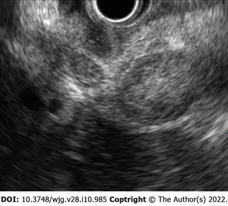
Figure 3 A 69 -year-old woman presented with abdominal pain and jaundice 12 mo after surgery for high-grade serous ovarian cancer.Endoscopic ultrasound from the duodenal bulb revealed numerous metastatic lymph nodes obstructing the bile duct by extrinsic compression. Endoscopic biliary drainage was performed, but the patient died 1 mo later.
Another report found stent occlusion in 50 % of lymph node metastasis cases vs 24 % in primary biliary tract cancer[231 ]. While the authors of the study suggested a limited role for metallic stents in biliary obstruction caused by metastatic lymph nodes, no other studies demonstrated a significant difference from primary cancers. Currently, metallic stents are widely used for this purpose. Bilateral stenting in high-grade inoperable strictures leads to lower re-intervention rates without sacrificing technical success or increasing adverse events[232 ].
Biliary obstruction due to lymphoma can occur from primary hepato-pancreato-biliary lymphoma as well as secondary lymphoma directly extending from abdominal lymph nodes. Most involve B-cell non-Hodgkin’s lymphomas (NHL), which cause 1 %-2 % of all MBOs, with a disproportionately high number of reports from diffuse large B-cell lymphoma[197 ,198 ]. MBO occurs in less than 2 % of NHL and about 0 .5 % of Hodgkin’s lymphoma[198 ,199 ]. Almost 1 % of NHL patients present initially with MBO[200 ].
CT of pancreatic metastases are generally hypervascular with well-defined margins[14 -16 ]. The hypointensity on T1 -weighted and hyperintensity on T2 -weighted imaging make them difficult to differentiate from primary adenocarcinoma on MRI[77 ]. They can also mimic neuroendocrine tumors on contrast MRI, due to their early contrast enhancement. Diagnosis can be made with high accuracy by EUS-FNA, with immunohistochemistry usually positive for pan-cytokeratin, CD10 , EMA, and PAX-8 and negative for synaptophysin, chromogranin, and beta-catenin (nuclear staining)[78 ].
Thyroid
Thyroid cancer most commonly metastasizes to the lung and bone. Pancreatic metastases can occur in papillary, follicular, and medullary carcinoma[233 ]. In a review of 24 reported cases in English and Japanese, the average delay from thyroid cancer diagnosis to pancreatic metastasis is 7 years. Most were solitary lesions, with half arising in the pancreatic head. Biliary obstruction due to metastatic papillary thyroid cancer has been reported[234 ]. EUS-FNA is commonly used for diagnosis, and solitary metastases are generally resected with good results in selected cases[235 ].
Gynecological cancers
About 8 % of ovarian cancer patients have metastases at diagnosis, but 22 % ultimately develop metastases after a median interval of 44 mo after diagnosis[236 ]. There are 17 reported cases of pancreatic metastases[237 ]. As BRCA1 and BRCA2 mutations are common risk factors for both ovarian and pancreatic cancers, pancreatic lesions should be evaluated by EUS-FNA to rule out the possibility of double cancers[238 ]. While MBO generally results from metastatic lymph nodes (Figure 3 ), obstructive jaundice due to metastasis to the major papilla has been reported[239 -241 ]. Distal pancreatectomy has been reported as an option for cytoreductive surgery in advanced ovarian cancer in a study of six patients with pancreatic metastases[242 ].
Less than ten cases of pancreatic metastasis from squamous cell carcinoma of the cervix have been reported[243 ]. On the other hand, biliary obstruction most often results from external bile duct compression, particularly in the porta hepatis[244 ]. Cases of periampullary and lymph node metastases requiring percutaneous drainage have been reported[120 ,245 ]. Endoscopic drainage has only been reported in one case to date[244 ].
You can guess what the sanitation department had to say about Mrs. Katrinka s crane. Sorry, ma am. We don t pick up 60 foot tall cranes. Old couches5, old tables, old refrigerators, and old washing machines are fine. Large, 60 foot tall cranes are not fine.
There are only two reports of pancreatic metastasis of endometrial carcinoma, with no reported obstructive jaundice[246 ,247 ]. While endometrial carcinoma is generally discovered early, caution is required for the papillary serious and clear cell types due to their tendency for early metastasis[246 ].
Urological cancers
While there are no more than ten reports of prostate cancer metastasizing to the pancreas, an autopsy study found pancreatic metastases in 1 .4 % of cases[248 -251 ]. Diagnosis with immunohistochemical staining for prostatic-specific antigen and successful ERCP have been reported[248 ].

Table 2 Relative frequency of pancreatic metastases by modality/procedure
Pancreatic metastases were reported in four urothelial carcinoma cases, of which two presented with obstructive jaundice[252 ]. Lesions could generally be diagnosed by EUS-FNA, with the help of immunohistochemistry. Bile duct wall metastasis has been reported from the aggressive micropapillary variant of bladder carcinoma, which was successfully drained by ERCP[253 ].
I ducked down behind the shoe mirror as they headed toward the golf ball section. Would they buy the Tour Edition Titleists? Probably not without help. I dashed down the club display aisle6 and slipped behind the mountain of shimmering7 red and gold boxes.
Esophageal cancer
There are more than ten reports of esophageal squamous cell carcinoma metastasizing to the pancreas[54 ,254 -259 ]. Such metastases can be synchronous or metachronous and may present as isolated metastases. Four cases underwent distal pancreatectomy among other treatments, with no short-term recurrence[255 -258 ]. One case presented as a cystic lesion which was diagnosed by EUS-FNA[259 ]. Five caused MBO, while lesions of all single case reports were located in the pancreatic body or tail. There is one report of MBO from recurrent esophageal cancer after esophagectomy which was first treated by ERCP and subsequently retreated by EUS-guided hepatico-gastrictubestomy when duodenal invasion precluded repeat ERCP[260 ].
Pure primary squamous cell carcinoma is a rare but important differential diagnosis, of which at least 54 cases have been reported[261 ]. Slightly over half are located in the head and 55 % have metastases at the time of diagnosis. MBO caused by metastatic squamous cell lung carcinoma has also been reported,requiring investigation for a lung primary[262 ].
Other primaries
There are isolated reports of pancreatic metastases from almost every malignancy, including meningioma, hemangiopericytoma, tonsillar squamous cell carcinoma, adenoid cystic carcinoma,hypopharyngeal carcinoma, thymoma, malignant pleural mesothelioma, pulmonary primitive neuroectodermal tumor, nephroblastoma, gastrointestinal stromal tumor, adrenocortical carcinoma, and testicular teratoma[263 -275 ].

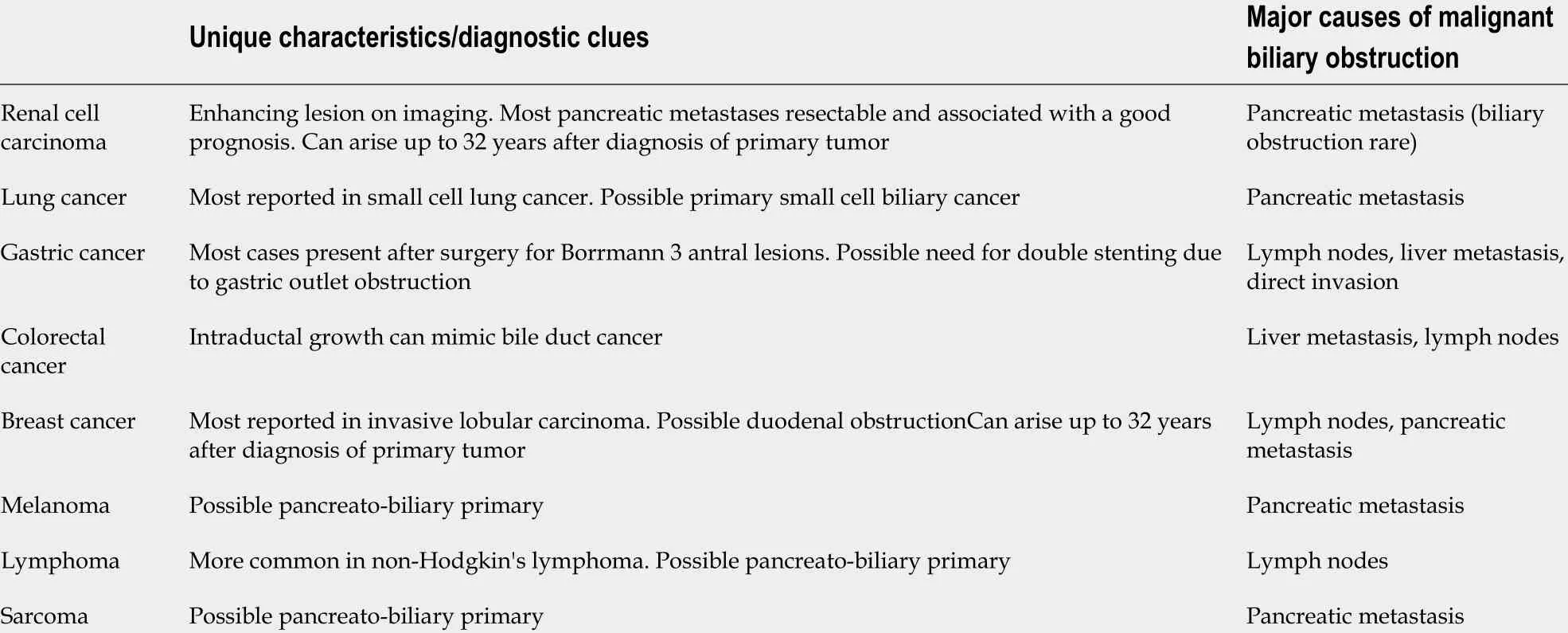
Table 3 Characteristics of malignant biliary obstruction caused by various primary malignancies
CONCLUSlON
Metastases from various primaries can cause MBO. The reported incidence pancreatic metastases vary across studies, depending on the selected modality (Table 2 ). While most can be diagnosed by EUS-FNA and treated by percutaneous or endoscopic drainage, factors specific to each primary should be kept in mind (Table 3 ). Surgery may be indicated in isolated metastases. There may be hope for long-term survival if systemic therapy can be resumed after biliary drainage or after margin-free resection of isolated metastases.
FOOTNOTES
Author contributions:Okamoto T wrote the manuscript.
Conflict-of-interest statement:The author has no financial disclosures or conflicts of interest to declare.
Open-Access:This article is an open-access article that was selected by an in-house editor and fully peer-reviewed by external reviewers. It is distributed in accordance with the Creative Commons Attribution NonCommercial (CC BYNC 4 .0 ) license, which permits others to distribute, remix, adapt, build upon this work non-commercially, and license their derivative works on different terms, provided the original work is properly cited and the use is noncommercial. See: https://creativecommons.org/Licenses/by-nc/4 .0 /
Country/Territory of origin:Japan
ORClD number:Takeshi Okamoto 0000 -0001 -9719 -0282 .
S-Editor:Chang KL
L-Editor:A
P-Editor:Chang KL
 World Journal of Gastroenterology2022年10期
- World Journal of Gastroenterology的其它文章
- Comment “Asymptomatic small intestinal ulcerative lesions: Obesity and Helicobacter pylori are likely to be risk factors”
- Gut dysbiosis and small intestinal bacterial overgrowth as independent forms of gut microbiota disorders in cirrhosis
- Diagnostic performance of endoscopic classifications for neoplastic lesions in patients with ulcerative colitis: A retrospective case-control study
- Loss of LAT1 sex-dependently delays recovery after caerulein-induced acute pancreatitis
- Feasibility of therapeutic endoscopic ultrasound in the bridge-to-surgery scenario: The example of pancreatic adenocarcinoma
- An update on the diagnosis of gastroenteropancreatic neuroendocrine neoplasms
