The lymphatic system: a therapeutic target for central nervous system disorders
Jia-Qi Xu,Qian-Qi Liu,Sheng-Yuan Huang,Chun-Yue Duan,Hong-Bin Lu,,Yong Cao,,Jian-Zhong Hu,
Abstract The lymphatic vasculature forms an organized network that covers the whole body and is involved in fluid homeostasis,metabolite clearance,and immune surveillance.The recent identification of functional lymphatic vessels in the meninges of the brain and the spinal cord has provided novel insights into neurophysiology.They emerge as major pathways for fluid exchange.The abundance of immune cells in lymphatic vessels and meninges also suggests that lymphatic vessels are actively involved in neuroimmunity.The lymphatic system,through its role in the clearance of neurotoxic proteins,autoimmune cell infiltration,and the transmission of pro-inflammatory signals,participates in the pathogenesis of a variety of neurological disorders,including neurodegenerative and neuroinflammatory diseases and traumatic injury.Vascular endothelial growth factor C is the master regulator of lymphangiogenesis,a process that is critical for the maintenance of central nervous system homeostasis.In this review,we summarize current knowledge and recent advances relating to the anatomical features and immunological functions of the lymphatic system of the central nervous system and highlight its potential as a therapeutic target for neurological disorders and central nervous system repair.
Key Words: central nervous system;central nervous system injury;glymphatic system;lymphatic vessels;meninges;neurodegenerative disorders;neuroinflammatory diseases;vascular endothelial growth factor C
From the Contents
Introduction 1249
Literature Search Strategy 1249
Anatomical and Functional Features of Lymphatic Pathways for the Central Nervous System 1250
Advances in the Understanding of Central Nervous System Fluid Dynamics 1250
The Lymphatic System and the Immune-privileged Status of the Central Nervous System 1251
Targeting the Lymphatic System in Neurological Disorders 1251
Discussion 1254
Conclusion 1254
Introduction
Lymphatic vessels (LVs) are present throughout most body tissues and organs and have critical functions in mammalian physiology.They act as a drainage system,help maintain fluid homeostasis,and participate in immune responses.In mice,LV development begins at approximately embryonic day 9.5 (E9.5) mainly from a subpopulation of venous endothelial cells (ECs) (Wigle and Oliver,1999;Wigle et al.,2002).LVs grow via sprouting from pre-existing LVs.The lymphatic vasculature absorbs the interstitial fluid (ISF) through initial lymphatics and drains lymph to collecting lymphatics and lymph nodes (LNs).Phasic contractions within the collecting lymphatics allow for unidirectional lymph propulsion (Oliver et al.,2020) in an arterial pulse-dependent manner.Lymph is ultimately drained into the right and left subclavian veins at the confluence of the internal jugular and subclavian veins (Alitalo,2011).Genetic determinants,blood flow,and the extracellular matrix can all affect the fate of lymphatic endothelial cells (LECs),thereby contributing to the plasticity and organ-specificity of the lymphatic vasculature (Ma and Oliver,2017).Recent progress in the understanding of how lymphatics develop has revealed the crucial role of the homeobox transcription factor prospero-related homeobox 1 (PROX1) and the receptor tyrosine kinase vascular endothelial growth factor receptor 3 (VEGFR-3 or FLT4) in lymphangiogenesis (Wigle and Oliver,1999;Wigle et al.,2002;Srinivasan et al.,2014).Additionally,surface markers of LECs,including lymphatic endothelial hyaluronan receptor 1 (LYVE-1),chemokine (C-C motif) ligand 21 (CCL21),and the membrane glycoprotein podoplanin (PDPN),have also been identified (Petrova and Koh,2020).
Until recently,the brain was considered to be an immune-privileged organ mainly owing to the absence of LVs.As early as 1787,Paolo Mascagni,an Italian physician,described the presence of LVs in human dura mater in his anatomical record (Bucchieri et al.,2015).During the next two centuries,there were scattered reports supporting the existence of meningeal lymphatics(Lecco,1953;Andres et al.,1987).Using histological methods,F?ldi et al.(1966) described LVs surrounding the cranial nerves and blood vessels around the dura.A paradigm shift occurred when the presence of LVs in meninges was confirmed through advanced methods (Louveau et al.,2015a).Moreover,these vessels have now been suggested to be key contributors to immunerelated neurological disorders.A previous study has already described the functional role of LVs in the meninges surrounding the brain and the spinal cord (Da Mesquita et al.,2018a). As a drainage system for the CNS,meningeal LVs (MLVs) have specific characteristics and execute unique functions (Petrova and Koh,2018).Studies investigating lymphatic drainage have led to a better understanding of brain and spinal cord homeostasis.Additionally,they have contributed to elucidating the mechanisms governing the movement of fluids,immune cells,and macromolecules into and out of the CNS,as well as the etiology of neurological disorders.Here,we further discuss their implications in the identification of novel therapeutic targets for neurological disorders,particularly neurodegenerative and autoimmune diseases and traumatic injury.
Literature Search Strategy
In this non-systematic review,the PubMed database was searched for both original research articles and review articles published between January 2002 and April 2022.To cover studies that discussed the lymphatic system relating to the CNS,the following search terms were used: (“l(fā)ymphatic system” OR“l(fā)ymphatic vessel” OR “glymphatic system”) AND (“central nervous system”O(jiān)R “brain” OR “meninges” OR “spinal cord”).After reviewing the title,abstract,and keywords,articles that were potentially related to anatomical,immune-related,and functional studies of the lymphatic system for the CNS were included.Several older publications (before 2002) were also included in consideration of their relevance to the field.Data extraction focused on information about the implication of the lymphatic system in neurological diseases.
Anatomical and Functional Features of Lymphatic Pathways for the Central Nervous System
The existence of functional lymphatic vasculature in the CNS is normally determined using lymphatic endothelium-specific markers.Important studies by Louveau et al.(2015b) and Aspelund et al.(2015) described vessel-like structures that expressed LEC-specific markers in the meningeal layer of the mouse dura mater.Meanwhile,Mezey et al (2021) identified similar structures in the autopsy of human meningeal specimens.LYVE-1,PDPN,and VEGFR-3 were employed as markers to characterize the distribution of potential lymphatic vasculature in postmortem human brain tissue.Besides meninges,LVs were also found along or within the walls of vessels in various brain regions and the subarachnoid space.Markers of lymphatic endothelium were also detected among the fibers and fascicles of cranial nerves (Mezey et al.,2021).
The lymphatic system of the brain
The cerebrospinal fluid (CSF) is secreted by the choroid plexus in lateral cerebral ventricles and circulates from the lateral ventricles to the third and fourth cerebral ventricles,finally entering the subarachnoid space and cistern to be absorbed into the blood by the arachnoid villi (Damkier et al.,2013).This process raises two significant questions,namely,how does the exchange of material occur between the CSF and ISF,and where does the drainage fluid end up,veins or LVs?
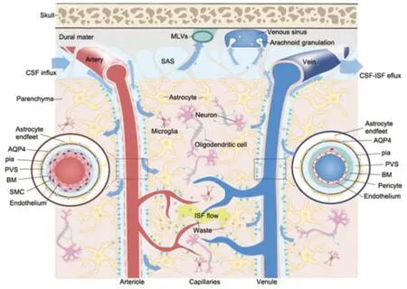
Studies have sought to describe the mechanisms underlying CSF-ISF exchange and ISF drainage from the CNS.The perivascular space (PVS) has long been suggested to be the conduit for the movement of CSF into or out of the brain (Rennels et al.,1985;Mathiisen et al.,2010).Iliff et al.(2012) were the first to propose the ‘glymphatic system’ to describe a paravascular pathway for fluid drainage involving glial cells.They found that subarachnoid CSF rapidly enters the brain parenchyma along the PSVs of penetrating arteries while perivascular ISF exits the brain via paravenous drainage pathways.Subsequently,imaging using intravital two-photon microscopy showed that perivascular CSF entered the brain via the periarterial space between smooth muscle cells (SMCs) and astrocyte end-feet.The employed tracers were observed to move inward along penetrating arteries and arterioles (maximally along large ventral perforating arteries of the basal ganglia and thalamus)to reach terminal capillary beds throughout the brain and outward along capillaries and parenchymal venules (primarily along medial internal cerebral and lateral–ventral caudal rhinal veins;Figure 1).The natural patterns of regional influx and tracer flow vary according to developmental stage (Munk et al.,2019).Besides,the astroglial aquaporin 4 (AQP4) water channel,which forms the subpial and subependymal glial limiting membrane,facilitates CSFISF exchange and promotes CSF flux into the brain parenchyma through the para-arterial space,and bulk ISF solute clearance from the parenchyma to the paravenous space (Iliff et al.,2012).
The subarachnoid CSF is believed to leave the cranium (i) via arachnoid granulation absorption,(ii) along the internal carotid artery,and (iii) along the perineural spaces of the olfactory and trigeminal nerves.Specifically,a dense lymphatic network has been identified within the nasal submucosa,with evidence showing that the CSF can drain to the deep cervical lymph nodes (dCLNs) (Kida et al.,1993;Goldmann et al.,2006;Papadopoulos et al.,2020).Approximately 15% to 30% of subarachnoid CSF is cleared across the cribriform plate,which projects into the nasal submucosa following the olfactory tracts (Bradbury and Cole,1980).The existence of CNS-draining LNs has led to the premise that CSF drainage flow requires LVs.CSF and ISF efflux to the dCLNs has been detected across mammalian species ranging from mice to humans,implying that it may be critical for the maintenance of fluid homeostasis (Bradbury and Westrop,1983;Boulton et al.,1998;Antila et al.,2017;Xue et al.,2020).Macromolecules were also cleared from the CNS parenchyma via MLVs (Aspelund et al.,2015;Louveau et al.,2015b).The MLV represents an additional exit pathway for fluid from the CNS into the systemic circulation.
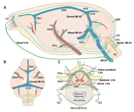
Aspelund et al.(2015) sought to identify the exact location of MLVs in the CNS using PROX1-GFP andVegfr3+/LacZreporter mice.The authors observed that MLVs were located in the dura matter at the base of the skull,mainly around the dural sinuses and middle meningeal arteries (Figure 2AandB).The mouse meninges are very small,which makes it hard to separate the meningeal layers,and may explain why the precise location of the MLVs in mice went undetected for so long.Also,MLVs develop postnatally,first appearing around the foramina in the basal parts of the skull and spinal canal,and then undergoing dynamic changes and sprouting along blood vessels and cranial and spinal nerves to various parts of the meninges surrounding the CNS.Antila et al.(2017) provided a detailed description of the anatomy of MLV development in mice.The authors reported that perisinusoidal MLVs developed from LVs located alongside the jugular veins and cranial nerves(IX and X).Like peripheral LVs,MLVs depend on VEGFR-3 signaling for their development.For example,K14-VEGFR-3-Ig transgenic mice display MLV aplasia,while VEGFR-3 signaling interruption affects CSF drainage functions.The expression of VEGF-C signals enwraps the blood vessels.The binding of VEGF-C to VEGFR-3 propels blood vessel growth and guides LV development and maintenance.Furthermore,Aspelund et al.(2015) reported that embryos lacking both VEGF-C alleles do not survive and that VEGF-C is essential for MLV development.Unlike with peripheral LVs,physical environmental properties might also affect the development and inhibit the sprouting of the meningeal lymphatic vasculature.
These studies have contributed to the unraveling of the fluid exchange pathways of the CNS,from the glymphatic pathway to draining LNs.These pathways are collectively referred to as the lymphatic system of the brain.However,many processes,including fluid entry into the MLVs and PVS extension into the subarachnoid space,take place in the meninges (Coles et al.,2017).Meanwhile,arguments and controversies remain on the cellular characteristics and developmental pattern of the lymphatic system (Bucchieri et al.,2015;Mestre et al.,2020).For instance,RNA-seq data has revealed that meningeal and peripheral LECs have distinct gene expression profiles(Louveau et al.,2018).LEC-specific gene set enrichment analysis,which included theLyve1,Pdpn,andProx1genes,did not identify differences between the two sets of LECs.However,alterations in multiple pathways related to the extracellular matrix,focal adhesion,angiogenesis,and response to endogenous and exogenous stimuli were observed in meningeal LECs when compared with peripheral LECs.In particular,genes involved in the regulation of lymphatic development and cell stiffness were differentially expressed in the meningeal LECs (Louveau et al.,2018).Meanwhile,LECs with unique phenotypes have been identified in different CNS locations;however,their role in the lymphatic system remains unclear.Izen et al.(2018) utilized whole-mount imaging to depict the postnatal MLV developmental pattern in mice.They found that from birth to postnatal day 13 (P13),MLVs extended alongside dural blood vessels,while between P13 and P20,they extended along with dural sinuses towards the olfactory bulb.Finally,the pituitary was found to be enriched with LVs,which likely contacted the lymphatic network in the nasal mucosa (Elham et al.,2020).
The lymphatic system around the spinal cord
In the early postnatal period,MLVs are observed almost simultaneously in the cervical,thoracic,and lumbar regions of the spinal cord (Figure 2C;Antila et al.,2017).Furthermore,the MLVs exit the spinal canal along the blood vessels and spinal nerves.Spinal MLVs have site-specific functional properties.For example,the LV pattern in spinal meninges was shown to change along the ventral-dorsal and cranial-caudal axes of the spine (Antila et al.,2017).As the LVs around the spinal cord have a delicate structure and are embedded deep within the spinal vertebral canal,they are difficult to visualize;consequently,little is known about them.To visualize the complex 3D morphology of LVs inside the spinal vertebral canal,Jacob et al.(2019) used the iDISCO+tissue preparation technique,which preserves intact vertebral bone structures for whole-mount immunostaining.They identified a dense lymphatic vasculature network that appeared to be predominantly restricted to the intervertebral spaces.The architecture of the vertebral LVs (vLVs) was found to be conserved along the vertebral column,and the vLVs were seen to be connected to the peripheral lymphatic system at intervertebral foramina along ventral nerve rami.Additionally,vLVs covering the dura matter of DRGs were observed to interact with the autonomous nervous system.Meanwhile,how vLVs connect to the peripheral lymphatic system differs among the cervical,thoracic,and lumbar vertebral levels (Jacob et al.,2019).
Advances in the Understanding of Central Nervous System Fluid Dynamics
In the CNS,several pathways are involved in the maintenance of CSF homeostasis.In the traditional routes,the molecular contents of the CSF are cleared via arachnoid granulations into the dural venous sinus,through the cribriform plate into the nasal mucosa,and via transporters and receptors present on the apical side of the choroid plexus epithelium (Weller et al.,2018).Research on the PVS or the glymphatic system forms the basis for understanding fluid management in the brain.Recent studies demonstrated that soluble tracers in the CSF can enter the brain via pial-glial basement membranes located between the layer of pia mater and astrocytes around cortical arteries;subsequently,the tracers drain out of the brain parenchyma along intramural peri-arterial drainage pathways (Albargothy et al.,2018;Owasil et al.,2020;Rasschaert et al.,2020).Carare et al.(2008) demonstrated that capillary and artery basement membranes act as ‘lymphatics of the brain’ for the drainage of fluid and solutes.Meanwhile,solute clearance along SMC basement membranes in the tunica media of arteries is referred to as intramural periarterial drainage.Albargothy et al.(2018) found that amyloid-β (Aβ) tracer and collagen IV co-localized in SMC capillary basement membranes,which indicate that Aβ was drained within the capillary basement membrane,thus questioning the function of the PVS for Aβ drainage.These observations indicate the need for an in-depth investigation of the function of arterial perivascular compartments.
The relative contribution of MLVs to CSF-ISF clearance among multiple post-glymphatic pathways needs further clarification using large-mammal models.The signal enhancement of tracers infused into ventricles can be observed using noninvasive imaging.An average initial signal increase at 15.9 ± 3.4 minutes has been reported in mandibular LNs (the subsequent draining LN of the dCLNs),significantly earlier than that for the venous signal(25.3 ± 2.8 minutes;Antila et al.,2017).This delay suggested that the active uptake of tracers occurred more rapidly via the lymphatic transport system.Additionally,signal enhancement was considerably higher in draining LNs than in saphenous veins (Antila et al.,2017).Through the intracerebroventricular administration of Evans blue dye,it was found that MLVs,and not nasal mucosa LVs,are the primary route for CSF drainage into the dCLN during the first 2 hours after administration (Louveau et al.,2015b).One study showed that the conditional ablation of MLVs nearly eliminated the drainage of intracerebroventricularly-injected microspheres to dCLNs (Antila et al.,2017).Furthermore,MLVs in the basal part of the skull (basal MLVs) showed a distinct structure from that previously reported for dorsal MLVs (Ahn et al.,2019).Basal MLVs,which run along the petrosquamosal and sigmoid sinuses,have larger diameters,a greater number of protruding capillary branches with blunt ends,and looser intercellular junctions than dorsal MLVs.They present collecting lymphatic-like structures,including more zipper-type junctions and lymphatic valves.However,the lack of SMC coverage makes them more like pre-collecting MLVs.The structure of basal MLVs and their proximity to the subarachnoid space render basal MLVs the ideal route for the uptake and drainage of CSF solute and macromolecules.
The spinal cord has an additional CSF flow pathway.Molecular tracers injected into one side of the spinal cord parenchyma at the thoracolumbar level were shown to accumulate in the ipsilateral paravertebral LV and mediastinal LN(Jacob et al.,2019).In vivoimaging revealed the CSF outflow routes in the spine.Tracers infused intracerebroventricularly indicated that the CSF flowed from the brain ventricles to the sacral spine within the central canal and accumulated in intervertebral regions.Moreover,the CSF outflow occurred along cranial nerves to extracranial LVs.On the dorsal side,tracers were drained into sacral LNs,while on the ventral side,collecting LVs along the axis of the spine drained tracers toward caudal mesenteric LNs.Tracers from both sides were ultimately drained to iliac LNs.Some of the LVs were also drained to the inguinal LNs.That CLNs still accounted for a large proportion of spinal CSF drainage suggested that these LN groups might play a key role in maintaining spinal cord homeostasis as well as in the occurrence of spinal cord disorders (Ma et al.,2019).
The Lymphatic System and the Immuneprivileged Status of the Central Nervous System
For years,the brain was thought to be an immune-privileged organ owing to the absence of a classical lymphatic drainage system in the parenchyma and the existence of the blood-brain barrier (BBB).In the CNS,antigens or antigen-presenting cells reach the CLNs via the cribriform plate,MLVs,or the rostral migratory stream and activate an immune response in peripheral LNs(Mohammad et al.,2014;Louveau et al.,2017).Networks rich in dendritic cells (DCs) and resident macrophages were reported in the vascular tissues and meninges of rats (McMenamin,1999).Meanwhile,Rustenhoven et al.(2021) demonstrated that the dural sinuses are a crucial interface for capturing and presenting CNS-derived antigens.Immune cells,such as T,B,and DCs,were found to be present in MLVs,indicating that the meningeal lymphatics participate in the drainage of immune cells from the CNS (Aspelund et al.,2015;Louveau et al.,2015b).The recognition of antigens drained through MLVs by antigen-presenting cells in the dural sinuses is an integral part of neuroimmune surveillance.Meningeal T cells are transported to the dCLN via a CCR7/CCL21-dependent pathway (Louveau et al.,2018).Resident microglia mainly induce a local adaptive immune response via the activation of infiltrating lymphocytes;consequently,immune surveillance is performed by long-lived macrophages in perivascular or subarachnoid spaces and meningeal DCs,which have access to the CSF-filled space (Papadopoulos et al.,2020).Activated T lymphocytes can also enter the CNS through the BBB,superficial brain barriers (pia mater and glia limitans),and the blood-CSF barrier (choroid plexus) (Engelhardt et al.,2016,2017).Hence,the role of MLVs in neuroinflammatory and autoimmune diseases becomes a major research direction in the future.
Signal transmission may account for the possible association between the draining of CSF by MLVs and the pathological immune environment.Esposito et al.(2019) proposed the existence of a “brain-to-CLN” signaling pathway in a rat model of stroke.The authors found that dynamic crosstalk between the CNS and systemic responses were channeled through the MLVs.The injured brain secreted VEGF-C into the CSF,thereby activating the proliferation of LECs and pro-inflammatory macrophages in CLNs.Neuroinflammation-related components in the CSF were also observed to induce lymphangiogenesis via VEGF-C/VEGFR-3 signaling (Hsu et al.,2019).Importantly,this signaling pathway from the CNS to the peripheral immune system was shown to be bidirectional,namely,the activated MLVs displayed a unique transcriptional signature and contributed to the invasion of the brain by autoreactive T cells (Louveau et al.,2018).However,the mechanism underlying how MLVs and LECs modulate the phenotype of immune cells remains incompletely understood.Finally,Liu et al.(2020c) proposed that lymphoangiocrine signals might,themselves,exert significant effects on the immune microenvironment of the CNS.
Targeting the Lymphatic System in Neurological Disorders
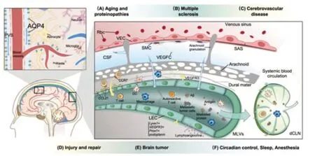
The lymphatic system of the CNS not only plays a key role in the maintenance of tissue homeostasis by recycling ISF but is also important in immune surveillance regulation and immune-cell recirculation.In the CNS,lymphatic vasculature dysfunction results in the disruption of the ISF balance,which has been implicated in multiple neurological disorders.The impairment of lymphatic system function can have a significant impact on neurological disease outcomes (Aspelund et al.,2015;Tarasoff-Conway et al.,2015;Engelhardt et al.,2017;Ma et al.,2017).In the following sections,we describe and provide further insight regarding the function and role of the lymphatic system of the CNS in neurological disorders (Figure 3).The regulation of lymphatic function might be a promising therapeutic target to prevent neurological disorders or delay their progression (Table 1).

Figure 1|Schematic illustration of the CSF-ISF flow in the glymphatic system.

Figure 2|Schematic illustration of the anatomical position of meningeal lymphatic vessels around the mouse brain and spinal cord.

Figure 3|The role of the lymphatic system in neurological diseases.

Table 1|Interventions targeting the lymphatic system in neurodegenerative disorders and central nervous system injury
Neurodegenerative disorders
The pathology of aging or proteinopathic neurodegenerative disorders,such as Alzheimer’s disease (AD),Parkinson’s disease (PD),glaucoma,frontotemporal dementia,and amyotrophic lateral sclerosis,is characterized by the insufficient clearance of neurotoxic proteins,including Aβ and tau(Boland et al.,2018).
The impairment of extracellular clearance by the lymphatic system of the brain has emerged as a key pathogenic mechanism in aging.Aged mice exhibited regression of dorsal MLVs and hyperplasia of basal MLVs.Their basal MLVs increased in size and became highly branched,leading to fewer lymphatic valves and zipper-type junctions,and more button-type junctions.Moreover,basal MLVs from aged mice showed a dysmorphic distribution of type IV collagen and reduced fluid transport.The observed lymphatic hyperplasia was regarded as a compensatory mechanism for the impaired function of the lymphatic system (Ahn et al.,2019).Additionally,lymphatic outflow from the CSF to blood and draining LNs was slower in aged mice (Antila et al.,2017).RNA-seq analysis of LECs revealed that genes related to the lymphangiogenic signaling pathway were differentially expressed in aged mice(Da Mesquita et al.,2018b).
Glymphatic system impairment exacerbated the pathogenic accumulation of Aβ and tau (Xu et al.,2015;Harrison et al.,2020).Wang et al.(2020)also identified an ocular glymphatic clearance route for Aβ.Intra-axonal Aβ was cleared from the retina and vitreous via the perivenous space and subsequently drained to cervical LVs.The fluid and waste clearance function of the pathway was dependent on the AQP4 channel and translaminar pressure.The AQP4 channel has been frequently targeted given its crucial role in pathological protein clearance (Valenza et al.,2019).AQP4 was demonstrated to function in the clearance of interstitial Aβ,suggestive of an association between brain lymphatics and AD progression (Tarasoff-Conway et al.,2015;Mader and Brimberg,2019).Impaired expression and function of AQP4 were demonstrated to be biomarkers for AD.Moreover,single nucleotide polymorphisms in theAQP4gene were correlated with Aβ neuropathology,disease progression,and cognitive decline (Chandra et al.,2021).In aged mice,a radiotracer clearance assay showed a reduction in CSF-ISF exchange efficiency,Aβ clearance rate,vessel wall pulsatility,and perivascular AQP4 polarization (Kress et al.,2014).Meanwhile,patients withAD displayed a significant decrease in CSF AQP4 levels,which was correlated with diminished levels of Aβ in the CSF (Arighi et al.,2019).Additionally,the expression of AQP4 was reported to be reduced in aging human brains,while a marked loss of perivascular AQP4 localization in the frontal cortex was observed in individuals with AD (Zeppenfeld et al.,2017).
Da Mesquita et al.(2018b) reported that MLV ablation promoted Aβ deposition in the meninges and aggravated Aβ accumulation in the brain parenchyma,while Wang et al.(2019a) showed that the ligation of dCLN also exacerbated AD-like phenotypes in mice.However,a study on postmortem meningeal tissue from patients with Alzheimer’s dementia did not detect Aβ deposition in MLVs,implying that MLVs were not Aβ-deposition sites (Goodman et al.,2018).An observational cohort study utilizing a novel MRI method showed that the glymphatic pathway and MLVs were impaired in aging human brains.The clearance of downstream putative MLVs was significantly related to the glymphatic system (Zhou et al.,2020).Using dynamic contrast-enhanced magnetic resonance imaging,Ding et al.(2021)demonstrated that patients with idiopathic PD or atypical Parkinsonian disorders exhibited reduced flow in the cerebral lymphatic system.The same method was used to measure the uptake of gadobutrol medium by MLVs and thereby quantify MLV flow.The signal intensity in the MLVs along the superior sagittal and sigmoid sinuses was significantly reduced in patients with idiopathic PD,and their dCLN perfusion was also delayed.Moreover,MLVs were observed to participate in the clearance of other abnormally accumulated proteins,such as tau and α-synuclein (Patel et al.,2019;Zou et al.,2019;Ding et al.,2021),suggestive of their clinical diagnostic value and their ability to restore lymphatic function in the treatment of neurodegenerative diseases.
Treatments for the lymphatic system are being investigated based on these mechanisms.Thrombolytic therapy,phosphodiesterase III inhibitors,anti-Aβ immunotherapy,and AQP4 activation have all been adopted to improve the glymphatic transfer of neurotoxic proteins to the circulation (Sun et al.,2018).Nevertheless,therapies that involve lymphatic intervention are still in their infancy.Seven days after the injection of VEGFR-3-specific recombinant VEGF-C into the cisterna magna,mice exhibited an increase in MLV diameter (Louveau et al.,2015b).A similar phenotype was observed in aged mice treated with AAV1-CMV-mVEGF-C.Moreover,1 month after AAV1/CMV/mVEGF-C treatment,aged mice that displayed impaired lymphatic function showed a significant increase in CSF tracer drainage into the dCLNs.Consequently,the rate of tracer influx into the brain parenchyma was also increased.The same therapeutic effect was obtained following the transcranial delivery of hydrogel-encapsulated VEGF-C peptide (Da Mesquita et al.,2018b).However,in AD mice with no apparent MLV dysfunction at a young age,the mVEGF-C treatment was inefficient,suggesting an upper limit for VEGF-C in lymphatic function.Recombinant VEGF-C was shown to promote tube formation in human LECsin vitro,while thein vivoinjection of 200 μg/mL recombinant human VEGF-C (rhVEGF-C) into the cisterna magna,every 2 days,four times in total,induced dural lymphangiogenesis,reduced the levels of soluble amyloid-β,and restored spatial cognition in AD model mice (Wen et al.,2018).Recently,the modulation of meningeal lymphatic function was shown to influence the outcome of passive immunotherapy in AD (Da Mesquita et al.,2021).The ablation of MLVs in a mouse model of amyloid deposition resulted in a decrease in the effectiveness of anti-Aβ monoclonal antibody therapy,whereas treatment with VEGF-C improved Aβ clearance.Additionally,the authors suggested that activation of the microglial inflammatory response might underlie the loss of treatment efficacy resulting from MLV ablation.
Neuroinflammatory diseases
In pathological states,including virus infections and autoimmune diseases,antigens from the CNS are drained to peripheral immune organs and activate lymphocytes,which then invade the CNS.Multiple sclerosis is an autoimmune neurological disease characterized by the infiltration of autoreactive T cells into the brain and subsequent neuronal damage (Liblau et al.,2013).CLNs have long been suggested as a site for activation of encephalitogenic T cells(Phillips et al.,1997).In an experimental autoimmune encephalomyelitis(EAE) mouse model of multiple sclerosis,the excision of CNS-draining LNs ameliorated chronic-relapsing EAE within the spinal cord (van Zwam et al.,2009).Fluorescent tracer injection and multiphoton intravital imaging revealed that MLVs could uptake and transport CSF constituents.The meningeal lymphatic subarachnoid extensions along the transverse sinus,superior sagittal sinus,and cerebellar ring presented multiple entry sites for fluids.The dCLNs and supraclavicular LNs were confirmed to be the major CNS-draining LNs.Importantly,CCR7+T cells have a close relationship with CCL21+LECs,suggesting that the CCR7-CCL21 pathway plays a primary role in meningeal T-cell migration.Additionally,MLVs were shown to serve as a route for the drainage of immune cells and macromolecules from the CNS.A transcriptomic analysis further highlighted the unique properties of meningeal LECs relative to peripheral ones,which might explain the limited lymphangiogenic ability of meningeal LECs under inflammatory stimulation.Moreover,the ablation of meningeal lymphatics using Visudyne (laseractivated) delayed the pathological process of EAE in mice.The transcriptomic profile of antigen-specific T cells isolated from dCLNs revealed that impaired lymphatic drainage prevented the formation of encephalitogenic T cells in dCLNs (Louveau et al.,2018).
Meanwhile,lymphatics near the cribriform plate and nasal mucosa are essential in multiple sclerosis.The nasal region is not considered a major exit route for CNS immune cells;instead,it is closely associated with chronic inflammation and later stages of disease development (Louveau et al.,2018).Neuroinflammation-induced lymphangiogenesis was reported to be dependent on VEGF-C/VEGFR-3 signaling.During EAE,VEGF-C is significantly downregulated.Newly formed LVs carried CD11b-and CD11c-positive immune cells.LECs near the cribriform plate also expressed CCR7+and were accompanied by increased numbers of CCR7+CD11c+cells.These observations confirmed that lymphangiogenesis occurred primarily in the cribriform plate.Accordingly,treatment with the VEGFR-3 tyrosine kinase inhibitor MAZ51 (10 mg/kg,once daily) reduced EAE severity by inhibiting lymphangiogenesis and decreasing the amount of autoimmune antigen drainage to the LNs (Hsu et al.,2019).
Neuromyelitis optica spectrum disorder is a neuroinflammatory,demyelinating disease of the CNS that most commonly affects the optic nerve and spinal cord.The presence of AQP4 IgG antibodies (AQP4-IgG) in serum represents the key determinant of this disease.AQP4-IgG is highly cytotoxic to astrocytes(Wu et al.,2019).Employing contrast-enhanced magnetic resonance imaging(Wang et al.,2021) observed that meningeal lymphatic flow was impaired in patients with acute neuromyelitis optica spectrum disorder.The contrastenhanced magnetic resonance imaging parameters of mLVs in the superior sagittal sinus were correlated with disease severity and were suggested to have potential as disease predictors.
CNS injuries
Mice subjected to traumatic brain injury (TBI) exhibit the loss of perivascular AQP4 polarization and impaired interstitial solute clearance.Aqp4knockout mice showed exacerbated post-traumatic neuroinflammation and cognitive deficits after TBI (Iliff et al.,2014).MRI revealed altered glymphatic clearance rates in limbic and olfactory bulbs after repetitive mild TBI (Christensen et al.,2020).Chronic glymphatic system dysfunction associated with repetitive brain trauma has been implicated in the subsequent development of chronic traumatic encephalopathy (Sullan et al.,2018).In mice subjected to TBI,treatment with omega-3 polyunsaturated fatty acids restored glymphatic clearance and reduced Aβ accumulation,thereby attenuating TBI-induced neurological impairment (Zhang et al.,2020).
After TBI,brain-to-CLN signaling largely depends on lymphatics.The suppression of glymphatic pathway activity prevented the delivery of TBI biomarkers (S100β,GFAP,and NSE) from the glymphatic system and dCLN to blood (Plog et al.,2015).The uptake of CSF by MLVs can also be largely impaired in TBI (Bolte et al.,2020).After 1 week of injury,mice with TBI exhibited increases in LYVE-1+percent area coverage,the number of capillary loops and sprouts,and LV diameter,and these parameters changed dynamically over time.After TBI,the disruption of lymphatic drainage may result from an increase in intracranial pressure (ICP).Pre-existing MLV dysfunction in aged mice also exacerbated post-injury inflammation and cognitive decline.Viral-mediated delivery of VEGF-C rejuvenated lymphatics and mitigated TBI-induced inflammation in aged mice.Additionally,aged mice with TBI that received VEGF-C treatment had significantly lower levels of Iba1,which were more similar to the levels of Iba1 immunoreactivity in young mice subjected to TBI.These results indicated that LV enhancement can mitigate TBI-driven inflammation (Bolte et al.,2020).In a mouse model of subarachnoid hemorrhage,the Visudyne-mediated ablation of MLVs led to impaired brain perfusion and neurological function,as well as the aggravation of inflammation and apoptosis in early brain injury (Liu et al.,2021).In K14-VEGFR-3-Ig transgenic mice subjected to TBI,CD4+T-cell infiltration in the perilesional cortex and hippocampus was reduced,suggesting that MLVs are indispensable for the establishment of a proper neuroimmune response after TBI (Wojciechowski et al.,2020).
However,compared to its beneficial effects in TBI,the brain-to-CLN signaling via VEGFR-3 participated in systemic inflammation in other diseases.In a rat model of stroke,the rapid appearance of dye that had originated from the CSF or brain parenchyma in CLNs suggested that inflammatory signals had been sent from the brain to peripheral LNs (Esposito et al.,2019).After cerebral ischemia,LEC proliferation,phosphorylated VEGFR-3 in CLNs,and VEGF-C upregulation were all observed.Moreover,activated lymphatic endothelium could upregulate pro-inflammatory macrophage responses in CLNs.After a stroke,the blockade of VEGFR-3 via the intracerebroventricular injection of MAZ51 or lymphadenectomy mitigated both the inflammatory process and brain injury (Esposito et al.,2019).In mice models of photothrombosisinduced stroke,meningeal lymphangiogenesis was seen in the alymphatic zone,lateral to the sagittal sinus.Additionally,the impairment of MLVs in mutant mice with defective VEGFR signaling worsened the outcome of stroke in the animals (Yanev et al.,2020).Meanwhile,in a lysolecithin-induced focal demyelination spinal cord injury model,LV diameter and area were only significantly enhanced in mice treated with VEGF-C and lysolecithin together,while the VEGF-C-induced increase in vertebral LV area amplified the cytotoxic effect resulting from lysolecithin-induced injury (Jacob et al.,2019).The physiological role of LVs in injury and inflammation,and whether they primarily reduce ICP through draining or promote the spread of inflammation,remain to be investigated.
Other neurological diseases
The fluid drainage and waste clearance functions of lymphatics are also associated with edema and hydrocephalus (Simka et al.,2018;Reeves et al.,2020).Congenital impairment of glymphatic function or AQP4 loss are critical pathogenic mechanisms in idiopathic normal pressure hydrocephalus (Hasan-Olive et al.,2019;Jacobsen et al.,2020).Furthermore,the location of MLVs renders them suitable as clearance pathways in subarachnoid hemorrhage and subdural hematomas (Pu et al.,2019;Liu et al.,2020b).Small vessel disease (SVD) is one of the most common etiologies of dementia in the elderly and can further deteriorate to stroke,disability,and cognitive impairment(Brown et al.,2018).The function of the lymphatic system is impaired in SVD.Enlarged PVS visible in MRI and protein deposits are hallmark features of this condition (Mestre et al.,2017).PVS enlargement can be seen in SVDs such as cerebral amyloid angiopathy and hypertensive arteriopathy (Martinez-Ramirez et al.,2018;Duperron et al.,2019).A large population-based cohort study found that a high burden of dilated PVS in basal ganglia and the hippocampus was associated with an increased risk of stroke and intracerebral hemorrhage(Duperron et al.,2019).Moreover,in hypertension,perivascular inflammation,pericyte dysfunction,BBB disruption,arterial stiffness,and decreased cerebrovascular pulsatility were reported to contribute to the obstruction and stagnation of fluid drainage (Mestre et al.,2017;Brown et al.,2018;Riba-Llena et al.,2018).
Dorsal MLVs presented significantly increased diameter and density in mice injected with glioma and melanoma cells,indicating that brain tumors can promote lymphangiogenesis.MLVs can serve as a tunnel through which tumor cells can metastasize toward dCLNs.Meanwhile,dorsal MLVs were shown to be the main route for the migration of DCs and other immune cells to dCLNs and the establishment of immune responses to brain tumors.Visudyne-mediated MLV ablation impaired the efficacy of anti-PD-1/CTLA-4 immunotherapy while overexpressing VEGF-C in tumor cells exerted the opposite effect in a CCL21/CCR7 signaling-dependent manner (Hu et al.,2020).The effect of treatments targeting the PD-1 checkpoint is also influenced by MLVs.The efficacy of monoclonal antibodies targeting PD-1,such as nivolumab,still can be enhanced in the treatment of glioblastomas(Wang et al.,2019b).For example,Song et al.(2020) revealed that the combination of anti-PD-1 antibody and VEGF could significantly suppress tumor growth and improve survival.Moreover,VEGF-C promoted antigen drainage and stimulated lasting and strong T-cell mediated immune responses.Finally,VEGF-C induced MLV lymphangiogenesis,leading to the generation of an anti-tumor environment that facilitated T-cell infiltration,priming,and recruitment (Song et al.,2020).
The glymphatic system is also implicated in various diseases.One study confirmed that glymphatic system impairment due to a reduction in AQP4 expression exacerbated hepatic encephalopathy (Hadjihambi et al.,2019).Hydrodynamic disorders,such as the retention of sodium and water in the body,that result from liver abnormalities,can lead to glymphatic system dysfunction (Gallina et al.,2019).Accordingly,cognitive dysfunction caused by substances that can injure the liver,such as alcohol,might be related to glymphatic system impairment (de la Monte and Kril,2014).Indeed,studies have shown that alcohol can have a dose-dependent effect on glymphatic system function (Lundgaard et al.,2018;Cheng et al.,2019).Multiple studies have demonstrated the risk of glymphatic system impairment during depression.For example,mice treated with chronic unpredictable mild stress exhibited AQP4 downregulation,Aβ accumulation,and reduced arterial pulsation (Wei et al.,2019;Liu et al.,2020a).
Sleep,wakefulness,and anesthesia are all closely related to the function of the glymphatic system.During wakefulness,the intensity of tracer signals in the periarterial spaces,subpial regions,and brain parenchyma was reported to be significantly lower than that during the sleep state.Additionally,ketamine/xylazine-induced anesthesia was shown to increase CSF influx.These results confirmed that sleep and anesthesia can stimulate lymphatic drainage and enhance waste clearance (Xie et al.,2013).Several studies have demonstrated that sleep deprivation can suppress CSF transport,which is associated with the accumulation of neurotoxic macromolecules and the progression of neurodegenerative disease (Achariyar et al.,2016;Shokri-Kojori et al.,2018;Piantino et al.,2019).The effects of sleep and anesthesia on glymphatic system function may be attributable to interstitial space enlargement.For example,in sleeping and anesthetized mice,the volume and tortuosity of the interstitial space were significantly increased(Xie et al.,2013).Meanwhile,Benveniste et al.(2017) discovered that the additional administration of dexmedetomidine when anesthetizing the rats with isoflurane induced a more efficient glymphatic clearance .Diabetesrelated sleep disturbance and cognitive dysfunction might also be caused by glymphatic system impairment,as evidenced by the fact that the diameter of PVS is reduced and waste clearance is impaired in type-2 diabetes mellitus(Jiang et al.,2017;Brzecka et al.,2021).The recording of the diurnal variation in the glymphatic system of mice led to the finding that CSF influx was under circadian control.The amount of CSF tracer influx into the brain during the day was approximately 53% more than that during the night,peaking at around mid-day when the mice were mostly asleep.The AQP4 intensity around the vasculature was also significantly higher during the day (Hablitz et al.,2020).This circadian control of the glymphatic system might also explain neural diseases caused by circadian misalignment (Depner et al.,2014).These studies have expanded the understanding of how circadian rhythms form and function and have also provided novel ideas regarding the occurrence of migraine,AD,and multiple other neurological disorders.
Discussion
MLVs and the perivascular drainage pathway together form a gradually elaborated lymphatic system for the CNS.The in-depth study of anatomical features has revealed the role of MLVs in CSF-ISF exchange,metabolite regulation,and immune cell migration.This harmoniously working ‘plumbing system’ helps maintain the delicate internal environment required for neuronal function.The lymphatic system-related accumulation of metabolites and toxic substances,as well as the imbalance and impairment of the immune response,can lead to the occurrence of several neurological conditions.Accordingly,the lymphatic system for the CNS has emerged as a novel therapeutic target for multiple neurological disorders.
The precise location of MLVs within the meningeal layers remains unclear.Their localization and contact with the subarachnoid space largely reflect the place where the absorption of macromolecule and immune cell occurs(Raper et al.,2016).Pathology data for human and nonhuman primates have indicated that at least some lymphatics are entirely contained within the dura (Absinta et al.,2017).Experimental/dynamic mathematical model combinations are still required to quantify the relative contribution of each CSF influx and efflux pathway to drainage.We propose the development of high-resolution intravital imaging tools to precisely visualize the structure of the meningeal lymphatic drainage system for the CNS.Meanwhile,given that meningeal LECs have gene expression patterns that are distinct from those of peripheral LECs (Louveau et al.,2018),additional research using genetic tools is required to determine the unique biological functions of meningeal LECs in the CNS.VEGF-C/VEGFR-3 signaling regulation controls LEC fate and ultimately affects LV development at the molecular level.
Although much is already known about the anatomy of perivascular fluid pathways and MLVs,relevant research interest is currently focused on their potential as therapeutic targets (Sun et al.,2018;Mestre et al.,2020).Targeted interventions can effectively influence the pathophysiology of neurological disorders,especially neurodegenerative diseases associated with the deposition of toxic substances (Rasmussen et al.,2018;Wen et al.,2018).Whether lymphatics can invade the brain parenchyma after an injury is still unknown.The observed co-expression of LYVE-1 and PROX1,and the assembly into putative pre-vessel structures by LYVE-1-expressing cells,have suggested that,in the adult rat spinal cord,mild lymphangiogenesis occurs after spinal cord contusion injury (Kaser-Eichberger et al.,2016).However,those phenomena need further elucidation regarding vessel structure and signatures.In zebrafish ischemic stroke models,MLVs were observed to rapidly grow into the injured brain parenchyma to drain the excess ISF and guide neovascular regeneration.The newborn LVs resulting from the response to ischemia assisted in tissue regeneration to a great extent.Moreover,once cerebrovascular regeneration was complete,the ingrown LVs were cleared via apoptosis,suggesting that they played only a transient role during injury (Chen et al.,2019).Although not yet confirmed in mammals,this finding revealed that the function of LVs consists of more than just the alleviation of edema during CNS injury.The expression of lymphatic markers was also detected in damaged mouse brain tissues after penetrating TBI (Meng et al.,2021).Recently,lymphoangiocrine signals secreted by LECs were identified as an essential component in cardiac repair (Liu et al.,2020c).The functions of LECs in CNS damage repair also merit further investigation.
Conclusion
Current evidence indicates that a strong relationship exists between the lymphatic system of the CNS and neurodegenerative,neuroinflammatory,and CNS injury disorders.These observations suggest that targeting the lymphatic system may represent a feasible therapeutic strategy for postinjury CNS repair.Notably,however,any invasive procedure can upset the delicate balance of the brain lymphatic system and interfere with the judgment of the actual physiological and pathological conditions of fluid flow,making experiments difficult to perform.The enhancement of lymphatic function via VEGF-C delivery has shown promising effects in promoting the clearance of neurotoxic substances and treating neurodegenerative disease.Lymphangiogenesis has also shown benefits in reducing ICP.VEGF-C intervention seemed to be mainly effective in aged or injured mice with preexisting lymphatic function impairment.In autoimmune diseases or stroke,the ablation of the lymphatic pathway could also mitigate inflammation.
Author contributions:JZH,HBL and CYD conceived and designed the manuscript.JQX performed the literature review and drafted the manuscript.QQL,SYH and YC designed the manuscript and performed the literature review.CYD critically edited the manuscript.All authors read and approved the final manuscript.
Conflicts of interest:The authors declare that they have no competing interests.
Open access statement:This is an open access journal,and articles are distributed under the terms of the Creative Commons AttributionNonCommercial-ShareAlike 4.0 License,which allows others to remix,tweak,and build upon the work non-commercially,as long as appropriate credit is given and the new creations are licensed under the identical terms.
- 中國神經(jīng)再生研究(英文版)的其它文章
- Neuro faces of beneficial T cells: essential in brain,impaired in aging and neurological diseases,and activated functionally by neurotransmitters and neuropeptides
- Profiling neuroprotective potential of trehalose in animal models of neurodegenerative diseases:a systematic review
- Cdk5 and aberrant cell cycle activation at the core of neurodegeneration
- Recent advancements in noninvasive brain modulation for individuals with autism spectrum disorder
- Vicious cycle of lipid peroxidation and iron accumulation in neurodegeneration
- Cell-based therapeutic strategies for treatment of spinocerebellar ataxias: an update

