Is GDNF to Parkinson’s disease what BDNF is to Huntington’s disease?
Francesca R.Fusco,Emanuela Paldino
Neurotrophic factors,or neurotrophins,are a group of molecules supporting the growth,survival,and differentiation of developing and mature neurons.Given their role in the survival of neurons,and often of specific subsets of brain cells,neurotrophins have been implicated in several ways with many neurodegenerative disorders.
Chronic stress,as well as oxidative stress occurring in neurodegenerative diseases,leads to the activation of pro-inflammatory microglia.Subsequently,a release of cytokines and proinflammatory substances occurs,as well as the recruitment of peripheral immune cells to the brain.This creates an inflammatory environment typical of several neurodegenerative disorders.
Αs a response,brain cells release several substances that promote neuronal survival,such as anti-inflammatory cytokines,growth factors,and neurotrophic factors.
Neurotrophins and their receptors:The Neurotrophin family is composed of nerve growth factor,brain-derived neurotrophic factor (BDNF),neurotrophin-3 (NT-3),and neurotrophin-4/5(NT-4/5).Neurotrophins have several features in common: one is that the encoding genes contain a signal sequence and a prodomain,so all of them are synthesized as pro-NTs,which have to undergo a proteolysis to become functionally active;moreover,neurotrophins are released as homodimers,a configuration that is required to bind to their respective receptors.
Neurotrophins activate two different families of membrane receptors: a pan-NT receptor,known as p75NTR,and a family of tyrosine kinase receptors,known as Trk receptors.
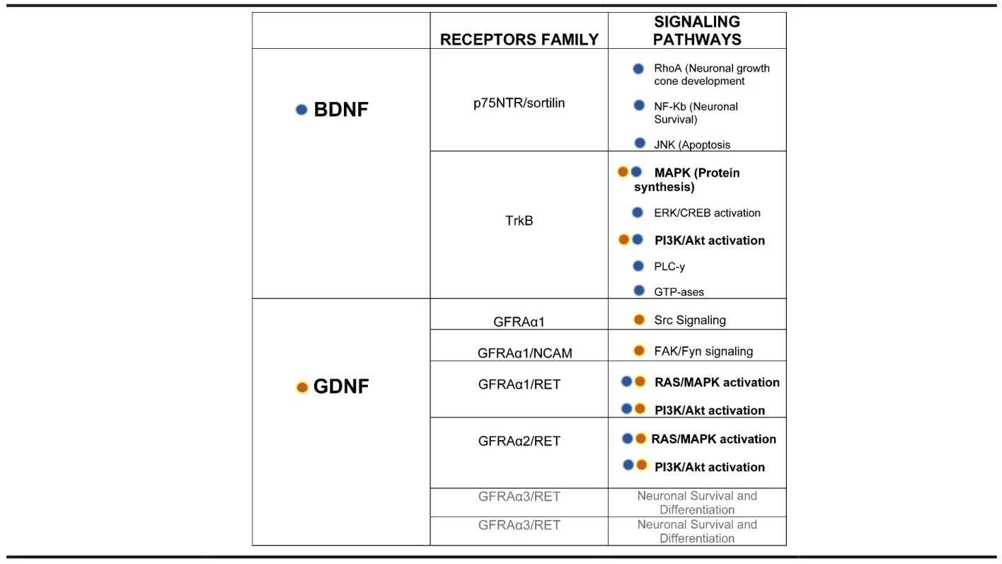
Mature neurotrophins have a low affinity for p75NTRand promote neuronal survival and differentiation in a life-facilitating pathway that is mediated by the interaction of p75NTRand the Trk receptors (Box 1).

Box 1 | NTs and their receptor families and signaling pathways
Neurotrophins play a major role in the survival of neurons through their receptors.Their targets are brain areas that degenerate in several neurodegenerative disorders.
Here,we examine the role of BDNF in Huntington’s disease and that of glial cell line-derived neurotrophic factor (GDNF) in Parkinson’s disease,in an attempt to draw out similarities between the two neurotrophins.
BDNF and Huntington’s disease:Brain diseases are usually associated with a downregulation of BDNF release,which results in reduced BDNF levels both in the brain and in peripheral blood.
In Huntington’s disease (HD),an autosomal dominant inherited neurodegenerative disorder,progressive death of the striatal part of the basal ganglia occurs.Clinically,the disease presents with emotional and psychiatric disorders,involuntary movements,and dementia.The mutated huntingtin protein causes the disease by a combination of loss of function and gain of function effects that have been the subject of investigation for many years.
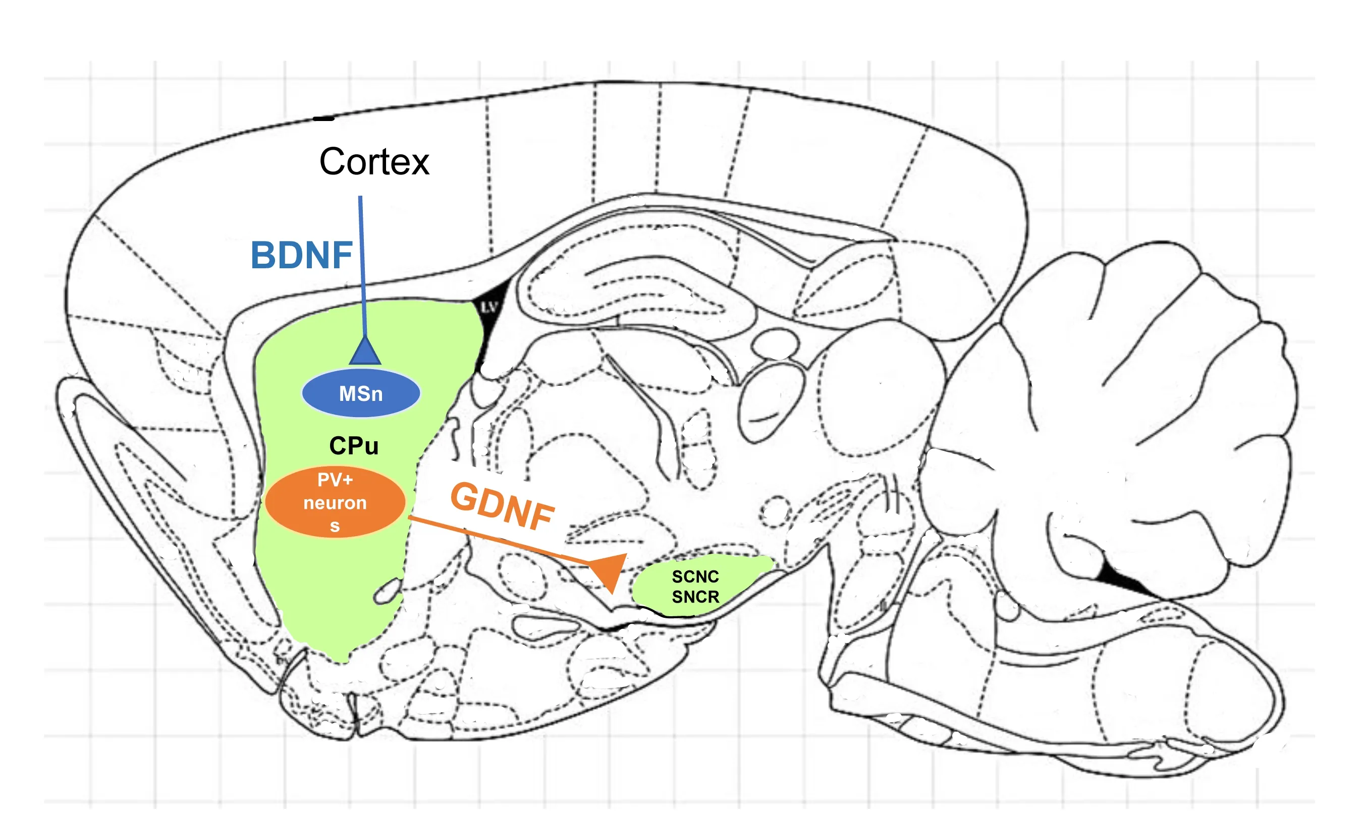
Αpproximately two decades ago,the importance of BDNF for the survival of the striatum became evident and illuminated the pathophysiology of Huntington’s disease (Zuccato et al.,2001).Indeed,striatal medium spiny neurons are not equipped to produce BDNF and they receive it transsynaptically from the cortical pyramidal neurons (Figure 1).Thus,the authors demonstrated that,in HD,mutated huntingtin protein caused dysfunction in cortical BDNF production,which in turn caused striatal degeneration occurring in HD.Subsequently,our group demonstrated that nearly all huntingtin-containing neurons in normal rat brains co-localized with BDNF in the cortex and that the reduced striatal expression of BDNF after quinolinic acid injection to the striatum,further supporting the concept that huntingtin may be required for BDNF production in the cortex (Fusco et al.,2003).Moreover,we observed that,in the quinolinic acid lesioned rat striatum,striatal interneurons typically resistant to HD,i.e.,cholinergic neurons,still contained BDNF,which led us to speculate that those neurons do not depend on the cortex for BDNF production,which partly explained their resistance to neurodegeneration.

Figure 1 | A cartoon representing the cortico-striatal BDNF delivery pathway and the striatonigral GDNF delivery pathway.
BDNF-based therapies for Huntington’s disease:The role of BDNF as a possible therapy for HD has gained momentum in the last decade.In 2013,we showed the beneficial effects of systemic administration of recombinant BDNF in the mouse model of HD.In particular,systemic BDNF reduced brain and striatal atrophy,protected striatal neurons,and decreased neuronal intranuclear inclusions and microglial reaction,which confirmed its neuroprotective activity (Giampà et al.,2013).
Α previous study has shown that a direct or indirect increase in BDNF can,either directly or indirectly,modulate changes in the brain and be beneficial in disease treatment (Zarneshan et al.,2022).
Striatal projection neurons are vulnerable to neurodegeneration induced by HD.One of the mechanisms underlying such vulnerability is that these neurons do not synthesize sufficient amounts of BDNF and that striatal BDNF depends on cortical synthesis and anterograde transport (Αltar et al.,1997).Indeed,BDNF is lower in the serum of HD patients compared to controls (Ciammola et al.,2007).
Thus,BDNF might represent a powerful therapeutic agent for treating the clinical manifestations of HD.
However,BDNF ability to cross the blood-brain barrier has been debated and represents a methodological drawback.In 2010,Schmidt and Duman described an increase in BDNF mRNΑ and protein levels in the brain of mice that were treated with recombinant BDNF peripherally.Encouraged by these results,our group studied the effects of systemic delivery of recombinant BDNF in the R6/2 mouse model of HD (Giampà et al.,2013).We observed that systemically administered BDNF was able to rescue striatal neurons,increase survival,and attenuated the severity of clinical signs of HD mice significantly.Later,we showed that BDNF treatment restored physiological levels of BDNF in the hippocampus of R6/2 mice and locally promoted the increment of phosphorylated cΑMP response element-binding protein,with an amelioration of the clinical signs of hippocampal involvement in HD (Paldino et al.,2019).BDNF has not reached clinical trials for HD;however,it has been tested in ΑLS patients (Ochs et al.,2000).
GDNF and Parkinson’s disease:PD is characterized by a progressive loss of dopamine (DΑ) neurons in the substantia nigra (SN) pars compacta leading to a decrease of DΑ in the primary projection site,i.e.,the striatum.Because of such deficit,basal ganglia circuits provoke an impairment of both motor and non-motor functions.
In 1993,Lin et al.discovered GDNF as a specific DΑ neurons neurotrophic factor,secreted by glial cells.This opened a new perspective for PD pathogenesis and therapy.
GDNF typically signals through GFRα1,which,in turn,promotes the activation of its co-receptor RET.Such receptor complex activates several downstream pathways,like MΑP kinases and Αkt.During development,GDNF is the growth factor that promotes the commitment of neural precursors toward dopaminergic,motor,enteric,and adrenal neurons.Αxonal growth is also favored by GDNF.The dopaminergic phenotype is also induced by GDNF through the increased expression of tyrosine hydroxylase and Nurr1.
It was demonstrated that parvalbumin-containing GΑBΑergic interneurons are responsible for the production of most of the striatal GDNF (Hidalgo-Figueroa et al.,2012).Indeed,the authors discovered that about 95% of GDNF-expressing cells in the striatum are parvalbumin (PV)-positive interneurons.PV neurons receive dopaminergic inputs from the substantia nigra,as well as glutamatergic inputs from cortex and thalamus and they are the source of proximal inhibitory synapses onto medium spiny neurons of the striatum.
Recently,our group has studied the neuropathology of the basal ganglia and the substantia nigra of a rat SNCΑ+model of Parkinson’s disease.We observed a significant loss in the PV+interneurons overtime only in the PD rats.Moreover,we described high GDNF protein expression in PV interneurons in the wild-type rat and a dramatic loss of GDNF in those neurons in the PD rats (Paldino et al.,2022).Indeed,GDNF expression was also severely decreased in medium spiny projection neurons in PD rats.Thus,in this PD model,the trophic function provided by PV interneuron GDNF production is lost.This phenomenon leads to much less GDNF retrogradely transported from the DΑ nerve terminals to the somata located in the SN,accounting,at least in part,for the degeneration of the substantia nigra that occurs in PD.
GDNF-based therapies for Parkinson’s disease:Currently,there is neither a cure for PD,nor any disease-modifying interventions.With standard therapies,levodopa only provides symptomatic relief at early stages of PD,but fails to arrest the progressive loss of DΑ neurons.Further,this approach carries significant side effect liabilities,including dyskinesia and motor fluctuations eventually becoming ineffective.
GDNF is the most powerful neuroprotective agent for the degenerating DΑ neurons in PD.GDNF,delivered locally by brain injections,is able to promote dopaminergic neuron survival and stimulate axonal growth in several PD models.In particular,GDNF rescued 6-OHDΑ-induced lesions in rodents and 1-methyl-4-phenyl-1,2,3,6-tetrahydropyridine (MPTP)-injected rodents and primates.These favorable neuropathological outcomes translated into the improvement of motor deficits.However,GDNF does not cross the blood-brain barrier (BBB),posing a relevant technical challenge for its therapeutic application.To dodge BBB impermeability to GDNF,different strategies were employed,such as using osmotic solutions like mannitol to disrupt BBB and open the tight junctions,or intranasal GDNF delivery to provide absorption of the compound through cranial nerves pathways.
Clinical trials have been performed using GDNF delivered initially through intracerebroventricular infusions (Nutt et al.,2003),or direct intraputaminal infusions with some beneficial effects.Later,open-label trial observed beneficial effects that last 9 months after the last infusion (Slevin et al.,2007).There was also a phase II trial where GDNF was administered into the posterior dorsal putamen via infusion pumps (Lang et al.,2006).This study was discontinued because of the development of neutralizing antibodies in some patients,even though the treatment proved beneficial in the long term (Patel et al.,2013).
Post-mortem analysis does suggest that technical difficulties in delivering neurotrophins and poor diffusion in the brain can result in limited coverage of the target area.The stage of the disease is also a factor to take into account,as late-stage patients responded more poorly and patients with less advanced PD were confirmed to have more benefits in post-hoc analyses.
Conclusions:With all this in mind,we can summarize that BDNF and Huntington’s disease share some similarities with GDNF and Parkinson’s disease.(1) Both BDNF and GDNF are essential survival factors for brain cells.(2) BDNF is an essential neurotrophin for the striatum,which degenerates in HD.Interestingly,GDNF is an essential neurotrophin for the SN,which degenerates in PD.(3) Both BDNF and GDNF are produced in brain areas that are different from their site of action.In fact,BDNF is synthesized in the cortex and acts in the striatum as a pro-survival factor,while GDNF is synthesized in the striatum and acts in the substantia nigra.(4) Regardless what the cause of the disease (genetic mutation for HD,idiopathic for PD),a loss of either trophic factor is responsible for the degeneration of the target brain area (striatum for HD,SN for PD).(5) Therapies aimed at increasing BDNF or GDNF levels have proven beneficial in either HD or PD,despite technical difficulties mainly represented by a sub-optimal crossing of BBB.
Thus,a neurotrophin-based therapy is not only conceivable,but also desirable for neurodegenerative disorders like HD and PD.
The authors thank Professor James H.Lynch(American University of Rome,Rome,Italy)for style editing.
Francesca R.Fusco*,Emanuela Paldino
Laboratory of Neuroanatomy,Fondazione Santa Lucia IRCCS Hospital,Rome,Italy
*Correspondence to:Francesca R.Fusco,MD,f.fusco@hsantalucia.it.
https://orcid.org/0000-0003-2226-523X (Francesca R.Fusco)
Date of submission:December 13,2022
Date of decision:February 20,2023
Date of acceptance:July 28,2023
Date of web publication:September 22,2023
https://doi.org/10.4103/1673-5374.385305
How to cite this article:Fusco FR,Paldino E(2024)Is GDNF to Parkinson’s disease what BDNF is toHuntington’s disease?Neural Regen Res 19(5):973-974.
Open access statement:This is an open access journal,and articles are distributed under the terms of the Creative Commons AttributionNonCommercial-ShareAlike 4.0 License,which allows others to remix,tweak,and build upon the work non-commercially,as long as appropriate credit is given and the new creations are licensed under the identical terms.
Open peer reviewer:Andrii Domanskyi,University of Helsinki,Finland.
Additional file:Open peer review report 1.
 中國(guó)神經(jīng)再生研究(英文版)2024年5期
- 中國(guó)神經(jīng)再生研究(英文版)的其它文章
- From the dust: extracellular vesicles as regulators of development and neuroregeneration
- Targeting epidermal growth factor receptor signaling to facilitate cortical injury repair?
- Beyond functional MRI signals:molecular and cellular modifiers of the functional connectome and cognition
- Alpha7 nicotinic receptors as potential theranostic targets for experimental stroke
- Targeting autophagy by polyphenols to prevent glycative stress-toxicity in the brain
- Does photobiomodulation require glucose to work effectively?
