Animal models of hepatitis E infection: Advances and challenges
Ze Xiang ,Xiang-Lin He ,Chuan-Wu Zhu ,Jia-Jia Yang ,Lan Huang ,Chun Jiang ,Jian Wu ,,Chinese Consortium for the Stuy of Hepatitis E (CCSHE)
a Zhejiang University School of Medicine, Hangzhou 310030, China
b Department of Infectious Diseases, The Fifth People’s Hospital of Suzhou, Suzhou 215007, China
c Department of Infection Management, The Affiliated Suzhou Hospital of Nanjing Medical University, Suzhou Municipal Hospital, Gusu School, Nanjing Medical University, Suzhou 215008, China
d Department of Clinical Laboratory, The Affiliated Suzhou Hospital of Nanjing Medical University, Suzhou Municipal Hospital, Gusu School, Nanjing Medical University, Suzhou 215008, China
Keywords: Hepatitis E virus Animal models Pathogenesis Prevention Treatment
ABSTRACT Hepatitis E virus (HEV) is one of the leading causes of acute viral hepatitis worldwide.Although most of HEV infections are asymptomatic,some patients will develop the symptoms,especially pregnant women,the elderly,and patients with preexisting liver diseases,who often experience anorexia,nausea,vomiting,malaise,abdominal pain,and jaundice.HEV infection may become chronic in immunosuppressed individuals.In addition,HEV infection can also cause several extrahepatic manifestations.HEV exists in a wide range of hosts in nature and can be transmitted across species.Hence,animals susceptible to HEV can be used as models.The establishment of animal models is of great significance for studying HEV transmission,clinical symptoms,extrahepatic manifestations,and therapeutic strategies,which will help us understand the pathogenesis,prevention,and treatment of hepatitis E.This review summarized the animal models of HEV,including pigs,monkeys,rabbits,mice,rats,and other animals.For each animal species,we provided a concise summary of the HEV genotypes that they can be infected with,the crossspecies transmission pathways,as well as their role in studying extrahepatic manifestations,prevention,and treatment of HEV infection.The advantages and disadvantages of these animal models were also emphasized.This review offers new perspectives to enhance the current understanding of the research landscape surrounding HEV animal models.
Introduction
Hepatitis E virus (HEV) is one of the leading causes of acute viral hepatitis worldwide [1].At least 20 million HEV infections occur each year,including more than 3 million symptomatic cases,resulting in approximately 60000 deaths [2].Although most of HEV infections are asymptomatic,some patients have the symptoms of acute hepatitis E (AHE),especially pregnant women,the elderly,and patients with preexisting liver diseases,who often experience anorexia,nausea,vomiting,malaise,abdominal pain,and jaundice [3].Hepatitis E may become chronic in immunocompromised individuals,such as those undergoing organ transplants or infected with human immunodeficiency virus (HIV).The results of liver enzyme test are also various;some patients show no abnormalities,while others present only mild increase [4].Furthermore,HEV infection can also cause extrahepatic manifestations [5].
HEV is a quasi-enveloped,single-stranded RNA virus [6-9].It belongs to the Hepeviridae family,and there are four main genotypes of HEV.HEV genotypes 1 and 2 can only infect humans.They are mainly transmitted by water and fecal-oral transmission and widely spread in resource-poor areas with poor sanitation in Asia and Africa [10].HEV genotypes 3 and 4 are the major zoonotic genotypes that are transmitted foodborne through infected animal products,mainly in industrialized countries [11].There are multiple subtypes of HEV genotype 3: subtypes 3a and 3c,and recently,subtypes 3b and 3m were identified in Italy [12].HEV genotypes 5 and 6 were found in wild boars in Japan [13],and genotypes 7 and 8 were discovered in camels [14].
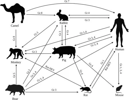
Overall,HEV exists in a wide range of hosts in nature,and most strains can spread across species (Fig.1).HEV strains from many animal species can infect humans,including pigs,boars,rabbits,rats and camels [14-18].Therefore,the research and establishment of HEV animal models is very crucial,which can help us study HEV transmission,clinical symptoms,extrahepatic manifestations,and therapeutic strategies.In this review,we summarized HEV animal models that have been constructed so far and analyzed their utility in future research on HEV.

Fig.1. HEV transmission among different hosts.HEV: hepatitis E virus.Gt: genotype.
HEV animal models
Pig
Pigs are the natural hosts of HEV genotypes 3 and 4.Pig HEV was first identified in the USA in 1997 and became the first animal strain of HEV discovered [19].Phylogenetic analysis showed that wild boar HEV sequences belonged to genotype 3 and clustered within subtypes 3a and 3c.HEV genotype 3 can be transmitted from infected wild boars to domestic pigs and wild boars through animal-to-animal contact [20].
HEV strains from other animal species can infect pigs.Rabbit HEV genotype 3 was proven to successfully infect pigs by intravenous injection [21].HEV genotypes 3 and 4 strains isolated from human were also able to successfully infect specific-pathogen-free(SPF) pigs [22,23].Besides,pig HEV genotype 3 is a pathogen of foodborne zoonotic diseases in developed countries.Humans can be infected with HEV by ingestion of meat and meat products prepared from HEV-infected pigs and wild boars [24,25].A study showed that pigs were sensitive to HEV infection,suggesting that pigs are easy to create experimental HEV models [26].The HEV pig model is of great importance for investigating the transmission,clinical symptoms,extrahepatic manifestations,and therapeutic strategies of HEV.
HEV is transmitted through blood transfusion [27].HEVpositive blood donations are detected in up to 0.14% in the Netherlands [28].Therefore,it is necessary to find a reliable method to inactivate the virus in blood samples to block transmission.D?hnert et al.mixed the intermediates of stabilized von Willebrand Factor/Factor VIII (VWF/FVIII) with liver homogenates from HEV-infected pigs and pasteurized at 60 °C for 3,6,and 10 h [29].According to the established model,the pigs were then inoculated with these samples.They analyzed fecal virus shedding,viremia,antibody production,and virus distribution in tissues.The results showed that pasteurization of HEV-positive plasma for 10 h at 60 °C can effectively reduce HEV infectivity,resulting in VWF/FVIII plasma products with an appropriate margin of safety for HEV,which may provide an effective method to solve the infection caused by HEV-positive blood donation and plasma products.
The chronic HEV pig model simulating immunocompromised patients was developed by administering immunosuppressive drugs to pigs,including cyclosporine,azathioprine,and prednisolone.Serum cytokine levels and cell-mediated immune responses were studied.It was found that interferon-γ-specific CD4 T-cell responses were reduced in immunosuppressed pigs during the acute phase of infection,but tumor necrosis factor (TNF)-αspecific CD8 T-cell responses were increased during the chronic phase of infection.This study suggested that active suppression of cell-mediated immune responses may cause chronic HEV infection [30].
The pig animal model helps us understand the extrahepatic manifestations of hepatitis E.HEV RNA was detected in the small intestine,lymph nodes,colon,and liver after infection of SPF pigs with porcine HEV and US-2 human HEV strains [31].Tian et al.showed that quasi-enveloped and non-enveloped HEVs could cross the blood-brain barrier (BBB).HEV RNA was detectable in the brain and spinal cord of pigs infected with HEV,and histological lesions were also observed,which revealed the underlying mechanism of HEV-related neuro-invasion [32].Besides,HEV infection can also cause pathological changes in pancreatic tissues.Jung et al.performed the virological and serological analyses of blood and stool samples using the miniature pig HEV genotype 3 model [33].The histopathology of the pancreas was analyzed,and immunohistochemistry was used to analyze cell death pathways and immune cell characterization in inflammatory lesions.They observed high levels of TNF-αand TNF-α-related apoptosis-inducing ligand in acinar cells throughout the pancreas,suggesting that HEV infection may lead to pancreatic necrosis.This study provided a method for the distribution of HEVin vivoand the pathogenesis associated with the resulting cell injury.
In swine HEV models,the prior injections of different HEV strains or their derived antigens have been proven to partially protect against homologous and heterologous HEV infections,which provides new idea into the development of HEV vaccine.Sanford et al.inoculated different groups of pathogen-free pigs with Nterminal truncated open reading frame 2 (ORF2) capsid antigens derived from swine,rat and avian HEV strains.In 4 weeks after vaccination,the animals were intravenously challenged with a genotype 3 mammalian HEV,followed by viremia,shedding of the fecal virus,and liver histological lesion analysis.The results showed that the pigs vaccinated with truncated recombinant capsid antigens from the three animal strains induced strong anti-HEV IgG responses,but these antigens provided only partial crossprotection against mammalian HEV genotype 3 [34].In another study,pigs in the three treatment groups were each inoculated with genotype 3 swine HEV,and 12 weeks later,challenged with the same genotype 3 swine HEV,genotype 3 human HEV,and genotype 4 human HEV,respectively.The control group was inoculated and challenged with phosphate-buffered saline (PBS).After challenges,viremia and fecal virus shedding were not detected,suggesting that prior infection with genotype 3 swine HEV prevented animals from developing viremia and shedding of fecal virus after challenges with homologous and heterologous genotypes 3 and 4 HEV.It was revealed that a vaccine derived from a genotype of HEV or an animal strain of HEV is sufficient to induce protective immunity against other HEV genotypes,and this finding is important when designing future HEV vaccine strategies [35].
Pigs have many similarities with humans in physiology and immunology.Domestic pigs are suitable and sensitive models for HEV genotype 3 infections since they are sensitive to human HEV genotype 3.However,HEV pig models have the disadvantage of high breeding cost and large body size that make them difficult to handle.Chinese Bama miniature pigs were found to be susceptible to HEV genotypes 3 and 4,and they can be used as an alternative to the standard pig models for the study of zoonotic HEV infection [36],which can compensate for the shortcomings of the standard pig models.Furthermore,it should be noted that pigs infected with genotypes 3 and 4 of HEV do not exhibit all the clinical manifestations associated with the virus.Consequently,this limits their ability to accurately replicate hepatic disease or induce pregnancy mortality,thereby restricting their usefulness in pathogenicity experiments.
Monkey
Natural HEV infection in monkeys has been reported.Serum tests in several countries have shown that nonhuman primates can be naturally infected with HEV [37-40].Most nonhuman primates are not natural reservoirs of HEV but are sensitive to experimental HEV infection,such as chimpanzees,cynomolgus,rhesus monkeys,owl monkeys,African green monkeys,Tamarin,and squirrel monkeys [41-43].
Monkeys can be infected across species by different HEV genotypes.For example,HEV genotype 1 from human can successfully infect rhesus and cynomolgus macaques and chimpanzees [44].A macaque monkey infected with rabbit HEV was observed to have elevated liver enzymes,seroconversion,viremia,and viral shedding in fecal specimens.The comparison of the genome sequence of the inoculum with the complete genome sequence of HEV transmitted in macaques showed 99.8% nucleotide identity [45].Experimental infection of two rhesus monkeys with swine-HEV showed seroconversion to anti-HEV antibodies and presence of viremia [46,47].HEV genotype 5 constructed by a reverse genetics system was able to infect cynomolgus monkeys,suggesting that HEV genotype 5 has the zoonotic potential [48].Cynomolgus macaques were infected after inoculation with filtered fecal samples containing BcHEV-GP(HEV genotype 8 isolated from Chinese Bactrian camels).In infected cynomolgus monkeys,viremia,viral urine and fecal excretion,elevated liver enzymes and seroconversion were observed,and anti-HEV IgM and IgG were detected [49].Yang et al.injected the V-105 strain of rat HEV into cynomolgus and rhesus macaques,and they observed serum conversion in all vaccinated monkeys and isolated rat HEV RNA in feces.Rat HEV was subsequently isolated from cynomolgus and rhesus monkeys,which can successfully infect nude mice and Sprague-Dawley rats.This suggested that rat HEV can replicate in monkeys,and that the infectious virus would then be released into feces.These results indicated that rhesus and cynomolgus monkeys are susceptible to rat HEV,revealing the possibility of zoonotic infection of rat HEV [50].Monkeys have been proven to be useful in animal models.Hence,the creation of a monkey model can be used to study the clinical symptoms,extrahepatic manifestations,and vaccines.
Monkeys can be used to study chronic HEV infection.Cynomolgus monkeys were treated with tacrolimus and infected with a Brazilian HEV genotype 3 strain isolated from naturally infected pigs.Three chronically HEV-infected monkeys showed microscopic features corresponding to chronic hepatitis in the absence of fibrosis and parenchymal cirrhosis [51].
In addition,due to the high similarity between monkeys and humans in terms of morphological structure and nervous system,monkeys have unique advantages as animal models of HEV in studying the extrahepatic manifestations of HEV on the nervous system.Zhou et al.inoculated monkeys with HEV particles and detected viral RNA and protein in brain tissue.Patients with HEVassociated neurological disease can excrete the virus into the cerebrospinal fluid,indicating direct infection of their nervous system.Therefore,HEV is neurotrophic in both monkeys and possibly humans,which challenged the existing perception of HEV as a pure hepatotropic virus and suggested that HEV infection should be considered in the differential diagnosis of idiopathic neurological diseases [52].HEV RNA and antigens were detected in the testes and epididymis of HEV-infected rhesus macaques.Testicular immunoblotting demonstrated the presence of HEV in spermatogonia with decreased testicular cell populations and primary spermatocyte and Sertoli cell numbers.The smooth muscle,tail-ciliated cells,and luminal sperm of the epididymis were also positive for HEV antigens.The presence of HEV RNA and antigens strongly suggested that HEV can replicate in the testes and epididymis,which may cause male infertility [53].Besides,cells fluorescently labeled with HEV antigen were detected in biopsies of bone marrow samples obtained from three monkeys with AHE and one with chronic hepatitis E,as well as from naturally infected monkeys.Doublestranded RNA was detected in bone marrow cells from acutely and chronically infected animals.This finding suggested that bone marrow cells could serve as a secondary target for the persistence of HEV and led to the hypothesis that HEV could be transmitted by bone marrow transplantation [54].
Monkeys are a good model for studying the efficacy of the HEV vaccine and for assessing the antigenicity of HEV.Im et al.proposed that a major structural protein expressed byEscherichia colicould prevent experimental HEV infection in primates.Monkeys inoculated with the purified peptide did not have detectable HEV in plasma samples after HEV infection and did not show evidence of HEV seroconversion [55].These results suggested that immunization with bacterially expressed peptides can prevent experimental infection of primates with homologous HEV strains [55].The monkeys were treated with alum-absorbed T1-ORF2 capsid protein derived from HEV strain T1 (genotype 4).After HEV genotype 1 and genotype 4 injections,immunized monkeys were found to be protected from infection and hepatitis,with shorter periods of elevated alanine aminotransferase (ALT) level and fecal shedding [56].In addition,the monkeys immunized with a purified peptide of the whole ORF3 protein of human HEV genotype 4 exhibited reduced viral titers and duration of viremia and fecal shedding after infection with HEV genotype 1 or genotype 4 and showed protection against hepatitis [57].Furthermore,Zhang et al.demonstrated that two doses of a candidate vaccine containing recombinant HEV capsid protein expressed in insect cells can partially protect rhesus monkeys from hepatitis E following intravenous challenge 6 or 12 months after vaccination [58].Li et al.also found that HEV 239 vaccine formulation partially protected against the infection and completely protected against hepatitis [59].
Besides,after treated rhesus macaques with depleting anti-CD8αmonoclonal antibodies M-T807R1,Bremer et al.challenged untreated and M-T807R1 treated rhesus macaques with HEV genotype 3 isolated from a chronically HEV-infected patient [60].They found that liver infiltration of functional CD8+T cells coincident with HEV clearance in untreated rhesus macaques,and that a 1-week delay in HEV clearance in CD8+T-cell depleted rhesus macaques.The findings indicated that HEV may be susceptible to multiple arms of the immune response that can act independently to terminate infection [60].
The comparison of nucleotide composition and codon usage bias between HEV genotypes and animal hosts showed that nonhuman primates and humans share the same large and highfrequency codons as HEV.The codon usage bias of nonhuman primates is closest to that of zoonotic HEV genotypes,suggesting that the cellular environment of nonhuman primates may be more suitable for viral infection than that of pigs and rabbits.However,monkeys and other nonhuman primates as model animals are expensive,difficult to operate experimental procedures,and have some problems in experimental ethics,restricting the study of monkeys as animal models of HEV.Moreover,most nonhuman primates are not natural reservoirs of HEV,so their use as model animals to observe the course and clinical manifestations of HEV infection is limited.
Rabbit
Rabbit HEV was first discovered in 2009 [61].A variety of rabbit HEV strains have been reported,demonstrating that rabbits are natural hosts for HEV.The serological survey on rabbit HEV infection was conducted in Italy.The results revealed an anti-HEV antibody seroprevalence of 3.40% in 206 farmed rabbits and 6.56%in 122 pet rabbits [62].Another study from Germany showed that 37.1% of hares were detected with HEV RNA [63].Besides,in a 2017 survey investigating the prevalence of rabbit HEV in Korea,rabbit HEV was found in two of six rabbit farms and 17 of 264 rabbit fecal samples [64].
HEV genotype 1 from human can successfully infect rabbits [65].It is possible to successfully establish models of infection with human HEV genotype 4,swine HEV genotype 4 and rabbit HEV genotype 3 infection models in rabbits and observe typical signs of acute hepatitis,including viremia/antigenemia and fecal virus/antigen shedding [65-67].Notably,rabbits are susceptible to zoonotic rabbit HEV genotype 3 infection and can exhibit characteristics of acute or chronic HEV infection similar to those of patients and may be suitable for monitoring the long-term dynamics of chronic HEV infection in humans in an immunocompetent setting [68,69].Liu et al.successfully established a rabbit model infected with swine-derived HEV genotype 4 isolated in China,which provided new insights into the pathogenicity of swine-derived HEV genotype 4 in rabbits and zoonoses of different hosts [70].The experimental HEV genotype 3 infection of European rabbits with a wild boar-derived strain also led to seroconversion within four to five weeks post inoculation [71].Rabbits are also cross-species infected with HEV genotype 8 found in Bactrian camels.Zhang et al.inoculated Japanese white rabbits with HEV genotype 8 and observed that viral RNA was present in fecal specimens from rabbits inoculated with HEV genotype 8 and that anti-HEV IgG antibodies were present in their serum samples [72].Antibodies raised in HEV genotype 8-infected rabbits did not protect them from the rabbit HEV challenge,suggesting a difference in antigenicity between HEV genotype 8 and rabbit HEV [72].The rabbit model is important in studying the effects of HEV on pregnancy,extrahepatic manifestations,and HEV vaccines and antiviral drugs.
HEV has been reported to increase maternal mortality in infected women,and hepatitis virus infection during pregnancy can also lead to adverse pregnancy outcomes,including miscarriage,stillbirth,fulminant liver failure,and ruptured fetal membranes [73].HEV infection was modeled in six pregnant rabbits.Two of the six pregnant rabbits were aborted and three died,which showed that vertical transmission was associated with HEV replication in the placenta [74].Li et al.constructed an HEV model in pregnant rabbits,and pregnant rabbits infected with HEV showed signs of HEV infection,including fecal viral shedding,elevated ALT/aspartate aminotransferase (AST) and histopathological changes,and adverse pregnancy outcomes,and resulted in vertical infection.HEV 239 vaccine was also shown to be effective in preventing rabbit HEV infection in pregnant rabbits,and anti-HEV antibodies persisted in newborn rabbits [75].These results revealed that women of childbearing age should be vaccinated against HEV before pregnancy to avoid the risk [75].Besides,rabbits inoculated with homologous rabbit HEV isolate (CHN-BJrb14 HEV strain) exhibited viremia,fecal viral shedding,and liver histopathology showing chronic inflammation and some degree of fibrosis [69].The association between viral dose and the development of chronic hepatitis should be further investigated.
Rabbits are considered a useful model for exploring the pathogenesis of hepatitis E,particularly extrahepatic manifestations from HEV.HEV infection can cause a range of neurological syndromes,including neuropathic myasthenia gravis,cerebral ischemia or infarction,seizures,encephalitis,and acute combined facial and vestibular neuropathy [76].Tian et al.observed neuropathological changes in rabbits infected with HEV,particularly viral encephalitis associated with perivascular lymphocyte cuffs and microglia nodules.They proposed to study the role of tight junction proteins in the integrity of BBB during HEV infection thus understanding the pathogenesis of HEV-induced central nervous system injury [77].
The rabbit model is also a good model for evaluating prophylactic vaccines and possible antiviral drugs.He et al.induced immune suppression in rabbits using cyclosporine A and inoculated them with rabbit-derived HEV genotype 3 and human-derived HEV genotype 3 or 4.Chronic HEV infection was successfully established in immunocompromised rabbits,exhibiting typical features of liver fibrosis [78].Ribavirin effectively cleared HEV infection in immunocompromised rabbits.Meanwhile,researchers found that vaccination completed before immune suppression provided complete protection against both HEV genotypes 3 and 4 infections,but vaccination during immune suppression only provided partial protection,and the efficacy did not increase with increasing vaccine dose [78].Rabbits immunized with recombinant rat HEV capsid protein were protected against HEV genotype 3 infection [79].In another study,Cheng et al.vaccinated rabbits with 0,10,and 20μg of the candidate HEV vaccine p179 followed by infection with isolated HEV strain H4-NJ703.All rabbits at a dose of 20μg were protected from HEV infection [67].The efficacy of different doses of the HEV 239 vaccine was evaluated in rabbits.The results showed that two doses of 10μg HEV 239 induced the best immune response in rabbits compared to two doses of 20μg and 30μg [80].Besides,HEV 239 vaccine was proven to protect pregnant rabbits from adverse pregnancy outcomes,and maternal anti-HEV protective antibodies can be transferred to pups [81].In addition,Liang et al.used a rabbit model for acute HEV infection and evaluated the effect of different doses of sofosbuvir against HEV genotypes 3 and 4.The results showed that sofosbuvir exhibited anti-HEV effects in a rabbit model and was more effective in eliminating AHE when used in combination with ribavirin [82].
Rabbit HEV is closely genetically and antigenically related to other mammalian HEV strains,and HEV-infected rabbits may serve as a useful natural animal model for studying the pathogenesis of HEV.In addition,rabbits infected with rabbit HEV may provide a suitable parallel animal model,especially in the medically important pregnancy setting,where experimental infection of pregnant rabbits with rabbit HEV can result in high mortality and HEV vertical transmission,with localized hepatocyte necrosis seen when the liver is observed under the microscope [83].No other animal models for enhancing maternal disease have been found.However,it should be acknowledged that rabbits cannot reliably be infected with human HEV strains,which poses limitations to their utility as animal models for HEV research.
Mouse
The lack of effective small animal models of HEV has hindered the study of HEV pathogenesis and the discovery and development of specific anti-HEV drugs.As HEV becomes more prevalent and more strains that are infectious to humans are discovered,there is a great need for suitable small animal models.HEV infection in human liver chimeric mice has been intensively studied,and these models can be used for the study of viral pathogenesis as well as for the identification and preclinical evaluation of new antiviral compounds [84,85].
Infection of primary human hepatocyte-repopulated UPA/SCID/Beige mice with HEV genotype 1 and genotype 3 established an effective model of HEV infection [86].Histopathological changes in the liver and spleen and elevated liver enzyme levels were observed in BALB/c nude mice inoculated with porcine HEV genotype 4,and HEV antigen was detected in different extrahepatic organs [87].BALB/c mice can also be infected with rabbit HEV by strong feeding method and contact exposure inoculation [88].The construction of HEV mouse models is important in studying the effect of HEV on pregnancy,extrahepatic manifestations,and HEV antiviral drugs.
Yang et al.assessed the effect of uterine damage caused by HEV infection on pregnant outcomes.The results showed that HEV replicates in the uterus and causes miscarriage and infertility [89].Severe damage was found in the uterus of HEV-infected mice that experienced miscarriage,with thinning of the endometrial thickness,increased inflammatory response,and increased apoptosis [89].These findings have important implications for understanding the pathogenesis of HEV during pregnancy.In addition,Yang et al.constructed a gestational mouse HEV model by injecting HEV into female mice at different periods after mating in healthy mice.A higher miscarriage rate was found in the middle of gestation [90].
The application of mouse models allows the study of HEV replication outside the liver.Indirect immunofluorescence and reverse transcription-nested polymerase chain reaction (RT-nPCR) showed that HEV antigen and HEV RNA were detected in the liver,spleen,kidney,jejunum,ileum,and colon of BALB/c nude mice inoculated with porcine HEV genotype 4 [87].In BALB/c mice inoculated with porcine HEV genotype 4 RNA constructed with reverse genetics,HEV RNA negative strands were again detected in the liver,spleen,and kidney [91].Besides,Situ et al.constructed a BALB/c mouse model by intravenous injection of human-derived HEV genotype 4 and observed positive signals for HEV antigen in the testis,epididymis,and seminal vesicles [92].Impaired sperm quality,disruption of the blood-testis barrier,and reduced spermatozoa suggested that HEV infection causes testicular damage and thus affects infertility.
Sari et al.inoculated human-liver chimeric mice with wild-type HEV or ORF3-null mutated HEV.The wild-type HEV established a persistent infection in the humanized mice,whereas the mutant HEV lacking ORF3 failed to sustain the infection,despite transient replication in the liver which was eventually cleared.These results demonstrated that ORF3 is necessary for HEV fecal shedding and persistent infection.Therefore,targeting ORF3 could be a viable treatment strategy for HEV infection [84].
The mouse models are good ones for anti-HEV drug studies.Silvestrol was administered to humanized mice infected with HEV genotype 1,which exhibited anti-HEV activity by blocking HEV replication in different subgenomic repeats in a dose-dependent manner at low nanomolar concentrations.A significant decrease in fecal HEV RNA levels was observed in experimental mice after administration,and this compound could be considered for future therapeutic strategies in immunosuppressed patients with chronic hepatitis E [93].PEGylated interferon alpha (PegIFN-α) was administered to humanized mice infected with HEV genotype 1 as well as genotype 3.The rapid viral clearance of PegIFN-αwas demonstrated in HEV genotype 1.A significant increase in intrahepatic interferon-stimulated gene transcript levels was also observed.It was shown that HEV genotype 1 and genotype 3 infections will not trigger an innate intrahepatic immune response and remain highly sensitive to PegIFN-αin immunocompromised humanized mice [94].However,PegIFN-αhas serious side effects that hinder the use of IFN-αin immunocompromised transplant recipients and increase the risk of acute rejection in HEV-infected solid organ transplant recipients.PegIFN-λwas identified as an alternative therapeutic option by Sari et al.[95],and they found that HEV antigen and HEV RNA were completely cleared after administration of PegIFN-λto HEV-infected liver-humanized mice.Importantly,PegIFN-λwas well tolerated and no systemic side effects were recorded.
The mouse models are also used for vaccine research.HEV ORF2 has been extensively studied as a vaccine candidate.Deshmukh et al.found that complete ORF2 and a small neutralizing epitopecontaining region were immunogenic in mice,which may be used as DNA and DNA-prime-protein-boost vaccines [96].In addition,HEV 239 protein in different preparations was also used as a vaccine candidate by Khateri et al.,and was injected subcutaneously in BALB/c mice and then evaluated for induced cellular and humoral immunity [97].Although the different formulations of HEV 239 vaccines were not significant compared to each other,they showed significant humoral and cellular responses compared to controls.
Human liver chimeric mice are capable of constructing various HEV models such as human HEV genotype 3,genotype 4,and rabbit genotype 3,which have the advantages of lower cost,ease to handle,and shorter life cycle.Also,human liver chimeric mice infected with human-derived HEV genotype 3 can develop 100 %chronic HEV infection,mimicking the infection process in solid organ and bone marrow transplant recipients.HEV in these mice is preferentially shed in the bile and feces,which corresponds to the secretion pattern seen in humans [85].Therefore,human liver chimeric mice are a suitable model for future HEV transmission and pathobiological studies.Although the chimeric mouse model can reproduce chronic HEV infection,human HEV strains does not naturally infect mice,and the interaction of innate pathways of human cells with the adaptive immune pathways of mouse cells cannot be fully reproduced in a chimeric mouse model [98].These limitations should be considered when conducting research on HEV chimeric mouse models.
Rat
Rat HEV was first discovered in Germany in 2010 and is widely present and spread in Asian and American countries [99,100].Rat HEV belongs to HEV species C genotype 1 (HEV-C1) [101],and rat HEV has only 50%-60% nucleotide sequence identity with HEV strains in HEVPaslahepevirus balayaniand is genetically separated from human HEV [102].
HEV from wild rats was able to infect Wistar rats and seroconversion and viral excretion were observed [103].Sridhar et al.immunosuppressed rats with a combination of drugs (prednisolone,tacrolimus and mycophenolate).Then rats were challenged with human-and rat-derived HEV-C1 strains or a human-derived HEVPaslahepevirus balayanistrain.The results showed that a highdose immunosuppression regimen consistently prolongs humanand rat-derived HEV-C1 infection compared to low-dose immunosuppression treatment and transient infection in immunocompetent rats.This protocol developed a reliable immunosuppressed rat model of chronic hepatitis E that closely resembles the characteristics of human chronic hepatitis E [104].Rats inoculated with human fecal suspensions containing HEV genotype 1 were infected and exhibited viral shedding,viremia,and histopathological changes in the liver,spleen,and lymph nodes [105].Jian et al.infected rats by intraperitoneal injection of genotype 4 HEVpositive porcine fecal suspension,and HEV RNA was detected in most tissues and organs after infection,while pathological changes of chronic hepatitis were observed in the liver,demonstrating that porcine HEV can infect Sprague-Dawley rats,and therefore the rat model of porcine HEV infection is suitable for studying HEV infection in small animal models [106].Debing et al.used the rat HEV strain LA-B350 to infect thymus-free nude rats [107].The cDNA clone pLA-B350 was constructed,and the infectivity of its capped RNA transcript was confirmedin vitroandin vivo[107].
Rats can be used as animal models to study HEV therapeutic strategies.Ribavirin was proven to effectively inhibit the replication of LA-B350 in thymus-free nude rats,confirming the applicability of the rat model in a HEV infection and antiviral study [107].Schlosser et al.used a wild boar-derived HEV genotype 3 strain to infect Wistar rats and monitored viral replication and shedding,as well as humoral immune responses [71].HEV RNA and anti-HEV antibodies were detected in one-eighth of the rats.In addition,researchers inoculated eight immunosuppressive dexamethasonetreated rats both intravenously and orally to assess the effect of immune status on infection dynamics and shedding.Interestingly,treatment with immunosuppressive dexamethasone of rats did not enhance susceptibility to HEV infection,but resulted in a complete absence of seroconversion and replication of RNA in all inoculated rats [71].
Rats have been shown to be susceptible to boar HEV genotype 3,porcine HEV genotype 4,and human HEV genotype 1 and can serve as a good small animal model for HEV infection studies [71,105,106].Furthermore,the effective inhibition of viral replication in HEV LA-B350-infected thymus-free nude mice by ribavirin and the effect of dexamethasone on the infection profile of Wister rats revealed the potential of rat models for HEV antiinfection and antiviral studies [71,107].However,studies related to HEV rat model are relatively few,so more efforts are needed in the future.Furthermore,rats and even nude rats showed resistance to experimental infection with HEV genotypes 1-4.This indicates that rats are not suitable animals for model for studying human HEV [108,109].
Other animal models
In addition to the major HEV animal models mentioned above,several other animals can be used as HEV models to help us understand HEV more thoroughly.Avian HEV was first isolated from chickens in 2001 [110],while phylogenetic analysis showed that the virus has 60% sequence similarity to human HEV [111].SPF chickens can naturally be infected with avian HEV through fecaloral transmission [112].Vaccine studies using the HEV chicken models have been fruitful.The chickens were immunized using a recombinant ORF2 capsid protein from avian HEV and then challenged with avian HEV.None of the test chickens immunized with the avian HEV capsid protein had detectable viremia,fecal viral shedding,or observable severe hepatitis lesions,indicating that the avian HEV ORF2 capsid protein allows chickens to develop protective immunity against avian HEV [113].However,chickens are not susceptible to human or porcine HEV and the clinical signs of infection observed do not match those of humans,making the study of chickens as HEV animal models limited.
Ferrets can also be used for HEV studies.Significant elevations in fecal and serum RNA,seroconversion,and ALT were observed in ferrets after HEV infection,further exhibiting three modes of infection: subclinical infection,acute hepatitis,and persistent infection [114].However,the low similarity between ferret and human HEV strains suggests that ferrets may not be suitable for studies to investigate human HEV vaccines and antiviral drugs.

Table 1Study on extrahepatic manifestations in different animal models.

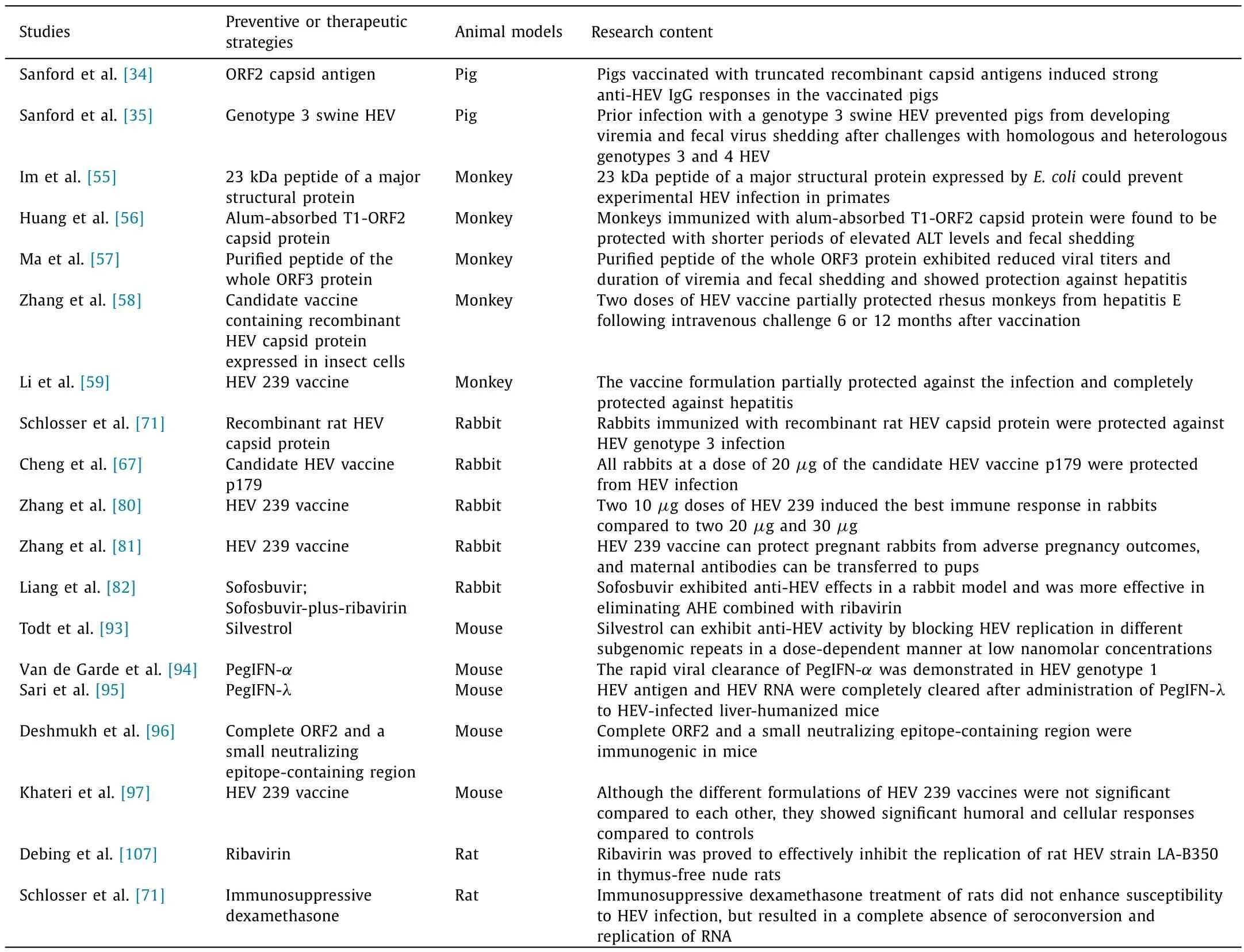
Table 2Study on preventive or therapeutic strategies in different animal models.
Zhang et al.inoculated Mongolian gerbils with HEV genotypes 1,3,4,5,7,and 8,rabbit HEV and rat HEV,and showed that HEV genotype 5 and rat HEV replicated efficiently in Mongolian gerbils in the same manner as HEV genotype 4.Mongolian gerbils are not only susceptible to HEV belonging to HEVPaslahepevirus balayani,but also to rat HEV belonging to HEV-C1,and thus Mongolian gerbils can be used as a small animal model for cross-protection experiments between HEVPaslahepevirus balayaniand HEV-C1 [115].Additionally,HEV genotype 4,genotype 5,and rat HEV were successfully transmitted to Mongolian gerbils by oral inoculation,and Mongolian gerbils are broadly susceptible to HEV,with the degree of susceptibility depending on the genotype [115].Besides,Shi et al.successfully inoculated Mongolian gerbils with genotype 4 strain swine HEV [116].They found that HEV can damage the BBB and replicate in brain and spinal cord,revealing the role of HEV in neurological diseases [116].Hence,this animal may be useful for HEV studies.
Furthermore,HEV has been found in bats [117].HEV antibodies have also been detected in goats,horses,and dogs,but HEV RNA has not been formally detected in these animals.These animals may serve as new models for HEV research in the future.
Studies on extrahepatic manifestations,preventive or therapeutic strategies in different animal models are listed in Tables 1 and 2.
Conclusions
More and more HEV strains and hosts are being discovered,and research on HEV is gaining attention,but more work is needed to study the molecular mechanisms,pathogenesis,and host immune response.HEV,as one of the major causes of viral hepatitis,can cause acute or chronic hepatitis,and lead to high mortality in pregnant patients.As a zoonotic disease,HEV can be transmitted across species in multiple hosts,forming multiple naturally occurring animal models capable of infecting human-associated HEV genotypes and mimicking clinical symptoms and extrahepatic manifestations.
This review summarized multiple HEV animal models and described their progress in HEV transmission,clinical symptoms,extrahepatic manifestations,and therapeutic strategies.These animal models have played an important role in providing new insights into HEV,but they also have issues such as cost and ethical limitations.Further research on animal models of HEV is urgent,especially those related to extrahepatic manifestations of HEV,anti-HEV drugs and vaccine studies.It is also important to develop animal models of HEV that are easier to manipulate,less costly,and closer to human HEV infection.
Acknowledgments
None.
CRediT authorship contribution statement
Ze Xiang:Conceptualization,Writing -original draft.Xiang-Lin He:Conceptualization,Writing -original draft.Chuan-Wu Zhu:Data curation,Writing -original draft.Jia-Jia Yang:Data curation.Lan Huang:Formal analysis.Chun Jiang:Writing -review &editing.Jian Wu:Conceptualization,Funding acquisition,Writing -review &editing.
Funding
This study was supported by grants from the National Natural Science Foundation of China (8 2272396) and the Fundamental Research Funds for the Central Universities (2 26-2022-00061).
Ethical approval
Not needed.
Competing interest
No benefits in any form have been received or will be received from a commercial party related directly or indirectly to the subject of this article.
 Hepatobiliary & Pancreatic Diseases International2024年2期
- Hepatobiliary & Pancreatic Diseases International的其它文章
- Editors
- Information for Readers
- Meetings and Courses
- Liver transplantation and liver resection as alternative treatments for primary hepatobiliary and secondary liver tumors: Competitors or allies?
- Laparoscopic anatomical liver resection of segment 7 using a sandwich approach to the right hepatic vein (with video)
- Severe liver injury and clinical characteristics of occupational exposure to 2-amino-5-chloro-N,3-dimethylbenzamide: A case series
