Preparation and evaluation of PEGylated phospholipid membrane coated layered double hydroxide nanoparticles
Mina Yan,Zhaoguo Zhang,Shengmiao Cui,Xuefei Zhang, Weijing Chu,Ming Lei,Ke Zeng,Yunhui Liao,Yihui Deng*, Chunshun Zhao,**
aSchool of Pharmacy,Shenyang Pharmaceutical University,103 Wenhua Road,Shenyang 110016,China
bSchool of Pharmaceutical Sciences,Sun Yat-sen University,132 Waihuan East Road,Guangzhou Higher
Education Mega Center,GuangZhou 510006,China
cGuangdong Pharmaceutical University,280 Waihuan East Road,Guangzhou Higher Education Mega Center, GuangZhou 510006,China
Preparation and evaluation of PEGylated phospholipid membrane coated layered double hydroxide nanoparticles
Mina Yana,b,Zhaoguo Zhangb,Shengmiao Cuic,Xuefei Zhangb, Weijing Chub,Ming Leib,Ke Zengb,Yunhui Liaob,Yihui Denga,*, Chunshun Zhaob,**
aSchool of Pharmacy,Shenyang Pharmaceutical University,103 Wenhua Road,Shenyang 110016,China
bSchool of Pharmaceutical Sciences,Sun Yat-sen University,132 Waihuan East Road,Guangzhou Higher
Education Mega Center,GuangZhou 510006,China
cGuangdong Pharmaceutical University,280 Waihuan East Road,Guangzhou Higher Education Mega Center, GuangZhou 510006,China
A R T I C L EI N F O
Article history:
Received 13 May 2015
Received in revised form 11
September 2015
Accepted 13 September 2015
Available online 25 September 2015
Preparation
In vitroevaluation
Layered double hydroxides
Phospholipid membrane
Size distribution
Zeta potential
The aim of the present study was to develop layered double hydroxide(LDH)nanoparticles coated with PEGylated phospholipid membrane.By comparing the size distribution and zeta potential,the weight ratio of LDH to lipid materials which constitute the outside membrane was identifed as 2:1.Transmission electron microscopy photographs confrmed the core-shell structure of PEGylated phospholipid membrane coated LDH(PEG-PLDH) nanoparticles,and cell cytotoxicity assay showed their good cell viability on Hela and BALB/ C-3T3 cells over the concentration range from 0.5 to 50 μg/mL.
?2016 The Authors.Production and hosting by Elsevier B.V.on behalf of Shenyang Pharmaceutical University.This is an open access article under the CC BY-NC-ND license (http://creativecommons.org/licenses/by-nc-nd/4.0/).
1.Introduction
Layered double hydroxides(LDHs)constitute a broad family of lamellar solids which in the last decades have deserved an increasing interest because of their applications in different felds, such as catalysts[1],traps for anionic pollutants[2],and fame retardants[3].In recent years,LDHs are gaining increasing attention as carriers for drug/gene delivery[4].They are sometimes named as anionic clays due to the similarities shared with cationic clays,or hydrotalcite-like materials,as derived from the natural hydroxycarbonate of Mg and Al[5].
LDHs are a class of anionic lamellar compounds made up of positively charged brucite-like layers with an interlayer gallery containing charge compensating anions and water molecules [6].The LDH composition can be expressed in a general formula [M2+1?xM3+x(OH)2](A?x·nH2O),where M2+and M3+can be most divalent and trivalent metal ions and A?any type of anions.Many studies demonstrated that anticancer drugs[7–9]and genes[9–11] could be successfully intercalated into the LDH layers.
As a nanocarrier,LDH shows many advantages over other delivery systems[4].First,they tend to exhibit low cytotoxicity,even at a high dose.In addition,LDH is easily degraded in the acidic environment,thus endowing it an advantage of biodegradability,which is also responsible for their endolysosome escaping behavior.However,positively charged LDH nanoparticles allow avid association with the negatively charged plasma proteins,which adversely infuences their pharmacokinetic behavior and reduces their blood residence time. Besides,we observed that they can easily cause death during intravenous injection.
Phospholipids have long been perceived as safe materials tocomposedrugdeliveryvehiclesbecauseoftheirsuperiorbiocompatibility.They could cover on the surface of other solid nanoparticles[12],and several lipid-coated hybrid carriers have beendeveloped[13].Veryrecently,wereportedaPEGylatedphospholipid membrane coated LDH(PEG-PLDH)delivery system with a core-shell structure[14].This new composite system showed enhanced therapeutic effcacy and survival rate when comparedtonakedLDHnanoparticlessincethepositivecharges were shielded by phospholipid membrane.DOPA was chosen as a membrane material because it readily forms liposomes [15],and has a negatively charged phosphatidic acid headgroup which aids in the shielding of positively charged LDH.A PEG–lipid conjugate was also included in the leafet lipids to prolong the circulation time[12].The pharmacokinetic study andin vivoantitumor activity of PEG-PLDH nanoparticles have been investigated;here we report the preparation,properties,as well asin vitrocell cytotoxicity of this novel delivery system.
2.Materials and methods
2.1.Materials
Dioleoylphosphate(sodium salt)(DOPA),distearoylphosphatidylethanolamine-[methoxy(polyethylene glycol)-2000] (DSPE-PEG2000)and cholesterol(Chol)were purchased from Avanti Polar Lipids(Alabaster,AL,USA).Dilauroylphosphatidylcholine(DLPC),dimyristoylphosphatidylcholine(DMPC), dipalmitoylphosphocholine(DPPC)and distearoylphosphocholine(DSPC)were purchased from Genzyme(Cambridge, MA,USA).Methotrexate(MTX)was provided by Amresco(USA). HeLa and BALB/C-3T3 cells purchased from the Laboratory Animal Center of Sun Yat-sen University(Guangzhou,China) were cultured with DMEM medium(Sigma-Aldrich,USA) supplemented with 10%calf serum(Life Technologies,USA).
2.2.Preparation of LDH and LDH-MTX nanoparticles
LDH nanoparticle suspension was prepared with a quick precipitation and subsequent hydrothermal treatment[16,17].In brief,3.0 mmol of MgCl2and 1.0 mmol of AlCl3were dissolved in 10 ml deionized water.This salt solution was then rapidly added to a basic solution(30 ml)containing 6.0 mmol of NaOH within 5 s to generate the precipitate of LDH.After being stirred for 10 min in N2stream at room temperature,the precipitate was collected via centrifugation and further washed twice.Henceforth,the washed precipitate was manually dispersed in 20 ml of deionized water and placed in a 25 ml autoclave with Tefon linen,followed by hydrothermal treatment at 100°C in an oven to get the suspension of LDH nanoparticles.To achieve methotrexate loaded LDH(LDHMTX)nanoparticles,0.1 mmol of MTX was dissolved in NaOH before the quick precipitation step.
2.3.Preparation of PEG-PLDH and PEG-PLDH-MTX nanoparticles
PEG-PLDH or PEG-PLDH-MTX nanoparticle suspension was prepared by self-assembly.Lipid materials were dissolved in CHCl3and dried under a N2stream.A trace amount of chloroform was removed by keeping the lipid flm under a vacuum.The lipid flm was hydrated with PBS(pH 7.4)to obtain an empty liposome suspension.LDH or LDH-MTX nanoparticle suspension was added to the liposomes.The mixture was sonicated (in a water bath)using a laboratory ultrasonic cleaning machine (SB-5200DTN,Ningbo Scientz Biotechnology Co.,Ltd.Zhejiang,China)at 250 W.
2.4.Size and zeta potential
The average hydrodynamic diameter,polydispersity index(PdI) and zeta potential of the nanoparticles were determined by dynamic light scattering using a Malvern Zetasizer Nano ZS90 (Malvern Instruments,Worcestershire,UK).The temperature of the cell was kept constant at 25°C.The zeta potential was calculated from the electrophoretic mobility using the Smoluchowski equation.Samples of the prepared complexes were diluted in distilled water and were measured at least three times.Size results are given as intensity distribution by the mean diameter with its standard deviation.
2.5.Transmission electron microscopy(TEM)
The particle morphology of LDH and PEG-PLDH was confrmed by usingTEM.The samples were put on carbon formvar coated grids,negatively stained with uranyl acetate 1.5%,and observed using a JEOL JEM-1400 instrument(JEOL,Japan) (120 kV).
2.6.Cell culture
Cells were cultured in DMEM supplemented with 10%calf serum and antibiotics(streptomycin 100 U/ml,penicillin 100 U/ml)at 37°C in humidifed air with 5%CO2.
2.7.Cell cytotoxicity assay
The cytotoxicity of free MTX,LDH-MTX and PEG-PLDH-MTX nanoparticles on HeLa or BALB/C-3T3 cells was examined via cytotoxicity assay.Briefy,cells were seeded in 96-well plates at a density of 1×10⁴cells per well and incubated for 24 h.Then, the cells were treated with serial concentrations of free MTX, LDH-MTX or PEG-PLDH-MTX nanoparticles.After incubation for certain time,10 μl of dimethyl thiazolyl diphenyl tetrazolium bromide solution(5 mg/ml)was added to each well and incubated for 4 h.Finally,the medium was replaced with 150 μl of dimethylsulfoxide and the optical density was determined with a microplate reader at a wavelength of 570 nm in triplicate.
3.Results and discussion
3.1.Preparation of LDH and PEG-PLDH nanoparticles
Layered double hydroxides(LDHs)were prepared by coprecipitation and subsequent hydrothermal treatment. Liposomes was achieved by using thin-flm hydration method. Fig.1 showed the schematic of the self-assembly of PEG-PLDH nanoparticles.The driving force of self-assembly was the electrostatic interaction between negatively charged segments of DOPA and positively charged surface of LDH nanoparticles.
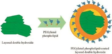
Fig.1–Schematic of self-assembly of PEG-PLDH nanoparticles.
3.2.Effect of hydrothermal treatment time on LDH nanoparticles
Hydrothermal treatment time has potential infuence on the particle sizes and surface charges of LDH nanoparticles.Fig.2 shows that after 4 or 6 h of heat-treatment at 100°C,the LDH particles become very uniform,with a narrow particle size distribution.However,too short treatment time(2 h)leads to much bigger particles,indicating the presence of some degrees of aggregation.It seems that 2 h of heat-treatment is not enough to disperse all aggregates into individual LDH crystallites.
3.3.Optimization of the preparation conditions for PEG-PLDH nanoparticles
3.3.1.Sonication time
The sonication time in preparation method was investigated at 0,1,5,10 and 15 min.As shown in Fig.3,the size distribution became very uniform after 10 min of sonication.
3.3.2.Weight ratio of LDH to lipid materials
DOPA,DSPC,DSPE-PEG2000and Chol in the molar ratio of 30:27:3:20 were used to form the lipid membrane.The LDH/ lipids weight ratio was investigated at 0:1,0.5:1,1:1 2:1 4:1,8:1, 16:1 and 32:1.With more and more positively charged LDH nanoparticles added into the system,the zeta potential increased from?41.8 mV to?14.4 mV(Fig.4).Meanwhile,the average size of PEG-PLDH nanoparticles decreased as the LDH/ lipids weight ratio increased from 0:1 to 2:1.Positively charged LDH nanoparticles could interact with negatively charged DOPA so that the phospholipid membrane coated tightly on the surface on LDH nanoparticles.As a result,the particle size decreased when LDH nanoparticles mixed with empty liposomes. However,when the LDH/lipids weight ratio further increased from 2:1 to 32:1,the average size increased with it.This may be caused by the exposure of positively charged LDH nanoparticles which would attract negatively charged PEG-PLDHnanoparticles around them.A proper ratio of LDH to lipid materials was very important when preparing PEG-PLDH nanoparticles.Therefore,2:1 was used as the optimum weight proportion between LDH and liposomes.
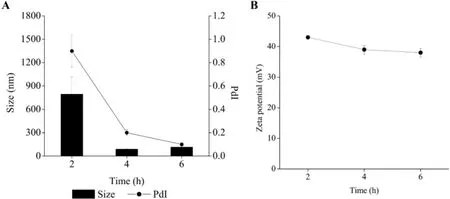
Fig.2–Effect of hydrothermal treatment time on(A)particle size,polydispersity index and(B)zeta potential of LDH nanoparticles(mean±standard deviation,n=3).
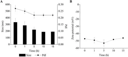
Fig.3–Effect of sonication time on(A)particle size,polydispersity index and(B)zeta potential of PEG-PLDH nanoparticles (mean±standard deviation,n=3).
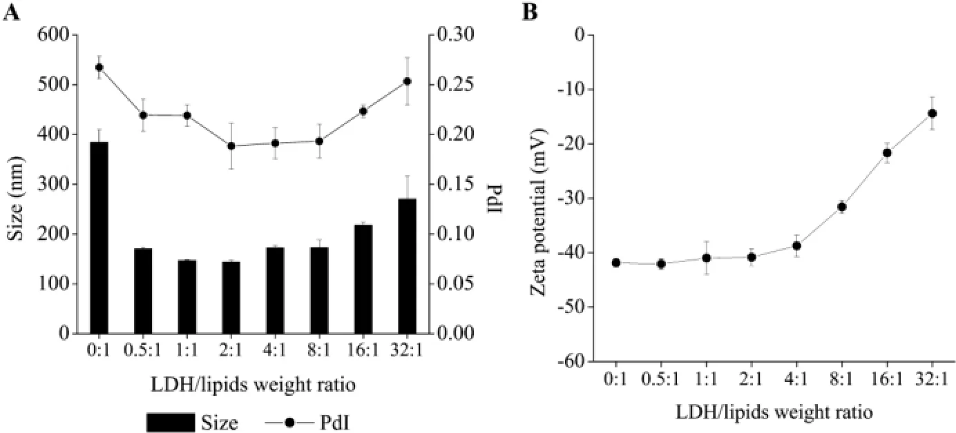
Fig.4–Effect of LDH/lipids weight ratio on(A)particle size,polydispersity index and(B)zeta potential of PEG-PLDH nanoparticles(mean±standard deviation,n=3).
3.3.3.Molar ratio of DSPE-PEG2000to phospholipids
The amount of phospholipids was fxed at 8 μmol,in which DOPA was fxed at 3 μmol.DSPE-PEG2000was added in different amounts to achieve different molar ratios of DSPE-PEG2000to phospholipids(1:20,2:20,3:20,4:20,6:20 and 8:20).As shown in Fig.5,the particle sizes and PdIs were similar when the molar ratio of DSPE-PEG2000to phospholipids rises from 1:20 to 4:20. When the molar ratio of DSPE-PEG2000to phospholipids was higher than 4:20,the size distribution became uneven.The surface charge rises with the DSPE-PEG2000/phospholipids molar ratio increase.It was probably because DSPE-PEG2000could forma shielding layer on the outside of the membrane which could cover the negative charge of DOPA.As a result,4:20 was chosen to be the optimal DSPE-PEG2000/phospholipids molar ratio.
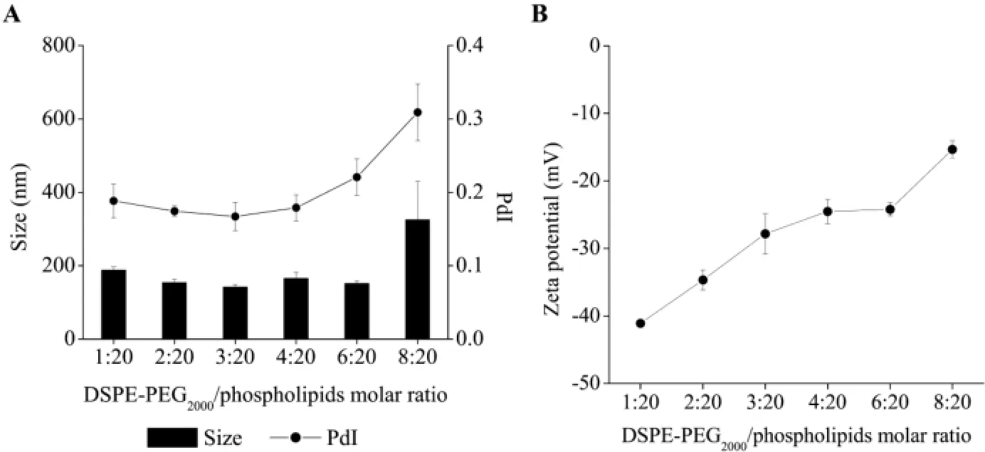
Fig.5–Effect of DSPE-PEG2000/phospholipids molar ratio on(A)particle size,polydispersity index and(B)zeta potential of PEG-PLDH nanoparticles(mean±standard deviation,n=3).
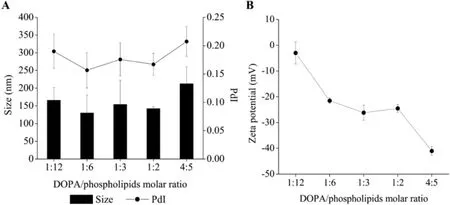
Fig.6–Effect of DOPA/phospholipids molar ratio on(A)particle size,polydispersity index and(B)zeta potential of PEG-PLDH nanoparticles(mean±standard deviation,n=3).
3.3.4.Molar ratio of DOPA to phospholipids
The amount of phospholipids was fxed at 8 μmol,and DOPA was added in different amounts to achieve different molar ratios of DOPA to phospholipids(1:12,1:6,1:3,1:2,4:5).Fig.6 showed that 1:2 was the optimum molar ratio of DOPA to phospholipids.Negatively charged DOPA molecules would repel each other when composing the phospholipid membrane.As a result, too much DOPA may lead to instability.However,the phospholipid membrane couldn’t interact closely with positively charged LDH nanoparticles without negatively charged DOPA.Therefore,the proper molar ratio of DOPA to phospholipids was a key point to obtain stable PEG-PLDH nanoparticles.
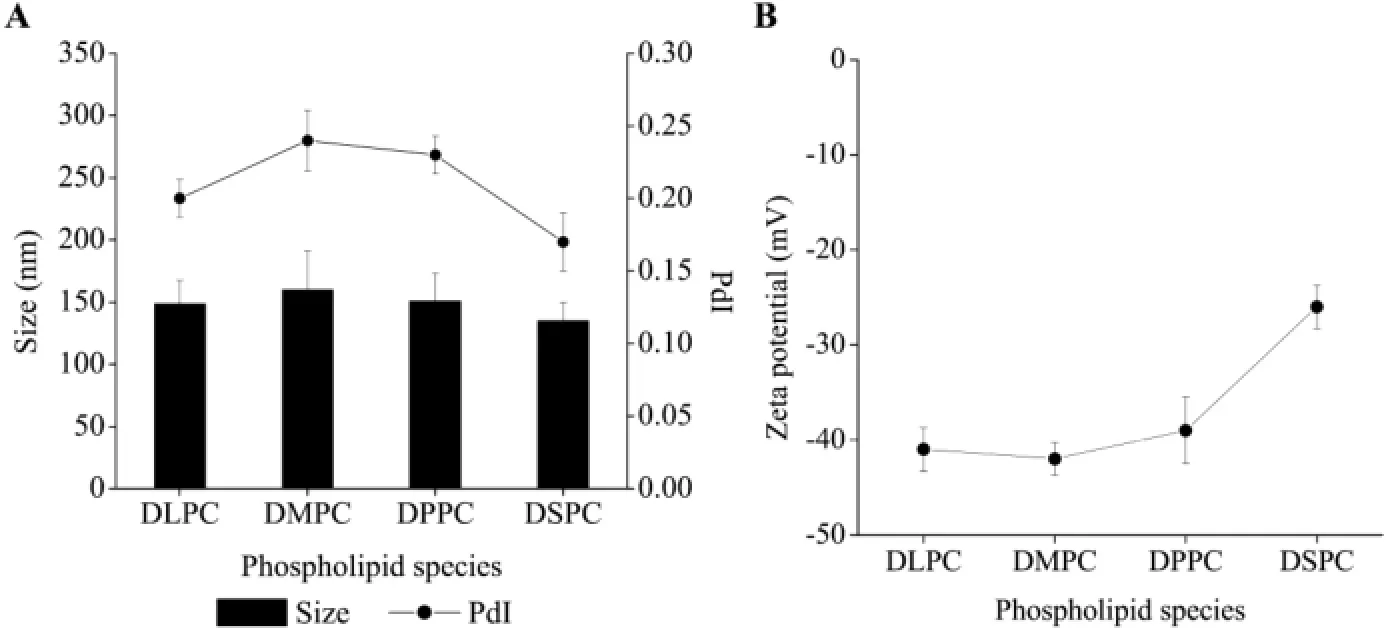
Fig.7–Effect of phospholipid species on(A)particle size,polydispersity index and(B)zeta potential of PEG-PLDH nanoparticles(mean±standard deviation,n=3).
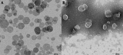
Fig.8–TEM images of(A)LDH and(B)PEG-PLDH nanoparticles.Scale bars indicate 200 nm.

Fig.9–The changes of(A)particle size,(B)polydispersity index and(C)zeta potential of LDH and PEG-PLDH nanoparticles during a month(mean±standard deviation,n=3).
3.3.5.Phospholipid species
The molar ratio of DOPA/DSPC/Chol/DSPE-PEG2000was fxed at 15:9:10:6,and DSPC was replaced by other three kinds of phospholipids(DLPC,DMPC and DPPC)in the same amount.Fig.7 showed that the particle size and PdI increased after DSPC was replaced by other phospholipids,and the zeta potential of PEG-PLDH nanoparticles decreased at the same time,indicating that DSPC was the optimal phospholipid.
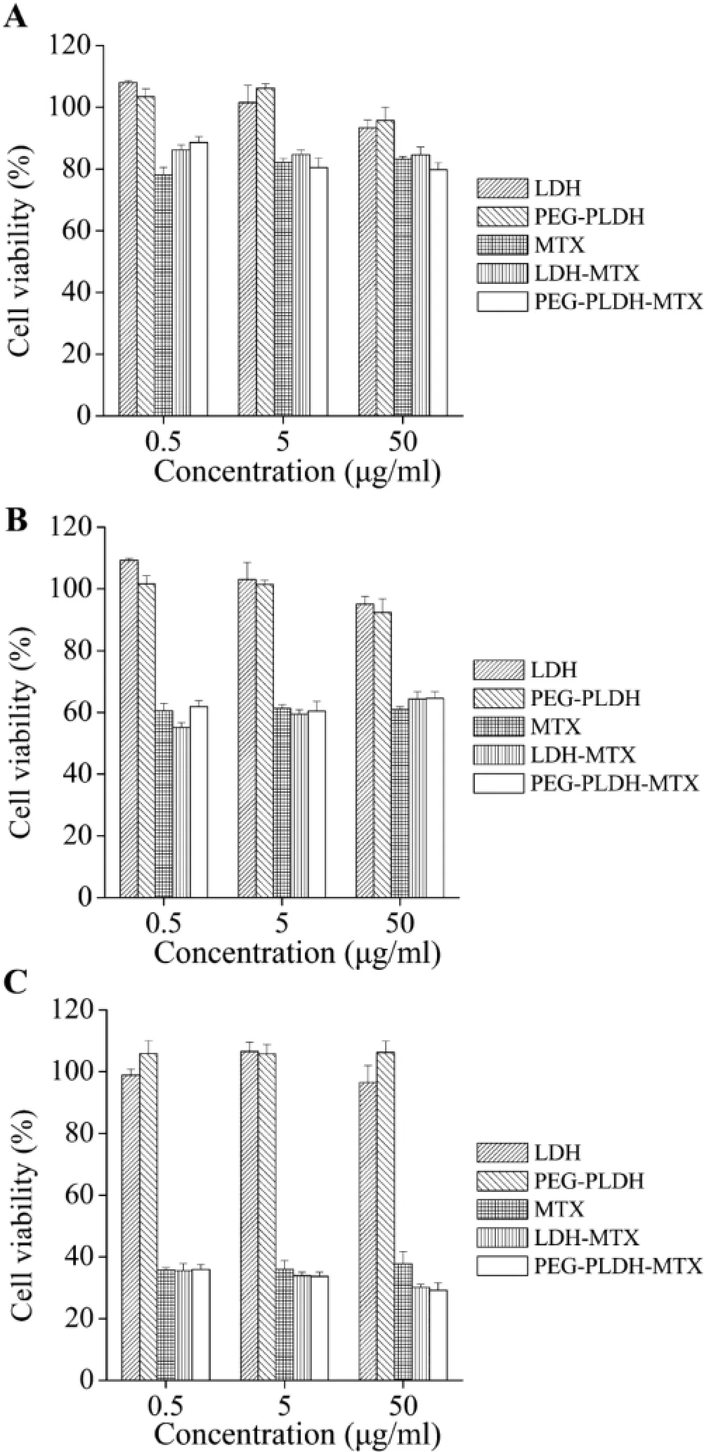
Fig.10–Cell viability of Hela cells after incubation with different formulations for(A)4 h,(B)24 h or(C)48 h (mean±standard deviation,n=5).
In summary,the optimal preparation method was determined as follows:LDH was heat-treated for 4 h;sonication time for PEG-PLDH was not less than 10 min;the weight ratio of LDH to lipid materials was 2:1;the molar ratio of DOPA/DSPC/Chol/ DSPE-PEG2000was fxed at 15:9:10:6.
3.4.Morphology study of nanoparticles
The particle morphology was investigated with transmission electron microscopy for LDH and PEG-PLDH.Fig.8 showed that the LDH nanoparticles were well-shaped in hexagonal form while the PEG-PLDH nanoparticles had a core-shell structure.
3.5.Stability study of nanoparticles
We investigated the storage stability of LDH and PEG-PLDH nanoparticles during 1 month of storage at 4°C.Fig.9 showed the particle size,PdI and zeta potential had little change during the one-month period,which suggested that both LDH and PEGPLDH nanoparticles had a high storage stability at 4°C.
3.6.Cell viability
In vitrocytotoxicity of LDH,PEG-PLDH,MTX,LDH-MTX and PEGPLDH-MTX nanoparticles was evaluated on HeLa and BALB/ C-3T3 cells.The MTX-free nanoparticles showed good cell viability over the concentration range used in this study,the cell viability was higher than 90%in all wells(Figs.10 and 11). As for MTX formulations,there is no obvious difference between free MTX,LDH-MTX and PEG-PLDH-MTX.
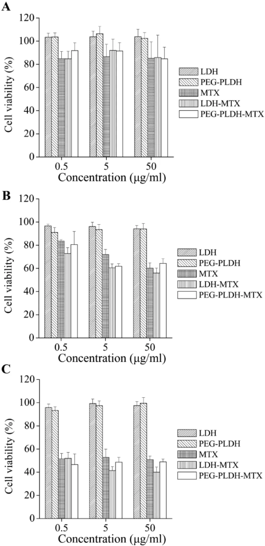
Fig.11–Cell viability of BALB/C-3T3 cells after incubation with different formulations for(A)4 h,(B)24 h or(C)48 h (mean±standard deviation,n=5).
4.Conclusions
PEG-PLDH nanoparticles were successfully manufactured in a laboratory-scale.The prepared nanoparticles have small particle size and uniform size distribution.During the preparation process,the sonication time,LDH/lipids weight ratio and DSPEPEG2000/phospholipids molar ratio were the three key points to achieve the good quality of PEG-PLDH nanoparticles.
Acknowledgments
We are grateful for the fnancial support from the National Natural Science Foundation of China(rant numbers 81302715/ H3008 and 81173003/H3008).
R E F E R E N C E S
[1]Liu X,Fan B,Gao S,et al.Transesterifcation of tributyrin with methanol over MgAl mixed oxides derived from MgAl hydrotalcites synthesized in the presence of glucose.Fuel Process Technol 2013;106:761–768.
[2]Drenkova-Tuhtan A,Mandel K,Paulus A,et al.Phosphate recovery from wastewater using engineered superparamagnetic particles modifed with layered double hydroxide ion exchangers.Water Res 2013;47:5670–5677.
[3]Jia C,Qian Y,Chen X,et al.Flame retardant ethylene-vinyl acetate composites based on layered double hydroxides with zinc hydroxystannate.Polym Eng Sci 2014;54(12):2918–2924.
[4]Kuo Y-M,Kuthati Y,Kankala RK,et al.Layered double hydroxide nanoparticles to enhance organ-specifc targeting and the anti-proliferative effect of cisplatin.J Mater Chem B 2015;3:3447–3458.
[5]Rives V,Del Arco M,Martín C.Layered double hydroxides as drug carriers and for controlled release of non-steroidal antiinfammatory drugs(NSAIDs):a review.J Control Release 2013;169:28–39.
[6]Bi X,Zhang H,Dou L.Layered double hydroxide-based nanocarriers for drug delivery.Pharmaceutics 2014;6:298–332.
[7]Gu Z,Wu A,Li L,et al.Infuence of hydrothermal treatment on physicochemical properties and drug release of anti-infammatory drugs of intercalated layered double hydroxide nanoparticles.Pharmaceutics 2014;6:235–248.
[8]Kim T-H,Lee GJ,Kang J-H,et al.Anticancer drug-incorporated layered double hydroxide nanohybrids and their enhanced anticancer therapeutic effcacy in combination cancer treatment.BioMed Res Int 2014;2014:193401.
[9]Li L,Gu W,Chen J,et al.Co-delivery of siRNAs and anti-cancer drugs using layered double hydroxide nanoparticles.Biomaterials 2014;35:3331–3339.
[10]Li B,Wu P,Ruan B,et al.Study on the adsorption of DNA on the layered double hydroxides(LDHs).Spectrochim Acta[A] 2014;121:387–393.
[11]Dong H,Parekh HS,Xu ZP.Enhanced cellular delivery and biocompatibility of a small layered double hydroxide–liposome composite system.Pharmaceutics 2014;6:584–598.
[12]Hadinoto K,Sundaresan A,Cheow WS.Lipid–polymer hybrid nanoparticles as a new generation therapeutic delivery platform:a review.Eur J Pharm Biopharm 2013;85:427–443.
[13]Roggers RA,Lin VSY,Trewyn BG.Chemically reducible lipid bilayer coated mesoporous silica nanoparticles demonstrating controlled release and HeLa and normal mouse liver cell biocompatibility and cellular internalization.Mol Pharm 2012;9:2770–2777.
[14]Yan M,Zhang Z,Cui S,et al.Improvement of pharmacokinetic and antitumor activity of layered double hydroxide nanoparticles by coating with PEGylated phospholipid membrane.Int J Nanomed 2014;9:4867–4878.
[15]Schmidt HT,Gray BL,Wingert PA,et al.Assembly of aqueous-cored calcium phosphate nanoparticles for drug delivery.Chem Mater 2004;16:4942–4947.
[16]Xu ZP,Stevenson G,Lu C-Q,et al.Dispersion and size control of layered double hydroxide nanoparticles in aqueous solutions.J Phys Chem B 2006;110:16923–16929.
[17]Xu ZP,Jin Y,Liu S,et al.Surface charging of layered double hydroxides during dynamic interactions of anions at the interfaces.J Colloid Interface Sci 2008;326:522–529.
*< class="emphasis_italic">Corresponding author.
.School of Pharmacy,Shenyang Pharmaceutical University,103 Wenhua Road,Shenyang 110016,China.Tel.:+86 24 2398 6316;fax:+86 24 2398 6316.
E-mail address:pharmdeng@gmail.com(Y.Deng).
**Corresponding author.School of Pharmaceutical Sciences,Sun Yat-sen University,132 Waihuan East Road,Guangzhou 510006,China. Tel.:+86 20 3994 3118;fax:+86 20 3994 3118.
E-mail address:zhaocs@mail.sysu.edu.cn(C.Zhao).
Peer review under responsibility of Shenyang Pharmaceutical University.
http://dx.doi.org/10.1016/j.ajps.2015.09.003
1818-0876/?2016 The Authors.Production and hosting by Elsevier B.V.on behalf of Shenyang Pharmaceutical University.This is an open access article under the CC BY-NC-ND license(http://creativecommons.org/licenses/by-nc-nd/4.0/).
 Asian Journal of Pharmacentical Sciences2016年3期
- Asian Journal of Pharmacentical Sciences的其它文章
- A novel LC–MS/MS assay for methylprednisolone in human plasma and its pharmacokinetic application
- Formulation and evaluation of co-prodrug of furbiprofen and methocarbamol
- Auricularia auricular polysaccharide-low molecular weight chitosan polyelectrolyte complex nanoparticles:Preparation and characterization
- Brain targeted delivery of paclitaxel using endogenous ligand
- Generic sustained release tablets of trimetazidine hydrochloride:Preparation and in vitro – in vivo correlation studies
- Effect of process and formulation variables on the preparation of parenteral paclitaxel-loaded biodegradable polymeric nanoparticles: A co-surfactant study
