Rare variant of pancreaticobiliary maljunction associated with pancreas divisum in a child diagnosed and treated by endoscopic retrograde cholangiopancreatography: A case report
Guang-Xing Cui, Hai-Tao Huang, Jian-Feng Yang, Xiao-Feng Zhang
Abstract
Key words: Pancreaticobiliary maljunction; Pancreas divisum; Endoscopic retrograde cholangiopancreatography; Variant; Communication; Children; Case report
INTRODUCTION
Pancreaticobiliary maljunction (PBM) is an uncommon congenital anomaly of the pancreatic and biliary ductal system, defined as a union of the pancreatic and biliary ducts located outside the duodenal wall[1]. Because of this anatomical anomaly, PBM has been frequently associated with cholelithiasis, cholangitis, pancreatitis[2], and increased risk of cholangiocarcinoma[3]. PBM has been classified into three types by Komiet al[4], according to the angle of the junction of the common bile duct (CBD) and pancreatic duct, dilatation of the common channel, and the running of the dorsal pancreatic duct. Types I and II have no pancreas divisum (PD). In type III, PD exists in all PBM patients with complete PD in types IIIa and b and incomplete PD in types IIIc1-3. According to the Komi classification of PBM, the CBD directly fuses with the ventral pancreatic duct in all types. PD occurs when the ventral and dorsal ducts of the embryonic pancreas fail to fuse during the second month of fetal development. It is the most common anatomical variant of the pancreas, in which ventral duct drains the minor part of the pancreas through the major papilla, whereas the dorsal duct drains the major part of the pancreatic juice through the minor papilla[5]. The coexistence of PBM and PD is an infrequent condition.
Here, we report an unusual variant of PBM associated with PD in a pediatric patient, in whom an anomalous communication existed between the CBD and dorsal pancreatic duct. Our case was hardly classified into Komi classification of PBM with the special anatomical variant. The patient was successfully diagnosed and managed by endoscopic retrograde cholangiopancreatography (ERCP). Written informed consent was obtained from his parents prior to the endoscopic therapy.
CASE PRESENTATION
Chief complaints
A boy aged 4 years and 2 mo was hospitalized for abdominal pain with nausea and jaundice for 5 d.
History of present illness
He had colic pain located in the right upper abdomen, without radiating to the back or paroxysmal attacks and with no confirmed exacerbating or relieving factors. He was sent to a local hospital after the first attack. Abdominal ultrasound indicated gallbladder muddy stones and CBD stones. After symptomatic treatment, he was referred to our institution for further diagnosis and therapy.
History of past illness and Personal and family history
Neither he nor his family had any past history of biliaro-pancreatic diseases or other abnormalities.
Physical examination upon admission
Physical examination revealed mild tenderness in the middle upper abdomen without rebound tenderness. Slight jaundice was observed in his sclera.
Laboratory examinations
Routine blood tests showed an inflammatory result: white blood cell count, 11.4 ×10⁹/L; neutrophils, 78.9%; and hypersensitive C-reactive protein (hs-CRP), 20 mg/L.Liver biochemical function tests indicated extrahepatic biliary obstruction: alanine aminotransferase, 78 U/L; aspartate aminotransferase, 55 U/L; γ-glutamyl transpeptidase, 210 U/L; alkaline phosphatase, 441 U/L; total bilirubin, 40.5 μmol/L; and direct bilirubin, 33.1 μmol/L. The level of serum amylase was elevated (135 U/L). The serum autoimmune antibody tests including IgG 4 were negative.
Imaging examinations
Abdominal ultrasound showed cholecystitis with cholestasis in the gallbladder,dilated middle-upper CBD with a diameter of 1.1 cm, and a strong echo in the lower CBD, indicating biliary stones. Computed tomography was not performed because of the radiation risk.
Preliminary diagnosis
These findings above supported a diagnosis of extrahepatic biliary obstruction caused by biliary stones, which is an indication for ERCP.
FINAL DIAGNOSIS
PBM associated with PD with a communication between the CBD and dorsal pancreatic duct; CBD stones with acute cholangitis
TREATMENT
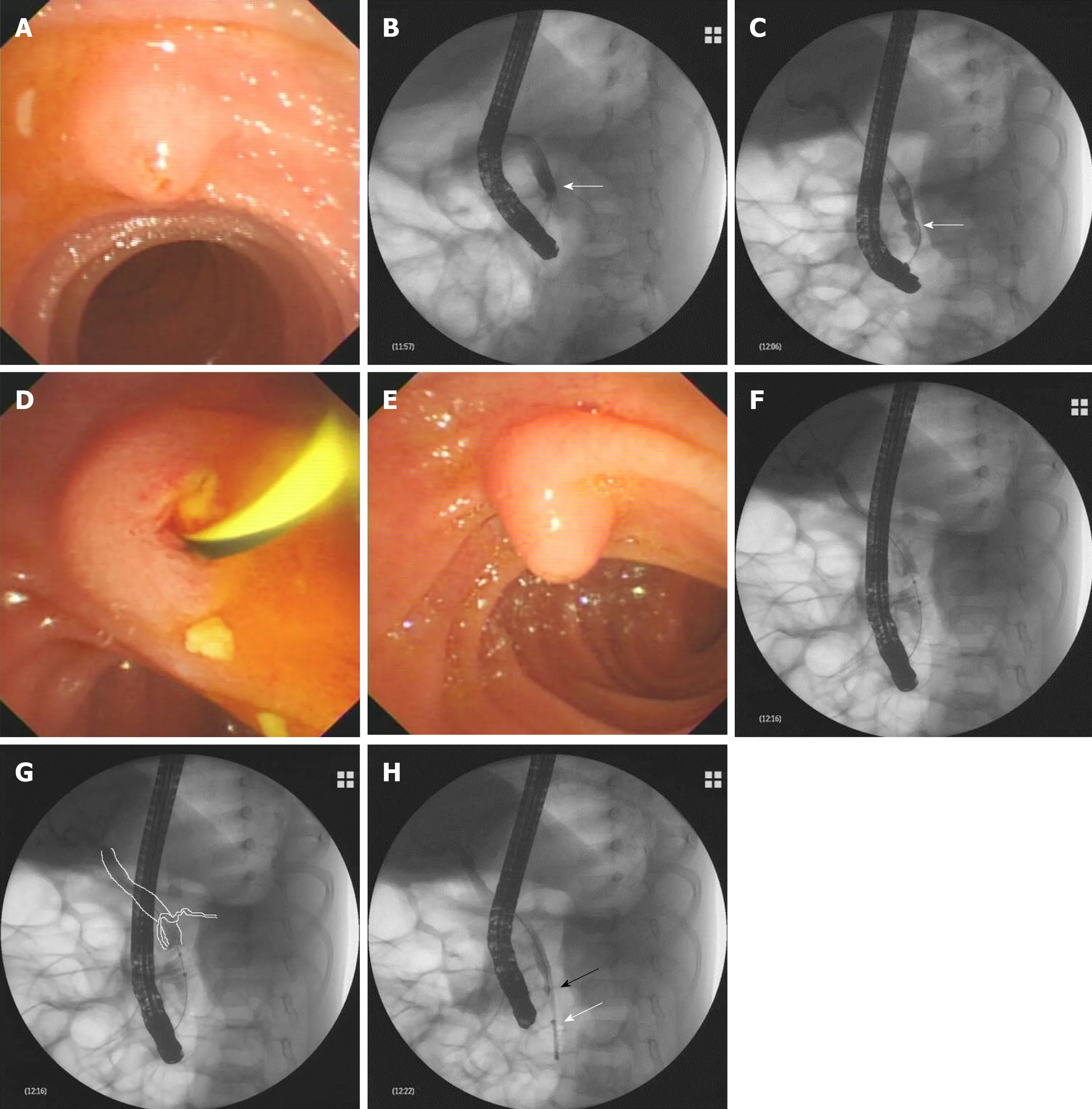
ERCP was performed to remove biliary stones. When the duodenoscope was advanced to the descending part of the duodenum, a hemispheric papilla with a villus-like opening was seen, which resembled the major papilla in both size and morphology (Figure 1A). This was wrongly considered to be the major papilla and cannulation was successfully carried out. X-ray examination after injecting a contrast agent into the papilla revealed a dilated CBD with a diameter of 0.9 cm, which was indicative of biliary stones (Figure 1B). However, the main pancreatic duct was not revealed. A minor endoscopic sphincterotomy was then performed, after which multiple small biliary stones were discharged from the papilla (Figure 1D). However,in a short distance beneath the papilla, another bigger papilla was detected, which was in fact the real major papilla (Figure 1E). The prior one was the minor papilla.After successful cannulation of the major papilla, the CBD was dilated with multiple filling defects, indicating biliary stones. However, the Wirsung duct was not observed. The middle-lower CBD was narrowed (Figure 1C). An endoscopic balloon was used to remove the biliary stones. During the process of pulling the balloon combined with injecting a contrast agent into the biliary tract, the dorsal pancreatic duct was unexpectedly revealed at the level of the middle-lower part CBD, which is rarely seen under normal conditions (Figure 1F and G). After clearing the CBD with the balloon, an 8.5 Fr 4-cm pigtail plastic pancreatic stent was placed in the biliary duct through the major papilla. Finally, the minor papilla was cannulated again and the guidewire was advanced into the CBD accompanying the biliary stent (Figure 1H), from which a communication between the CBD and dorsal pancreatic duct was created.
OUTCOME AND FOLLOW-UP
After the procedure, the child recovered uneventfully. Six months later, his biliary stent was removed after he had no symptoms and normal laboratory tests. In the following 4-year period with periodic telephone call and outpatient visits, the child grew up normally with no more attacks of abdominal pain.

Figure 1 Results of endoscopic retrograde cholangiopancreatography. A: During endoscopic retrograde cholangiopancreatography, a hemispheric papilla with a villus-like opening resembling the major papilla was seen. B-E: Successful cannulation of the papilla and a dilated common bile duct (CBD) detected by X-ray after injection of a contrast agent (B). Multiple small biliary stones were discharged from the papilla after minor endoscopic sphincterotomy (D). Beneath the papilla, the real major papilla was detected (E). After cannulation of the major papilla, the CBD was observed again, which was dilated with multiple filling defects (C), and at the level of the middle-lower CBD, narrowing was observed (arrow); F: During removal of the biliary stones with an endoscopic balloon, unexpectedly, the dorsal pancreatic duct was revealed at the level of the middle-lower part of the CBD; G: Schematic representation of the image of F; H: After clearance of the CBD with the balloon, an 8.5 Fr 4-cm pigtail plastic pancreatic stent was placed in the biliary duct through the major papilla (white arrow); the minor papilla was cannulated again; and the guidewire was advanced into the CBD (black arrow).
DISCUSSION
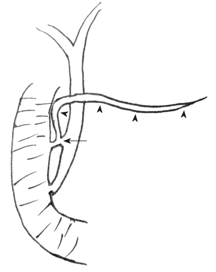
PBM is a congenital anomaly that occurs when the pancreatic and bile ducts are united outside the duodenal wall. In patients with PBM, the sphincter of Oddi functionally loses its effect on the union of the two ducts. Therefore, continuous reciprocal reflux between pancreatic juice and bile occurs, which can result in various pathological conditions in the biliary tract and pancreas[2]. Under normal circumstances, the hydrostatic pressure in the pancreatic duct is usually higher than that in the bile duct, which means that the pancreatic juice more frequently refluxes into the biliary duct than the pancreatic duct in PBM[6]. This might be an etiological factor in choledocholithiasis, inflammatory ductal epithelial changes, distal common bile duct strictures, and recurrent attacks of acute cholangitis. Additionally, PBM resulting in chronic inflammation of the bile duct is considered to be frequently related to biliary tract malignancy. PD occurs when the ventral and dorsal ducts of the embryonic pancreas fail to fuse during the second month of fetal development[5]. It is the most frequent congenital anomaly of the pancreas in which the dorsal and ventral pancreatic ducts drain separately into the duodenum. The dominant pancreatic juice is drained by the dorsal pancreatic duct through the minor papilla. Whether PD causes pancreatitis or other complications remains controversial. The co-occurrence of PBM and PD is an uncommon condition. Teruiet al[7]found that PD was detected in one of 71 cases of PBM, with an incidence rate of 1.4%. In the current study, as shown in the schematic illustration (Figure 2), our case had three pancreaticobiliary abnormalities: PBM, PD, and abnormal communication between the CBD and dorsal pancreatic duct.
Currently, the Komi classification for PBM has been widely accepted and utilized,which influences the selection of type of surgical procedure and prognosis after surgery, especially in patients with complicating cases like type IIIc3[4]. In the Komi classification, all terminal CBDs join the ventral pancreatic duct. Matsumotoet al[8]retrospectively analyzed 202 patients with PBM to develop a new concept of the embryonic etiology of PBM. They found no patients in whom the terminal bile duct was joined with the dorsal pancreatic duct, nor was there a communication between the CBD and dorsal pancreatic duct. However, not all PBM cases can be classified according to the Komi classification. A few complicated PBM cases with rare anatomical variants have been reported by a small number of researchers[9-11]. Parlaket al[9]reported a 42-year-old woman who underwent ERCP for recurrent biliary pain attacks. During ERCP, the dilated CBD was found to fuse to the dorsal pancreatic duct directly without common channel dilation. Therefore, they thought that it represented a new type of PBM that could not be included in the Komi classification. Zhanget al[10]reported four complicated PBM cases, in which the CBD also joined the dorsal pancreatic duct in a direct way. All four cases were female with the youngest aged 11 years. They were successfully treated with intraductal drainage by ERCP. McMahonet al[11]reported an anomalous communication between the dorsal pancreatic duct and CBDviaa small ventral pancreatic duct branch. This patient was a 30-year-old woman who suffered from chronic debilitating pain for several years. The patient's anomaly was indicated by magnetic resonance cholangiopancreatography (MRCP) with intravenous secretin administration. She received a Whipple pancreaticoduodenectomy combined with cholecystectomy. The aberrant ductal communication was confirmed by the resected specimen.
In our case, we found a communication between the CBD and dorsal pancreatic duct, which was similar to that reported by McMahonet al[11]. The little difference is that the communication in our case was located closer to the minor papilla. Although no definite communication was delineated by ERCP, the CBD was clearly observed by cannulation of both papillae. Moreover, the dorsal pancreatic duct was developed when removing the biliary stones with the balloon at the middle-lower level of the CBDviathe major papilla. This may have been caused by high-pressure injection of contrast agent into the dorsal pancreatic ductviathe communication between the CBD and dorsal pancreatic duct when the balloon was pulled down. We speculated that the communicating pancreatic duct was located at the middle-lower level of the CBD.As indicated earlier, all previously reported cases with this rare anomaly were female,with the youngest being aged 11 years[9-11]. However, in the present case, the child was male and aged 4 years.
To date, MRCP as a noninvasive approach is the first choice for diagnosis of pancreaticobiliary disorders. However, MRCP is limited in diagnosing the common biliopancreatic duct and biliopancreatic junction, compared with ERCP[12-14], even when secretin is used[15]. Diagnostic accuracy may be increased using 3D or dynamic MRCP with secretin stimulation[16]. For diagnosing patients with anatomical maljunction, ERCP remains the gold standard. In the current case, the patient was successfully diagnosed by ERCP.
PBM is generally recognized to be a risk factor for biliary tract malignancy[3]. Here,surgery is considered as radical treatment for patients with PBM. Timely surgical division of the biliary and pancreatic ducts is essential for patients with PBM to prevent free reflux of pancreatic juice into the biliary tract, regardless of the presence or absence of choledochal cyst[17-20]. ERCP is also a useful therapeutic option for patients with PBM, and it can be used to relieve acute biliary obstruction by removing biliary stones, implanting a biliary stent, or sphincterotomy[12-14]. It is helpful to plan the timing and choice of the appropriate surgical procedure. Samavedyet al[21]studied the potential benefit of ERCP in patients with PBM and found that 13 of 15 cases presenting with relapsing pancreatitis benefiting from endoscopic therapy. They assumed that ERCP was the logical first step to manage most symptomatic patients with PBM. Until now, only one similar patient with rare variant communication between the CBD and dorsal pancreatic duct has been reported, who underwent surgical treatment at age 30 years[11]. There is lack of therapeutic experience for such cases. In our case, given the factors of age, growth, and surgical trauma to the body,ERCP was chosen as initial therapy. During ERCP, an endoscopic balloon was used to remove the biliary stones and place a biliary stent through the major papilla. The child remains asymptomatic during 4 yr of follow-up. Furthermore, close long-term followup is needed to supervise the development of biliary malignancy.

Figure 2 Schematic representation of the pancreaticobiliary system. This child had three anomalies:Pancreaticobiliary maljunction, pancreas divisum (arrowheads indicating dorsal pancreatic duct), and abnormal communication between the common bile duct and dorsal pancreatic duct (arrow).
CONCLUSION
In summary, timely diagnosis and treatment of PBM associated with PD are important, especially when it is combined with aberrant communication between the CBD and dorsal pancreatic duct. We consider that ERCP is effective and safe in pediatric patients with PBM combined with PD, and can be the initial therapy to manage such cases. Considering the potential of PBM to develop into biliary malignancy, close follow-up is needed for small children after endoscopic therapy.Once the evidence of neoplastic degeneration is detected during follow-up, timely surgical therapy should be adopted.
ACKNOWLEDGEMENTS
This case had been presented as a clinical case presentation at UEGW 2018, Vienna,Austria.
 World Journal of Clinical Cases2019年9期
- World Journal of Clinical Cases的其它文章
- Coexistence of breakpoint cluster region-Abelson1 rearrangement and Janus kinase 2 V617F mutation in chronic myeloid leukemia: A case report
- Crizotinib-induced acute fatal liver failure in an Asian ALK-positive lung adenocarcinoma patient with liver metastasis: A case report
- Adult-onset mitochondrial encephalopathy in association with the MT-ND3 T10158C mutation exhibits unique characteristics: A case report
- Nerve coblation for treatment of trigeminal neuralgia: A case report
- Management of the late effects of disconnected pancreatic duct syndrome: A case report
- Sofosbuvir/Ribavirin therapy for patients experiencing failure of ombitasvir/paritaprevir/ritonavir + ribavirin therapy: Two cases report and review of literature
