
Retiform Hemangioendothelioma
2021-10-14 06:27:00JingWangandJianMinChang
國際皮膚性病學(xué)雜志 2021年3期
Jing Wang and Jian-Min Chang?
Department of Dermatology,Beijing Hospital,National Center of Gerontology,Institute of Geriatric Medicine,Chinese Academy of Medical Sciences,Beijing100730,China.
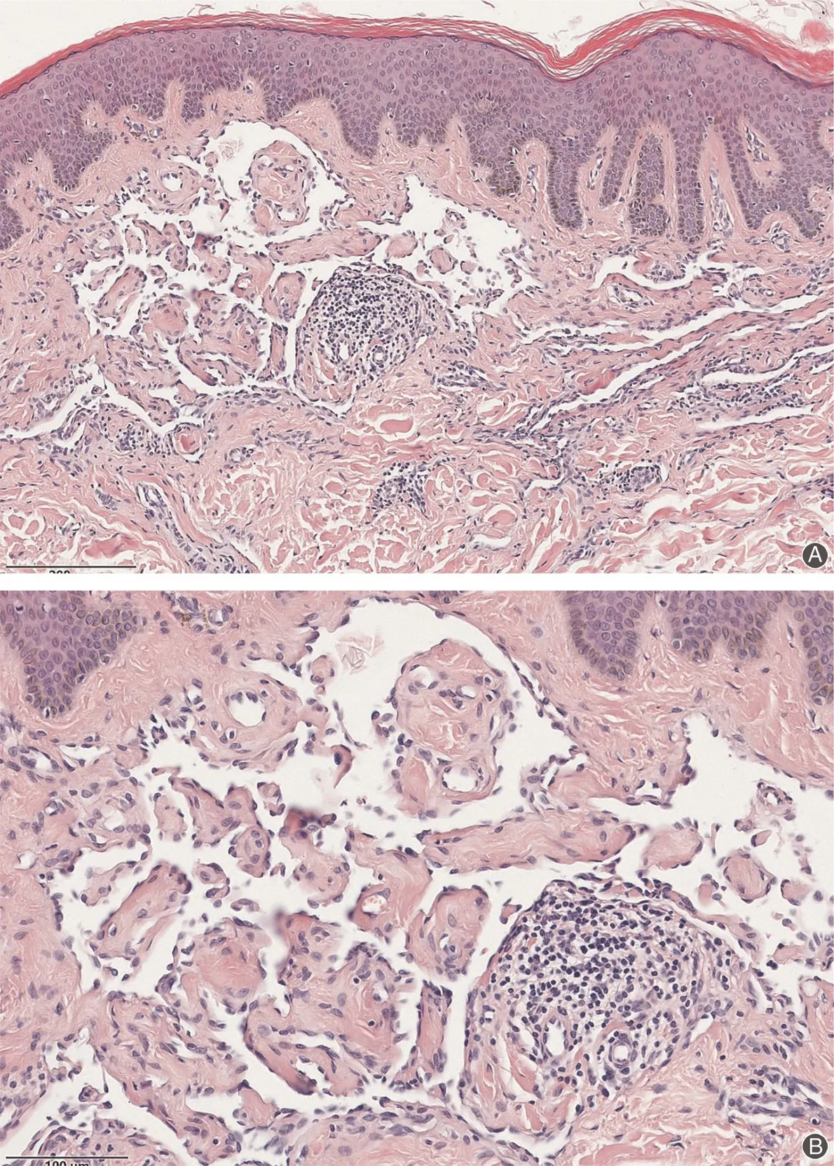
Figure1.Histopathological features of Retiform Hemangioendothelioma.The epidermis is normal.However,arborizing blood vessels are visible in the dermis.The vessels are lined by monomorphic endothelial cells,and interspersed lymphocytes can be seen in and around the blood vessels.(A)Hematoxylin-eosin,×100.(B)Hematoxylin-eosin,×200.
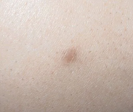
Figure2.Clinical presentation of Retiform Hemangioendothelioma.Asymptomatic,well-defined,solitary nodular lesion on the lower limb.No abnormality is present in the surrounding skin.
Histopathology
Histopathological examination is required to confirm the diagnosis of retiform hemangioendothelioma(RH)because RH does not have distinctive clinical features.Histopathological examination reveals arborizing blood vessels in the dermis,extending between the collagen bundles in a reticular fashion and flanked by lymphocytes and hyaline sclerosis.The vessels are lined by monomorphic endothelial cells with scanty cytoplasm and prominent protuberance of the nucleus.The cells can protrude to the vascular lumen like a matchstick or hobnail,and nuclear atypia and mitotic figures are generally absent(Fig.1).1Some interspersed lymphocytes can also be seen in and around the blood vessels.Immunohistochemical examination of RH shows positive expression of vascular endothelial cell marker CD34and weak positive expression of CD31and FVIII-related antigen.2This low-grade tumor recurs frequently,but the risk of metastasis is very low.
Clinical features
RH is an extremely rare variant of low-grade angiosarcoma that was first described in1994as a distinctive form of angiosarcoma.3It often occurs in young or middle-aged people and has an unclear pathogenesis.Its main clinical feature is an asymptomatic,slowly growing,well-defined solitary nodular or plaque-like lesion on either the extremities or trunk(Fig.2).2,4
The differential diagnoses for RH are angiosarcoma and targetoid hemosiderotic hemangioma.The histopathological features of angiosarcoma are very similar to those of RH.However,angiosarcoma often exhibits nuclear atypia and mitotic figures and the absence of a retiform pattern of blood vessels;additionally,it is more invasive than RH and has a higher risk of recurrence and metastasis.In contrast,targetoid hemosiderotic hemangioma is a superficial and limited lesion,and shoe nail-like vascular endothelial cells can only be seen in some areas.3These lesions can be identified by histopathologic examination.
About60% of patients with RH develop local recurrence with a tendency for local aggressive behavior.Previous reports have described regional lymph gland metastasis in a single patient and local spread to soft tissue in another patient.5Only one RH-related death has been reported to date.Surgical excision is the most effective treatment for a circumscribed lesion.6
- 國際皮膚性病學(xué)雜志的其它文章
- Mask on Followed by Gloves on:Do We Have a Choice?
- Mobilization of Melanocytes During NB-UVB Treatment of Vitiligo
- Consensus on the Diagnosis and Treatment of Melasma in China(2021Version)#
- Malignant Syphilis as an Initial Presentation of HIV Infection:A Case Report
- Treatment of Nevoid Basal Cell Carcinoma Syndrome by Surgery Combined With ALA-PDT:A Case Report
- Scrofuloderma:A Rare Case Report of Sequelae of Intestinal Tuberculosis