Protein Aggregation and Phase Separation in TDP-43 Associated Neurodegenerative Diseases*
SHILi?Jun ,XU An?Qi ,ZHU Li
(1)State Key Laboratory of Brain and Cognitive Science,Institute of Biophysics,Chinese Academy of Sciences,Beijing 100101,China;2)College of Veterinary Medicine,Northwest A&F University,Yangling 712100,China;3)College of Life Sciences,University of Chinese Academy of Sciences,Beijing 100049,China)
Abstract With the aging population increasing worldwide,neurodegenerative diseases are becoming a major public health crisis.TAR DNA?binding protein 43 (TDP?43) is one of the major components in the inclusion bodies containing aggregated proteins in affected patients with several types of neurodegenerative diseases,including amyotrophic lateral sclerosis (ALS),frontotemporal lobar degeneration (FTLD) and Alzheimer’s disease (AD).A large number of mutations in TDP?43 have been identified in familial cases of ALS.TDP?43 is an essential RNA/DNA binding protein critical for RNA?related metabolism,it shuttles between nucleus and cytoplasm,and undergoes phase transition to induce cytoplasmic and nucleoplasmic inclusion formation.Here,we summarized the recent advances in our understanding of protein aggregation and phase transition of TDP?43 in vitro and in vivo.Understanding the aberrant transition of TDP?43 will help identify potential therapeutic targets for neurodegenerative diseases.
Key words TDP?43 proteinopathy,amyotrophic lateral sclerosis (ALS),frontotemporal lobar degeneration (FTLD),amyloid aggregation,liquid?liquid phase separation(LLPS)
Transactive response DNA?binding protein 43(TAR DNA?binding protein 43,TDP?43) is ubiquitously expressed DNA? and RNA?binding protein that forms cytoplasmic inclusions in almost all cases of sporadic amyotrophic lateral sclerosis(sALS)and approximately 45% of frontotemporal lobar degeneration (FTLD) as well as other neurodegenerative diseases,such as Alzheimer’s disease (AD),Parkinson’s disease (PD) and chronic traumatic encephalopathy (CTE)[1].Cytosolic accumulation of TDP?43 is usually phosphorylated,ubiquitinated and truncated in the brains of patients with FTLD and ALS[2].TDP?43 toxicity is primarily driven by cleavage and accumulation of its C?terminal fragments (CTFs) which harbor most of the ALS?causing mutations[3?4].Both N terminal truncated proteins and short peptides generated from its prion?like domain (PrLD) in the C terminus of TDP?43 are indeed capable to form amyloid aggregation detected by fluorescence dye or electron microscopy(EM)[5?7].
As an RNA/DNA binding protein,TDP?43 shuttles between the nucleus and cytoplasm.During the shuttling,TDP?43 undergoes phase separation/transition in both nucleus and cytoplasm,dynamically associated with membraneless organelles including nuclear bodies (NBs),cytoplasmic stress granules(SGs) and processing bodies (P?bodies,PBs,related mRNA?protein granules),etc[8?12].Liquid?liquid phase separation(LLPS)of TDP?43 also needs the existence of PrLD[13].Various studies suggest that the dysfunction in nucleus?cytoplasm shuttling and liquid phase homeostasis trigger excessive cytoplasmic distribution/aggregation of TDP?43 protein,which contributes to the neurotoxicity and pathogenesis in neurodegenerative diseases including ALS,FTLD and AD[1,10].Prolonged retention of TDP?43 in the cytoplasm caused by loss of nuclear import and function of TDP?43 in the nucleus are neurotoxic,which ultimately causes neuronal cell death[10].However,it remains unknown for the detailed interplay between TDP?43 and other RNA binding proteins (RBPs),the roles of disease?associated mutations and other genetic modifiers,and the dysfunction induced by aggregation/phase separation.
In this review,we evaluate recent findings of amyloidogenesis of TDP?43,including the formation of amyloid aggregates by its C terminal PrLD and full?length protein.Recent hot?spot lies in the phase transition and formation of supramolecular assemblies of RBPs,especially FUS and TDP?43.We focus on some interesting researches about TDP?43 and aim to highlight the current published data on cytoplasmic and nucleoplasmic inclusion formation that links TDP?43 and neurodegeneration.We hope to provide reliable finding sources towards the development of early diagnostic and therapeutic tools for TDP?43 proteinopathy.
1 TDP-43 proteinopathy
In 2006,ubiquitinated TDP?43 inclusion was identified as the pathological protein in the most common subtypes of FTLD and ALS[2,14].TDP?43,encoded by the transactive response DNA binding protein (TARDBP) gene,is an essential conserved DNA/RNA binding protein involved in transcriptional regulation,RNA splicing,transport and mRNA stabilization[15?16].During the following decade,a large number of mutations have been identified in the human genes encoding RBPs such as FUS and other heterogeneous ribonucleoprotein (hnRNP) family members,involved in RNA homeostasis among patients affected by ALS and FTLD[17?19].These genetic studies provide strong support for the critical roles of nuclear RBPs in neuronal function and neurological diseases.
1.1 ALS
ALS,also known as Lou Gehrig’s disease,is the most common motor neuron disease (MND)characterized by progressive motor neuron degeneration in the brain and spinal cord.Approximately 5%-10% of ALS cases are familial(fALS),inherited in an autosomal dominant manner,and the majority of ALS cases are sporadic(sALS)[20].The Cu/Zn superoxide dismutase 1 (SOD1) gene is the first discovered ALS?associated gene and its mutations only account for 15%-20% of fALS and 3% of sALS[21].In the remaining cases of ALS,TDP?43 protein inclusion is found in the pathological tissues in the vast majority (>95%) of ALS patients(ALS?TDP)[2,4,18,22].
1.2 FTLD
FTLD,also called frontotemporal dementia(FTD),is the second most common cause of early?onset dementia (<65 y/o) after AD.It is characterized pathologically by atrophy of the frontal and temporal lobes of the brain.FTLD patients exhibit progressive changes in their personality and/or language since brain regions of the frontal and temporal lobe control behavior and cognitive function (e.g.language).The mutations of tau protein encoded gene,MAPT,are associated with FTLD[23].Pathologically,tau?positive inclusion accounts for 35% of FTLD cases (FTLD?tau) and the remaining FTLD cases are characterized as ubiquitinated inclusion (UBI) (FTLD?U)[22,24].TDP?43 protein was identified as the main component of UBIfor most frequent subtypes (45%) of FTLD(FTLD?TDP)[2,18,25?26].
1.3 ALS and FTLD are clinically and genetically overlapped
FTLD is accompanied by PD or MND symptoms in a significant proportion of patients[27?28],and cognitive and behavioral impairment is also observed in up to 75% of ALS patients[29].Thus,ALS and FTLD are clinically two groups of complex multigenic spectrum disorders linked by overlapping symptoms[30].In fact,ALS and FTLD are genetically overlapped,not only because of TDP?43 as a major pathological protein in ALS and FTLD,but most other genes whose mutations cause FTLD are also involved in ALS,includingFUS,C9ORF72,VCP,CHMP2B,UBQLN2andCHCHD10,etc[18,30?31].In this review,we only focus on TDP?43 associated neurodegenerative diseases and try to emphasize the common mechanism in their molecular and cellular pathological pathways.
1.4 TDP-43 proteinopathy is a group of clinicpathological spectrum disorders
Similar protein inclusions,such as ubiquitinated TDP?43,have been observed in ALS and FTLD[2,14],revealing that they share common molecular mechanisms leading to neurodegeneration[32].Except in ALS and FTLD,TDP?43 pathology has also been found in several other forms of neurodegenerative diseases,such as AD (up to 50% of patients)[33?36],dementia with Lewy bodies (DLB)[37?38],Pick’s disease (PiD)[14],corticobasal degeneration (CBD) or PD[39].Therefore,it is proposed that these TDP?43 inclusion involved clinic?pathological spectrum are collectively named as TDP?43 proteinopathy[40?41].
More than 40 dominant mutations inTARDBPthat encodes TDP?43 have been identified in rare cases of fALS[42?43],but only occasionally found in FTLD patients[22].Nevertheless,TDP?43 plays a crucial role in not only ALS and FTLD but also AD and other TDP?43 positive neurodegenerative diseases.Exploits of the common mechanism of these disorders will help to understand the molecular basis of TDP?43 proteinopathy.
2 The prion-like characteristics of TDP-43 accumulation
2.1 Aggregation and amyloid formation of TDP-43 with ALS and FTLD mutations in C terminal low complexity domain
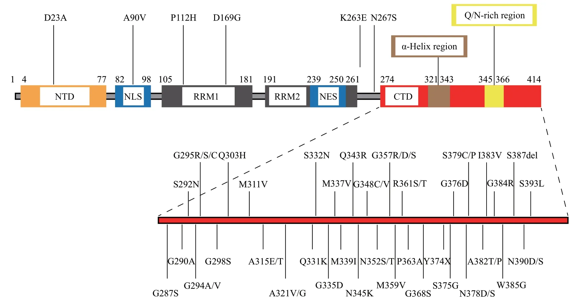
TARDBPis a highly conserved gene on chromosome 1 of the human genome[44]and is initially identified as a transcriptional repressor of HIV?1 gene expression[45].The encoded protein TDP?43 with 414?residue is composed of a folded N terminal domain (NTD),two RNA recognition motifs(RRM1 and RRM2) and an intrinsically disordered C terminal domain (CTD) with an α?helix region and a Q/N?rich region(Figure 1).
Nuclear magnetic resonance (NMR) studies showed that the NTD of TDP?43 adopts a well?folded structure[46?47]and is essential for its biological activity and aggregation pathology[48?49].Both the structures of RRM1 and RRM2 are solved by either X?ray crystallography[50]or NMR[51].The RRM motifs bind UG?rich segments of RNA and are critical for the physiological functions of TDP?43[51].Recent cryo?electron microscopy (cryo?EM) studies showed the amyloid fibrils formed by the entire low complexity domain (LCD) in the CTD of TDP?43 adopt different structural features from previously found fibrils generated from short proteins or peptides[52?53].

Fig.1 Primary structure of the 414-residue TDP-43 protein and the amino acid positions of ALS/FTLD-TDP associated mutations
2.2 TDP-43 has a prion-like domain
TDP?43 is a predominantly nuclear protein,with the nuclear localization sequence (NLS) located between the NTD and RRM1 while the nuclear export sequence (NES) hosted by RRM2,both of which regulate the shuttling of TDP?43 between the nucleus and cytoplasm[16,58?59].The disorganized C terminal region covering residues 274-414 (Figure 1) contains a low?complexity sequence abundant in Gly (G),Gln(Q) and Asn (N) and hosts the majority of ALS?associated mutations[18,57,60?62].This GQN rich C terminus shares 24.2% sequence identity with the N terminal yeast prion domain of Sup35[63?65].Thus,the CTD of TDP?43 has been proposed to be responsible for the prion?like spreading of ALS[63],which is also called the prion?like domain (PrLD)[5,61,64].Indeed,the CTD of TDP?43 plays a critical role in ALS and FTLD pathogenesis by the evidence collected in the past few years.
2.3 Both TDP-43 peptides in the PrLD and the intact TDP-43 PrLD form amyloid fibrils in vitro
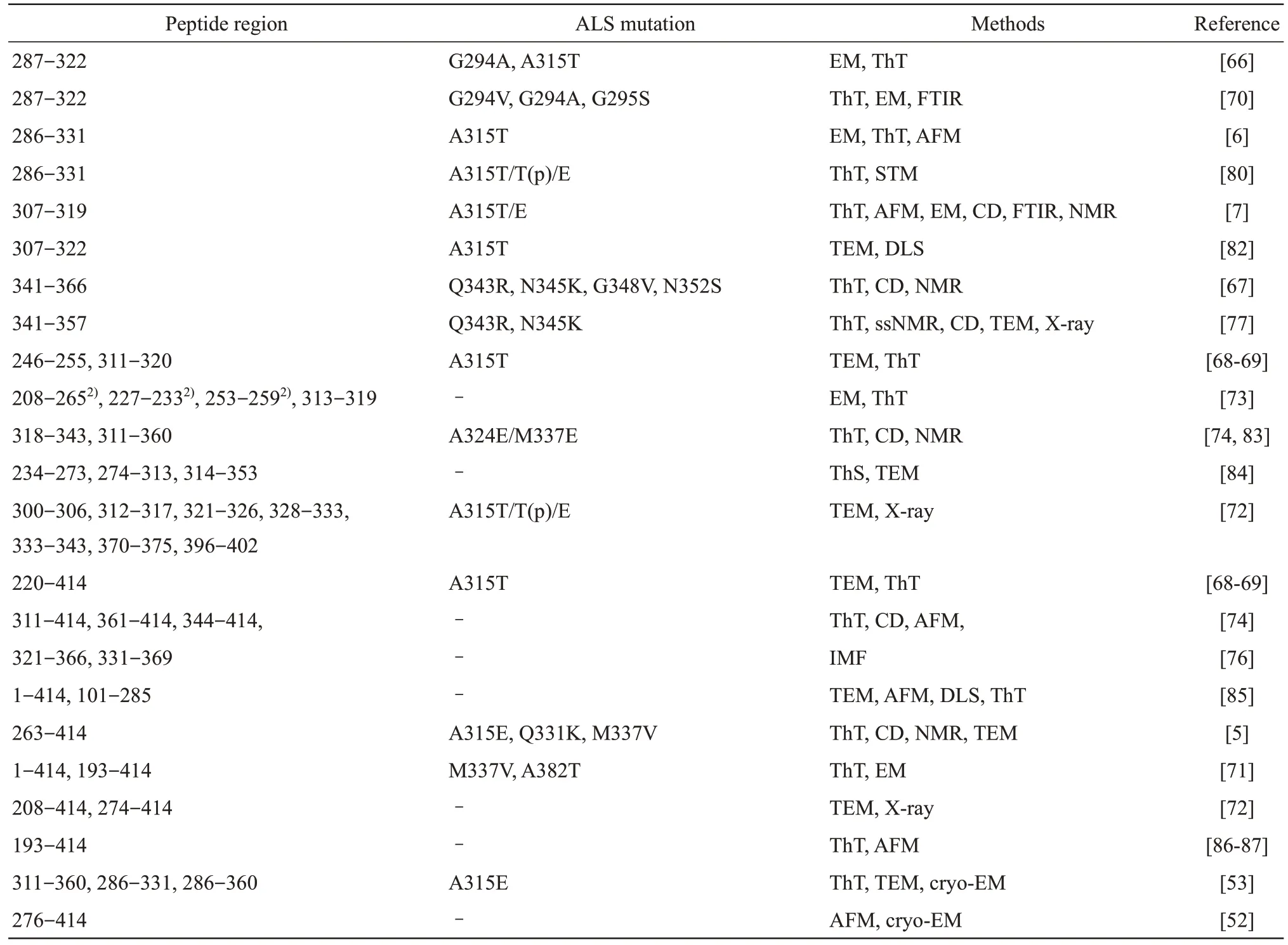
Short peptides derived from either RRM2 or the C terminal PrLD adopt fibril structures that indeed bind amyloid?specific dye,such as Thioflavin T(ThT)[6,7,53,66?72](Table 1).Specifically,the truncated form of RRM2 and Gly?rich region assembles into fibrilsin vitrorevealed by negative?staining of electronic microscopy (EM) and forms cytoplasmic inclusions in neuronal cells[73].The hydrophobic patch of residues 318-343 forms a helix?turn?helix by NMR analysis,which has a strong propensity to structurally transform into a β?sheet?rich structure and regular fibrils during aggregation[74].
In the Q/N?rich region of TDP?43,the peptide composed of residues 342-366 is mainly in disordered conformation revealed by circular dichroism (CD) and NMR.The pathological mutations (Q343R,N345K,G348V and N352S) have impact on the structure of this region[67].In addition,the peptide composed of residues 314-367 is initially unfolded and modeled as a β?hairpin structure but it converts into an amyloid?like structure which binds ThT[67,75?77].
Our study focuses on the Gly?rich 286-331 residue region which is in front of the above Q/N?rich segment and contains ALS?associated mutation A315T/E[6,78?79].This 46?mer peptide has a higher propensity to form a β?sheet structure and the A315T or A315E mutation is predicted to increase the probability to form β?sheets.Indeed these wild?type and A315T mutant peptides form amyloid fibrilsin vitrocharacterized by ThT binding fluorescence,SEM,atomic force microscopy (AFM) and scanning tunneling microscopy (STM)[6,80].Utilizing NMR analyses to dissect the atomic structure of shorter peptides in this Gly?rich region,the A315E?TDP?43 peptide of 307-319 residues indeed adopts an anti?parallel β conformation.This ALS?mutant A315E?TDP?43 peptide is capable of cross?seeding other TDP?43 peptides and amyloid?β peptidesin vitroand induces endogenous TDP?43 redistribution from the nucleus to the cytoplasm when added to cultured cells[7].
The full?length PrLD of TDP?43 is intrinsically disordered without any stable secondary structure,but it can assemble into amyloid oligomers rich in β?sheet structures with the presence of ALS?causing mutations (A315E,Q331K,M337V),nucleic acids interactions or the membrane environments[5].The self?associations for A315E and M337V mutants are significantly sped up by higher protein concentrations comparing with wild?type peptides of TDP?43 by CD,NMR and ThT fluorescence as well as intrinsic fluorescence.Both wild?type and three mutants form amyloid fibrils with the widths of fibrils ranging from 15 to 30 nm,similar to the fine structures detected in the TDP?43 inclusions in patient brain tissues[81].Residues 311-340 constitute the main region for interacting with both bicelle and dodecyl phosphocholine (DPC) micelle.While peptide containing residues M332-Q344 adopts well defined α?helix structure,peptide with residues M311-N319 assumes a well?defined Ω?loop structure with hydrophobic side chains of M311,F313,A315,F316,I318,all of which are pointed out to constitute a hydrophobic surface[5].This could explain the controversial structure of 318-343[74]and 307-319[7]overlapped at the residues of 318 and 319.
Based on the published data summarized in Table 1,it is clear that the region from 193 to 414,including RRM2 and the C terminal PrLD,plays a critical role in TDP?43 aggregation.In this region,peptides derived from RRM2 (such as 208-265,227-233,246-255,253-259,234-273) form amyloid fibrils.In the C terminal region (274-414),purified protein fragments (276-414,286-360,344-414,etc.) and even very short peptides from Gly?rich region of TDP?43 (such as 274-313,287-322,286-331,300-306,307-319,311-320,312-317,321-326,328-333,333-343,318-343,etc.) form fibril structures.On the other hand,peptides derived from Q/N?rich segment of TDP?43(such as 341-357,341-366,331-369,321-366,314-353,370-375) also can convert into amyloid?like structures.However,the cyro?EM structures of amyloid fibrils formed by the entire LCD of TDP?43(276-414)[52]differ from not only the cryo?EM structures formed by two relatively short fragments of TDP?43 LCD[53]but also the NMR structures of other short peptides.The obtained results are sometimes controversial and need more data for a clear conclusion.

Table 1 Regions for peptides or protein fragments forming amyloid fibrils in TDP-431)
2.4 TDP-43 protein forms amyloid-like aggregates in cell culture models and ALS or FTLD-U postmortem brain tissues
In many studies of cell culture models,TDP?43 protein displays similar characteristics to those of prion proteins[88?89].Full?length TDP?43 forms toxic amyloid oligomers in the forebrain of transgenic TDP?43 mice and FTLD?TDP patients,suggesting that TDP?43 oligomers may play a critical role in TDP?43 proteinopathy[85,90].Most importantly,using a higher resolution of EM with immuno?gold labeling,the brain sample of patients shows TDP?43 positive amyloid?like fibrils,which is the solid evidence for TDP?43 fibrils formation in TDP?43 proteinopathy including ALS and FTLD[7,81,91],as well as other neurodegenerative diseases[92].More evidence has been reported for amyloid fibrils formation generated by TDP?43 in tissues of patients with ALS and FTLD,which can be detected using ThS?specific fluorescence dye and optical microscopy[93?94].
Neuronal cytoplasmic inclusions in both FTLD?U and ALS tissues were stained positively by a specific antibody generated by the 25?ku fragment of TDP?43 but not normal TDP?43,suggesting that some inclusions in both FTLD?U and ALS contain caspase?derived C terminal fragments of TDP?43[3].A recent study with brain tissues of ALS?TDP patients showed that the C terminal GQN region plays a key role in amyloid formation[95]and the C terminal fragments of insoluble TDP?43 extracted from ALS?TDP or FTLD?TDP brains act as seeds to induce TDP?43 pathology in SH?SY5Y or neuronal and glial cells culture,including ubiquitination and phosphorylation of insoluble TDP?43 inclusions in the cytoplasm and propagation between cells[96?98].These studies not only provide the most convincing evidence for the prion?like properties of pathological TDP?43,but also suggest that the C terminal PrLD may play a key role in the progression of TDP?43 proteinopathy.
Together,the structural features of TDP?43 and above investigations from full?length and C terminal truncated forms of TDP?43 suggest that insoluble,aggregated TDP?43 presents prion?like properties that contribute to TDP?43 proteinopathy.Although recent studies strongly suggest the TDP?43 conformers could spread through the nervous system in a prion?like fashion,the prion?like behavior of TDP?43 still needs further characterization,including the detailed description of seeding,propagation and neuronal invasion mechanisms,and potential TDP?43 strain types[99?100].Nevertheless,understanding the prion?like properties of TDP?43 not only provides insight into the pathological mechanisms of FTLD and ALS but also helps in establishingin vivoandin vitromodels for drug development for TDP?43 proteinopathy in the future.
3 Phase separation/transition of TDP-43 protein and others
The majority of known ALS/FTLD?causing mutations cluster within the CTD of TDP?43 (Figure 1),which is common in RBPs.RBPs are crucial to the maintenance of RNA homeostasis inside cells.They play a key role in RNA metabolism,such as transcriptional regulation,RNA maturation and microRNA generation,etc.RBPs with LCDs that may drive proteins aggregation are associated with neurodegenerative diseases,which include TDP?43,FUS,hnRNPA1,hnRNPA2B1,TAF15 and EWSR[12].In addition,LCDs mediate protein and RNA interactions and undergo a process termed LLPS.Here,we compiled TDP?43 gathering events related to LLPS,which may help us to better understand the physiological and pathological aggregation of TDP?43.
3.1 Liquid-liquid phase separation
LLPS is a powerful mechanism that controls the spatial positioning and processing of molecules and is the driven force for the formation of membraneless organelles (MLOs) or the assembly of a large body of biomacromolecular condensates.These phase?separated condensates participate in a variety of cellular activities including but not limited gene expression and regulation in which RBPs are involved[101].MLOs behave like liquids and drip into a spherical structure during fusion,which can be optically distinguished as spherical micrometer?sized droplets[102?104].LLPS produces a variety of MLOs,containing not only proteins in the form of aggregates but other biomolecules that exist inside cells,which include granules or nuage in germ cells[105],SGs or PBs in the cytoplasm,and interchromatin granule clusters,NBs or Cajal bodies (CBs) in the nucleus,etc[12,15,106].
LLPS relies on weak multivalent interactions between long polymers and promotes delamination,including electrostatic,cationic?π,π?π,hydrogen bonding,and hydrophobic interactions[107?109],which usually occur in multiple folding domains or interaction motifs of proteins and between of them[110].The driving force of LLPS is reducing the free energy of the entire system by forming favorable energy interactions between biological macromolecules[111].LLPS also occurs between proteins and DNAs,through which innate immune signaling is activated[112],as well as between proteins and RNAs to form RNP droplets[113].For example,RBPs can form aggregatesviaweak multivalent interaction between intrinsically disordered regions (IDRs),which is believed to have instantaneous interactions and link proteins together to form a dynamic network[12,114].Simultaneously,the change of IDR sequence length and sequence pattern can adjust the tendency of protein phase separation[115].LLPS can be affected or modulated by the concentration of proteins,nucleic acids,salts,as well as the pH value,pressure,temperature and other environmental parameters[115?116].Under normal circumstances,the phase behavior of biomolecules can be regulated in many ways by the cell state,including protein mutation and post?translational modification (PTM),which may be associated with neurodegenerative diseases,e.g.FUS in ALS[117],TDP?43 in ALS and FTLD[9],and PrPCinduced Aβ oligomers in AD[118].
3.2 The aggregation and phase separation of TDP-43 in vitro and in vivo
As an important inducer of sALS,FTLD and AD,TDP?43 indeed undergoes a phase change in the pathological process.TDP?43 is a member of the hnRNP family.As shown in Figure 1,the NTD helps TDP?43 maintain normal functions,which plays an important role in the initial dimerization of TDP?43 and the recruitment of other RBPs.In addition,NTD is necessary for pathological aggregation and is involved in the formation of RNP particles[119].N terminal deletion or mutations of residues 6-9(involved in stabilizing the first β?sheet of the NTD)can cause diffuse cellular localization of TDP?43,and the N terminal Dix?like domain of TDP?43 plays a regulatory role in the formation of different types of aggregates[54,120].The RRM domains are also related to the aggregation of TDP?43.Studies have shown that oligonucleotides (such as 12 TG repeat DNA sequences) can prevent TDP?43 aggregationviathe combination of RRM and CTD regions,indicating that RRMs directly or indirectly participate in protein aggregating[121].
As predicted,the CTD (residues 274-414) of TDP?43 is essential for spontaneous aggregation,which includes two intrinsic disordered regions(IDR1 and IDR2) and a short conserved region (CR),meanwhile all three regions contain key sites that can form weak homopolymeric contacts promoting TDP?43 cohesion[1].Since CTD is crucial in the pathogenesis involving almost all ALS?related mutations,several mutations have been shown in this domain (Q331K,M337V,Q343R,N345K,R361S,and N390D,etc.) to increase the number of TDP?43 aggregates and promote aggregation toxicityin vivo,except that they accelerate the aggregation of TDP?43in vitro[122].LCD can easily induce the occurrence of LLPS through the interaction between the intermolecular α?helix/α?helix and the GQN?rich region.The substitution of glycine to helix?enhancing alanine in the LCD enhances the helical structure and further increases the interaction and/or phase separation[123].The primary sequence in the TDP?43 CTD is poorly conserved while the sequence composition and hydrophobic spacing are conserved[124].Furthermore,the CR domain in the middle of LCD is essential for the high?efficiency LLPS of full?length TDP?43,which is rich in alanine,methionine and leucine,and can promote the aggregation of TDP?43 in the nucleus andin vitro.The absence of the CR domain profoundly impacts the destruction of LLPS,the number of nuclear accumulations and the liquidity of TDP?43[125].
Studies on the self?association and droplet formation of TDP?43 show that the LLPS of TDP?43 is a balance between hydrophobicity and electrostatic force[13].Interestingly,a mutation failing to phase?separate at physiological concentrations still has the ability to mediate exon skipping,suggesting that LLPS of TDP?43 is not essential for its splicing function[124].Importantly,the LLPS of TDP?43 also leads to the formation of a variety of membraneless organelles in physiological or disease processing as discussed below.
3.3 TDP-43 is involved in the liquid droplet-like nuclear bodies
The mature TDP?43 protein needs to enter the nucleus through the NLS to perform its normal function,which leads to the possibility of TDP?43 aggregation in the nucleus.Similarly,the lack of NES also leads to a significant increase in nuclear TDP?43 depositions[126].Various cellular stresses trigger TDP?43 to form dynamic and reversible NBs that are observed in human somatic cells,mouse primary neurons,and fruit fly brains,suggesting TDP?43 shows an endogenous NB structure in a variety of cell types[10].NBs are membraneless nuclear structures that can concentrate specific nuclear proteins and RNAs to regulate nuclear function and homeostasis[127].The assembly of RNP particles in the nucleus is driven by LLPS[128],but the truncation of LCD does not affect the formation of TDP?43 NBs[8].Conversely,the absence of RRM1 and RRM2 significantly affects the morphology and structure of NBs.The loss of RRM1 results in the existence of large and circular NBs,while the loss of RRM2 results in the formation of a smaller network of NB structures[8].Besides,compared with RRM2,the deletion of RRM1 greatly reduces the inhibition of TDP?43 LLPS by total RNA because RRM1 has a longer Loop3 region than RRM2 which has a higher affinity for RNA[129].
Unlike misfolded protein aggregates,the turnover of TDP?43 NBs does not depend on the autophagy?lysosomal pathway,whereas it is sensitive to the changes in the nucleoplasmic microenvironment.Arsenite and leptomycin B (LMB)can simulate stress sourcesin vitroto induce endogenous TDP?43 to form NBs,and partly co?localize with paranuclear plaques,indicating that NB formation is a general mechanism of TDP?43 to cope with stress[8].However,abnormal phase transitions can cause TDP?43 to condense into irreversible pathological fibrils,which are associated with neurodegenerative diseases[32].The accumulation of TDP?43 in ALS patients is accompanied by a significant decrease in Hsp70 levels,which indicates that the pathological disorders of TDP?43 protein homeostasis may be related to molecular chaperones[130].Interestingly,a recent study has shown that Hsp70 has co?localization with TDP?43 NBs in which Hsp70 directly interacts with the CR domain of TDP?43 LCD through Hsp70 NTD to stabilize TDP?43 in the liquid phase and prevent its amyloid fibrosis.Meanwhile,another Hsp70 family protein Hsc70 is also recruited into TDP?43 NBs under stress[131].Another study found that Hsp70 chaperones RNA binding?deficient TDP?43 into anisotropic intranuclear liquid spherical shells under physiological conditions[11].The nuclear aggregation of TDP?43 is also regulated by lncRNAs (such as NEAT1)[8]and other proteins,as well as acetylation/deacetylation[11],but the formation and maintenance mechanism of TDP?43 NBs under pathological conditions still needs further exploration.
3.4 TDP-43 associates with cytoplasmic stress granules and P-bodies
Apart from specific protein:protein,protein:RNA and/or RNA:RNA undergo nuclear interactions,LLPS also drives compartmentalization in the cytoplasm,forming cytoplasmic RNA structures such as SGs and PBs.SGs and PBs are spherical RNP particles,simultaneously assembled on untranslated mRNA of decomposed polysomes in cells subjected to environmental stress.They usually contain untranslated polyadenylated mRNA transcripts,translation initiation factors,small ribosomal subunits and RBPs,etc[132].Both of SGs and PBs are functional by?products of RNA metabolism,which can exist independently and can be assembled or integrated with proteins that mediate splicing,transcription,adhesion,signal transduction and other cellular processes,thereby affecting cell metabolism and survival[133].Complex interactions are exhibited between SGs and PBs although they show distinguishable movement types.SGs fuse continually and hold relatively fixed in the cytoplasm,while PBs move quickly without changing their sizes.The PBs can be docked on the SGs intermittently and transiently,allowing RNAs and proteins to shuttle between them[134].In addition,SGs respond to the stress that hinders mRNA translation,inhibits translation initiation and consumes mRNA translation initiation factors,representing the local area where translation initiation is stalled.In contrast,PBs do not regulate the mRNA translation initiation but mediate mRNA decay,including nonsense?mediated decay(NMD) and RNA interference,representing the sites of mRNA degradation,translation inhibition,untranslated mRNA and RNA?binding proteins[133].The formation of SGs and PBs is a protective mechanism created by cells in response to stress,whereas the inappropriate phase transition of RNP particles may be a crucial factor in the pathogenesis of neurodegenerative diseases.
In the patients with ALS and FTLD,TDP?43 is depleted from the nucleus and accumulates in large cytoplasmic aggregates[135],which may be caused by disease?associated mutations that disrupt the nuclear transport kinetics of TDP?43,leading to increased cytoplasmic TDP?43 proteins and retention of TDP?43 RNAs in the nucleus[136].The altered TDP?43 protein/RNA ratio creates an environment for TDP?43 prone to aggregate,but the cellular pathways that promote the abnormal TDP?43 phase transition are still unclear.Current studies have shown that prolonged stress will produce some TDP?43?positive SGs,and the cytoplasmic accumulation of TDP?43 is associated with changes in SGs homeostasis[137].The high local concentration of TDP?43 in SGs is believed to aggravate the self?interaction of TDP?43 and the interaction of TDP?43 with other hnRNPs containing LCD domains,eventually proceeding to pathological inclusions[12].Regulation of SG components can reduce the formation and toxicity of TDP?43 inclusions in overexpression models.Similarly,the consumption of SG components mediated by antisense oligonucleotides targeting ataxin?2 increases survival and improves motor function in the TDP?43 rodent model[138].
Recent studies have shown that the formation of cytoplasmic TDP?43 aggregates can either be an indirect result of SGs assembly or be completely independent of this process[139?140].Some TDP?43 inclusion bodies also show gradually increased co?localization with PBs but less association with SGs in a yeast model[141].However,there is no consistent relationship between the ability to form SGs or PBs with the aggregation,toxicity,or abundance of TDP?43 protein.TDP?43 toxicity is also not strictly related to TDP?43 abundance or aggregation[141].
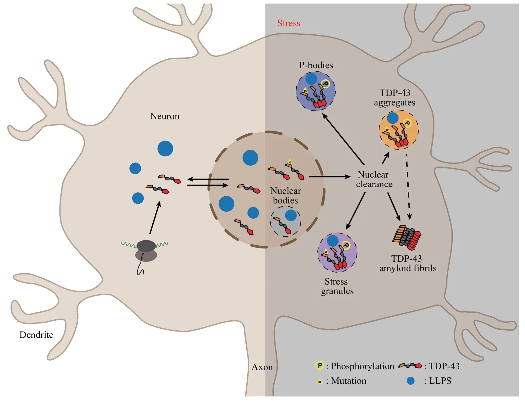
Since TDP?43 shuttles between nucleus and cytoplasm,performing its normal functions in neurons under physiological condition,its localization and status is complex especially when stresses are present,such as aging and disease?causing mutations.TDP?43 undergoes normal or abnormal phase transition in the nucleus and cytoplasm,forming nucleoplasmic NBs and cytoplasmic SGs or PBs,even amyloid fibrils,transiently or permanently,which may lead to neurotoxicity (Figure 2).Therefore,the correlation between TDP?43 localization and abnormal accumulation,and the connection between TDP?43 status and neurotoxicity deserve further investigation.

Fig.2 TDP-43 localization in neurons under physiological and stressful conditions
4 Diagnostic and therapeutic perspective for TDP-43 proteinopathy
TDP?43 is transported into the nucleusviaits NLS regions and functions as an RBP when it is synthesized in the cytoplasm.Misfolded forms of TDP?43 caused by a variety of ALS?related mutations in the cytosol should be degraded either by the ubiquitin?proteasome system (UPS) or by autophagy?lysosome pathway[142?143].Failure in the clearance of TDP?43 aggregates due to the reduced activities of abnormal UPS and autophagy caused by mutations or aging?altered condition will result in the accumulation of TDP?43 protein in ALS and FTLD[144].Recent studies indicated that mitochondria also play a role in the degradation of TDP?43 in cellular and animal models[91,145].Therefore,protein aggregation and phase separation of TDP?43 in neuronal cells are regulated by the orchestration of UPS,autophagy and mitochondria?associated degradation.
TDP?43 protein fragments can be detected in blood and cerebrospinal fluid (CSF) of ALS,FTLD?TDP and some AD patients caused by TDP?43 aggregation,and CSF TDP?43 seems to be mainly blood?derived[146?147].Therefore,blood?derived TDP?43 can be used as a diagnostics marker of these neurodegenerative diseases.However,due to the low absolute level,the quantitative analysis of TDP?43 in biological fluids requires a very sensitive immunoassay,preferably specific for pathological TDP?43[148].The current clinically used TDP?43 pathology?specific antibodies mainly target the serine residues near the C terminus of TDP?43 that can be phosphorylated[149].There are more than 100 kinds of antibodies that can be used for TDP?43 pathological detection,and more than 30 kinds of antibodies can be detected by immunostaining and Western blotting at the same time[146].Among these antibodies,most are polyclonal antibodies and are initially produced internally.To avoid the appearance of biased results,three kinds of antibodies are considered as“standard”antibodies and widely used for the study of TDP?43 proteinopathy.They are 10782 (aa203-209 and AA near N terminus),2E2?D3(aa205-222)and TIP?PTD?P01&?P02 (pS409/410,aa405-414/P)[150].In addition,researchers are also working to broaden the biomarkers of TDP?43 cases.For example,truncated STMN2 can be used as a marker of TDP?43 dysfunction in FTLD patients[151].
Regarding therapeutic approaches of ALS and FTLD,there are very few options currently.Since almost all patients affected by ALS or tau?negative FTLD share the presence of aggregated TDP?43 in their brains,it represents a very promising target to develop novel therapeutic options.Since TDP?43 dysregulation is also associated with up to 50%of AD cases[34,36]and most of the Aβ?targeted clinical trials have not succeeded[152],it is reasonable to propose that TDP?43 might be a potential target when we re?evaluate anti?Aβ therapeutic strategy for treating this most common and complex disease.Identification of small molecules or RNA that could prevent the transition of TDP?43 from liquid or granule states to solid aggregates might be one approach.These small molecule drugs may not only restore homeostasis of RBPs with PrLDs,but also enhance the activity of heat shock proteins against disease?linked RBPs with PrLDs,both of which will delay and alleviate the progression of these devastating diseases caused by aberrant phase transition of TDP?43 induced neurodegeneration.
- 生物化學與生物物理進展的其它文章
- 細菌的信號轉(zhuǎn)導系統(tǒng)及其在耐藥中的作用*
- CRBGP Inhibited The Activity of Glioma U251 Cells Through Suppressing FAK-AKT Pathway and The Secretion of Interleukin-6*
- Bradykinin Upregulated The Expression of Cyclooxygenase-2 in The Submucosal Plexus of Enteric Nervous System of Guinea Pig*
- 阿爾茨海默病體外診斷納米技術(shù)*
- α-Synuclein as a Diagnostic Marker and Therapeutic Target for Parkinson Disease*
- 甘丙肽對抑郁癥狀的調(diào)控作用及其機制的研究進展*

