Early experience with the novel glaucoma shunt device: Paul glaucoma implant in the Indonesian populations
Emma Rusmayani, Viona Viona2, Iwan Soebijantoro, Rini Sulastiwaty, Arini Safira Nurul Akbar, Muhammad Yoserizal, Zeiras Eka Djamal, Widya Artini Wiyogo
Abstract
?AIM:To investigate the safety and efficacy of Paul glaucoma implant (PGI) in the short-term follow-up period and share first experience with this novel aqueous shunt in Indonesian populations.
?METHODS:A total of 21 patients (22 eyes) with PGI implants from April 2022 to December 2022 and with at least a complete 2mo follow-up were retrospectively analyzed. The primary outcome measure was failure, defined as intraocular pressure (IOP) out of the target range of 21 mmHg or less than 20% reduction from baseline for 2 consecutive visits, other glaucoma surgeries required, or removal of the implant.
?RESULTS:The follow-up period was 2 to 6mo. The mean IOP reduction was 52.27±22.94%, with a range of 9% to 90%. The complete success rate was 59%, and patients with or without a history of glaucoma surgery had 50% and 59% of complete success rates, respectively. Complications of the surgery were diplopia (n=2), early hypotony (n=1), hyphema (n=1), and exposed tube (n=2).
?CONCLUSION:The complete success of the PGI implantation was 57%. No serious postoperative complications were found in our cases. One case of hypotony resolved in the early postoperative period.
?KEYWORDS:Paul glaucoma implant; glaucoma tube shunt; glaucoma drainage implants; glaucoma surgery; Indonesian populations
INTRODUCTION
Glaucoma is a major healthcare burden worldwide, and the risk of being affected by the condition increases as the aging process[1]. There are several options for treating glaucoma, such as the non-surgical and surgical treatments. The non-surgical treatments are medications to reduce intraocular pressure (IOP), and laser trabeculoplasty aimed to improve the eye’s drainage angle. Surgical treatments are options in advanced cases where medications or laser treatments may not be enough to control the IOP to prevent further disease progression[2-3].
In cases with uncontrollable IOP, aqueous tube shunts, also known as aqueous shunt devices or glaucoma drainage devices, are considered effective, even for patients with a higher risk of complications. The two most commonly used devices are the Ahmed valve (New World Medical Inc. Rancho Cucamonga, CA) and the Baerveldt implant (Abbott Medical Optics, Santa Ana, CA). The Ahmed valve has a built-in flow restrictor that helps prevent postoperative hypotony, but it can result in bleb encapsulation, which leads to insufficient IOP reduction and the need for additional treatments or surgery. The Baerveldt implant, on the other hand, requires the surgeon to create a temporary tube ligature to regulate flow during the healing process, which may result in hypotony if the healing around the plate for the flow regulation is inadequate. However, the Baerveldt implant provides excellent IOP control once the ligature is dissolved or removed due to its large end-plate surface area of 350 mm2[4-6].
The Paul glaucoma implant (Advanced Ophthalmic Innovations, Singapore, Republic of Singapore; PGI) is a novel aqueous shunt device designed by Professor Paul Chew from Singapore. It has several advantages compared to other existing devices. One of the benefits of the Paul implant is its smaller tube, with a diameter of 127 microns, which can be stented using 7/0 Prolene without the need for a Vicryl tie or Sherwood slits to prevent early hypotony[7]. Additionally, the silicone plate of the Paul implant is larger and more flexible than the Baerveldt shunt, making it more reliable in achieving controlled flow in the immediate postoperative period and reducing fluctuations in IOP[7].
Through this study, we aim to provide an overview of our experience with the newest glaucoma tube shunt, the Paul implant.We also aimed to report the safety and efficacy of the PGI in the short-term follow-up period and also to share our first experience with this novel aqueous shunt in Indonesian populations.
METHODS
This study was a retrospective study of all patients with PGI implants from April 2022 to December 2022 with at least a complete 2-month follow-up. The research adhered to the principles and tenets of the Declaration of Helsinki. Ethical clearance was reviewed by the local committee at both Jakarta Eye Center. Written informed consent was obtained from all patients prior to the surgery. Patients with uncontrolled IOP despite being on maximum glaucoma medication, having failed trabeculectomy, or using glaucoma tube shunts were eligible for inclusion in the study. The patients were classified as having either primary or secondary glaucoma. Secondary glaucoma caused by intraocular inflammation, post-keratoplasty, aphakia, neovascular, or a silicone-filled eye was also included.
SURGICAL TECHNIQUE
The PGI procedure is performed while the patient is under general anesthesia in the supine position. The surrounding skin and conjunctival sac are cleaned with 5% povidone-iodine and a corneal traction suture is placed using a vycril 7/0 suture to rotate the eye into down gaze. The superotemporal quadrant is exposed through conjunctival and Tenon’s peritomy and minimal cautery is applied to the scleral bed. The plate is then positioned beneath the recti, held in place with squint hooks, and sutured to the sclera with 8/0 Ethilon sutures, typically 8 to 10 mm from the limbus.
A 7/0 prolene stent is inserted into thetube to prevent early hypotony (Figure 1). The tube is cut in a beveled manner to ensure a small short length within the anterior chamber or posterior chamber, and the prolene within the tube is maintained in position at the tip of the tube. A scleral flap is made using a diamond blade, measuring approximately 4×3mm with a depth two third of scleral thickness. Tube placed to the anterior chamber or posterior chamber was gained using a 26-G needle, positioned parallel to the iris (Figure 2). The tube is inserted into the anterior or posterior chamber, and the entry site is checked for any peritubular leak. The scleral flap is then closed with a scleral graft to prevent future tube exposure (Figure 3). The tube secured with a 8/0 Ethilon box suture to prevent tube movement. The conjunctiva is closed using vycril 8/0 and subconjunctival antibiotic and steroid are administered. The patient is given prednisolone acetate 1% eye drops 6 times a day for 2 to 3mo and a quinolone antibiotic topically for 4wk. Immediately after the procedure, all glaucoma medications are discontinued. The Prolene stent can be taken out 2 to 3wk post-surgery during an outpatient slit lamp examination[7-8].
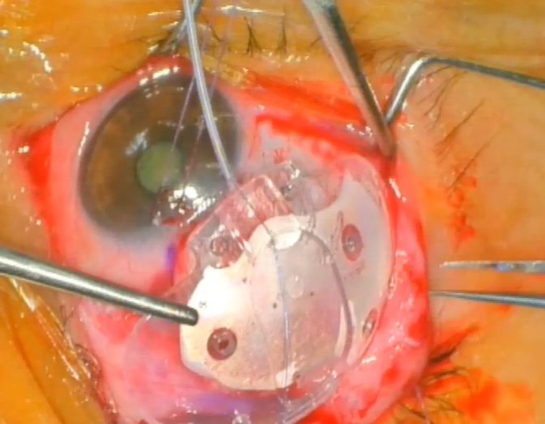
Figure 1 Intraoperative of Paul glaucoma implant with 7.0 Prolene stent.
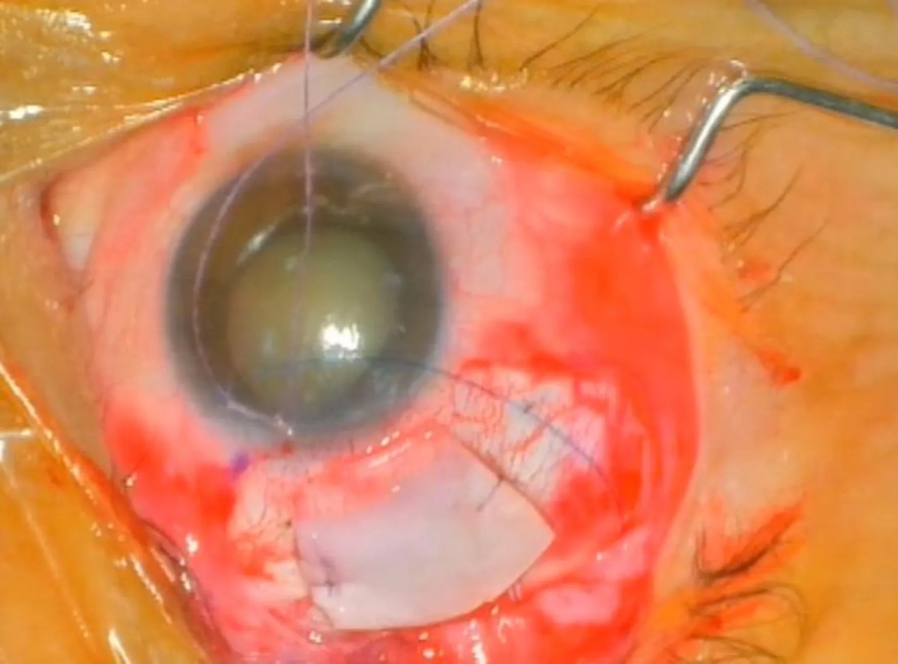
Figure 2 Single entry site to anterior chamber made using 26-G needle parallel to iris plane.
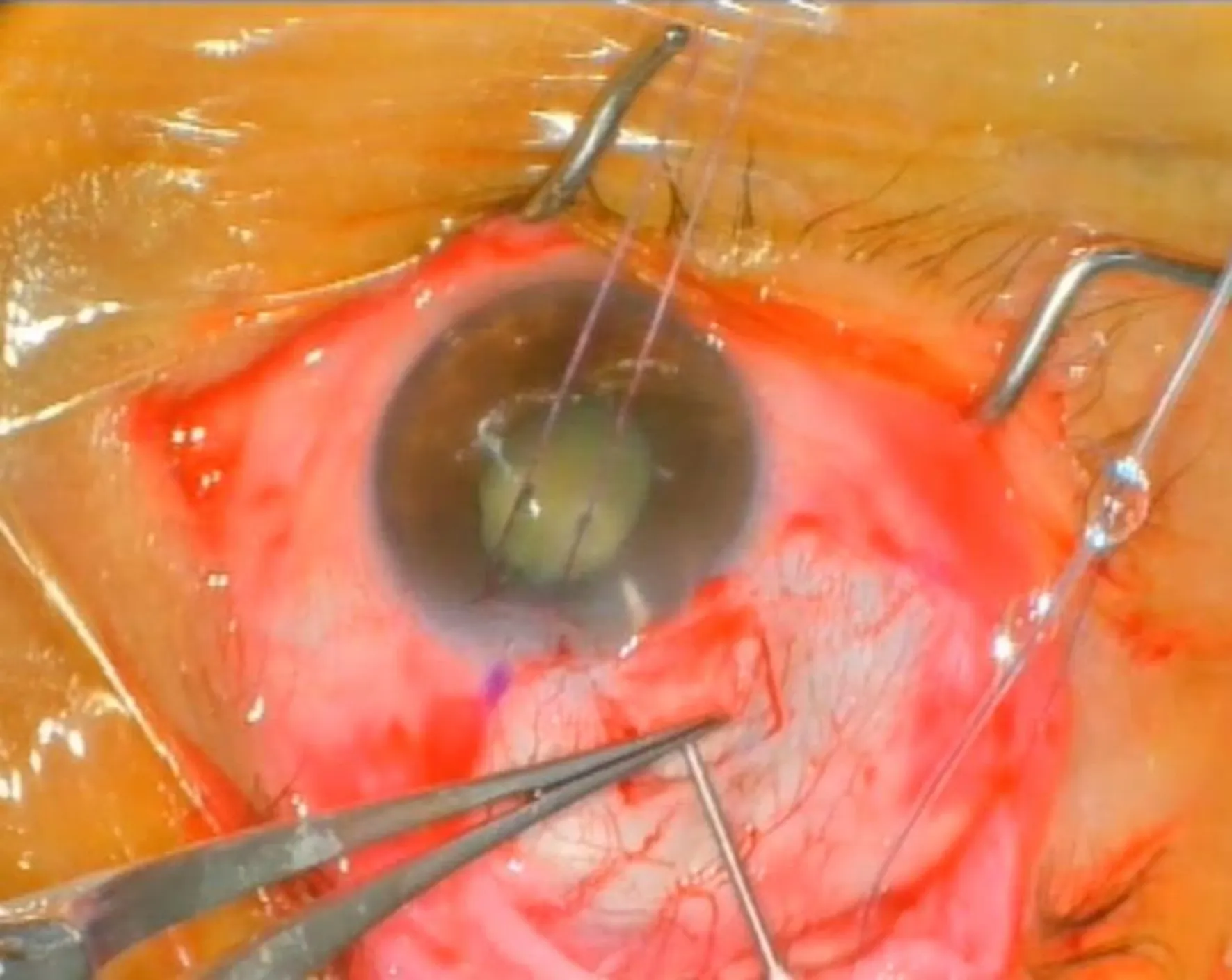
Figure 3 Scleral patch placed above the flap.
In our cases, we decided to remove the prolene stent earlier. In this study, we opted for an earlier Prolene stent removal at 3wk postoperative. In contrast to the conventional Baerveldt tube implantation which typically necessitates a minimum of 2mo due the tube was likely to open after a period of 5 to 7wk[9-10]. Furthermore, the PGI with its smaller lumen and reduced risk of hypotony, combined with our experience in managing patients with uncontrollable IOP and high IOP baseline, provided a viable option for earlier stent removal, as long as a deep anterior chamber was present[8].Prior research on PGI has yielded diverse outcomes, with varying timing for stent removal reported in different studies. Some studies removed the Prolene stent after a mean duration of 5.60±5.12mo, while others performed the removal at a mean of 4.76±2.90mo. Notably, in one study, the Prolene stent was removed after an average period of 8.4mo. Another study removed the second Prolene suture from all eyes with a mean time of removal. Regarding post-operative management, Prolene stent removal at 3mo or later was recommended, or if the IOP rises above 24 mmHg. In such cases, stent removal is preferable to reintroducing topical glaucoma therapy.Given these findings, we reached the decision to explore the possibility of earlier Prolene stent removal for patients experiencing IOP>24 mmHg for a minimum of 3wk postoperatively, taking into account the aforementioned factors[7-8,11-12].
OUTCOME MEASURES
The primary outcome measure success is defined as IOP less than 21 mmHg or more than 20% reduction from baseline for 2 consecutive visits, failure is defined as other glaucoma surgeries required (e.g.additional tube shunt, cyclodestructive procedure,etc.), or removal of the implant. Complete success was defined as no antiglaucoma medications needed to reach the target IOP. Partial success was IOP reached the target with additional antiglaucoma medications. Complications were defined as early and late-period complications. Serious complications were defined as endophthalmitis or needing a return to the operating room[4-6,8].
RESULTS
The baseline characteristics of all study participants (Table 1) with the number of eyes included in this study was 22 from 21 patients with a follow-up period of 2 to 6mo (Figures 4-6). All patients had an uncontrolled IOP despite maximum glaucoma topical medications. We included patients with a wide range of glaucoma severity. The baseline characteristics of the patients in this study are mentioned in Table 1. Preoperative IOP was 42.95±13.86 mmHg with 3.65±0.49 medications. Patients with secondary glaucoma were higher in this study population (n=15; 70%). There were 2 pediatric patients with a history of Ahmed tube implant and trabeculectomy(Figure 7). Additionally, there were 2 primary glaucoma cases with a history of failed trabeculectomy and Ahmed tube implant. Higher numbers of na?ve eyes (82%), which are 3 cases of primary open-angle glaucoma and 15 cases of secondary glaucoma. In the primary glaucoma cases, we opted for the PGI procedure instead of considering trabeculectomy. This decision was based on several factors, including the high baseline IOP, the severity of the cases, and the patients’ ability to undergo routine checkups. Additionally, we took into account the long-term risk of bleb infection in trabeculectomy patients[13-15]. IOP was measured in week 1, week 3, month 1, month 2, month 3, and month 6 follow-up period. In this study, posterior chamber placement of the tube was observed in two patients: one with secondary glaucoma associated with an Aphakic lens, and the other with secondary glaucoma related to iridocorneal endothelial syndrome (ICE). For the remaining cases, the tube was placed in the anterior chamber.
Mean IOP during the 1-week (n=22), 3wk (n=16), 1mo (n=18), 2mo (n=18), 3mo (n=10), and 6mo (n=5) follow-up period (Figure 8). The mean IOP were 13.52±9.67 mmHg in 1wk, 18.69±8.38 mmHg in 3wk, 21.15±8.14 mmHg in 1mo, 18.27±5.16 mmHg in 2mo, 20.50±12.86 mmHg in 3mo, and 25.20±21.39 mmHg in 6mo. In most cases in our study, the PGI stent was removed during the 3-week follow-up period (n=17, 77%), although some cases had it removed during the 1-month follow-up. Additionally, stent removal was also considered if the IOP exceeded 24mmHg. The range for the stent removal varied between 3 and 24wk in our cases. Patients with complete success (59%) were higher than those who needed anti-glaucoma medications observed in the postoperative follow-up period (Table 2). Patients with or without a history of glaucoma surgery had 50% and 59% of complete success rate, respectively. The mean IOP reduction was 52.27±22.94%, with range of 9% to 90%. In this study, the complete success was 59% (n=13), with 27% (n=6) observed in primary glaucoma cases and 32% (n=7) in secondary glaucoma cases. On the other hand, the partial success was found to be 41% (n=9), consisting of 18% (n=4) primary glaucoma and 23% (n=5) secondary glaucoma.
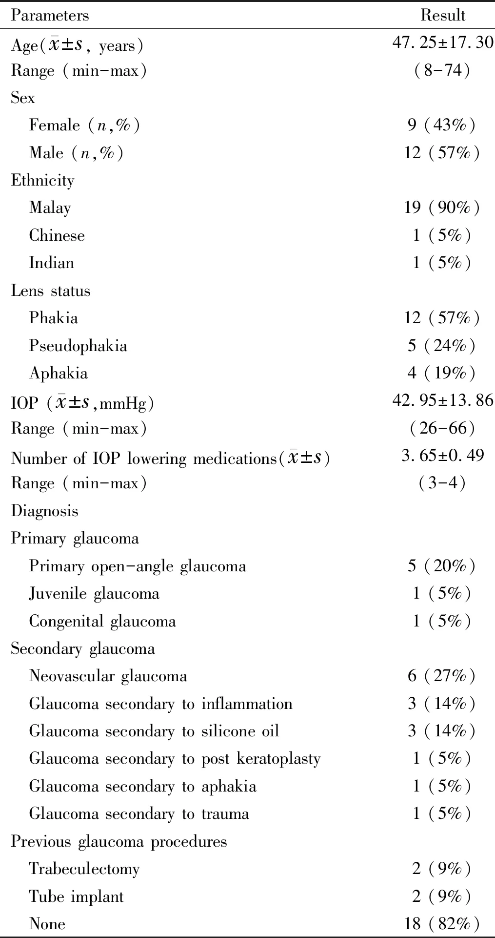
Table 1 Baseline characteristics
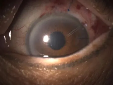
Figure 4 Paul glaucoma implant in the anterior chamber with 7/0 Prolene stenting in 1-day follow-up. The tip of the stent suture was made blunt using low-temperature cautery and placed superior or lateral to be easily visualized by the surgeon at the slit lamp.

Figure 5 Scleral graft placed above the scleral patch to prevent risk for exposure.

Figure 6 Paul glaucoma implant without Prolene stenting in 2wk of follow up.
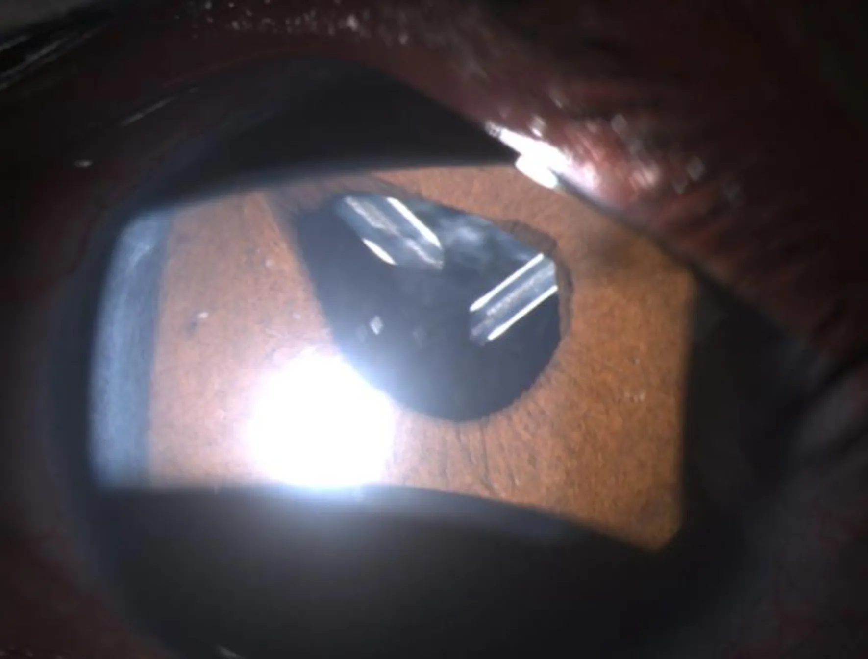
Figure 7 Comparison of lumen size of Ahmed tube and Paul glaucoma implant.
ComplicationsDiplopia was observed in two patients both in the early postoperative period (<3mo). Early hypotony was found in one case due to traction of the Prolene suture and able to be resolved in 2wk post-op follow-up period. One case of hyphema in neovascular glaucoma and resolve within 3wk. Two exposed tubes, both with pericardium, needed to be revised in the operating theatre. No cases of endophthalmitis were found.
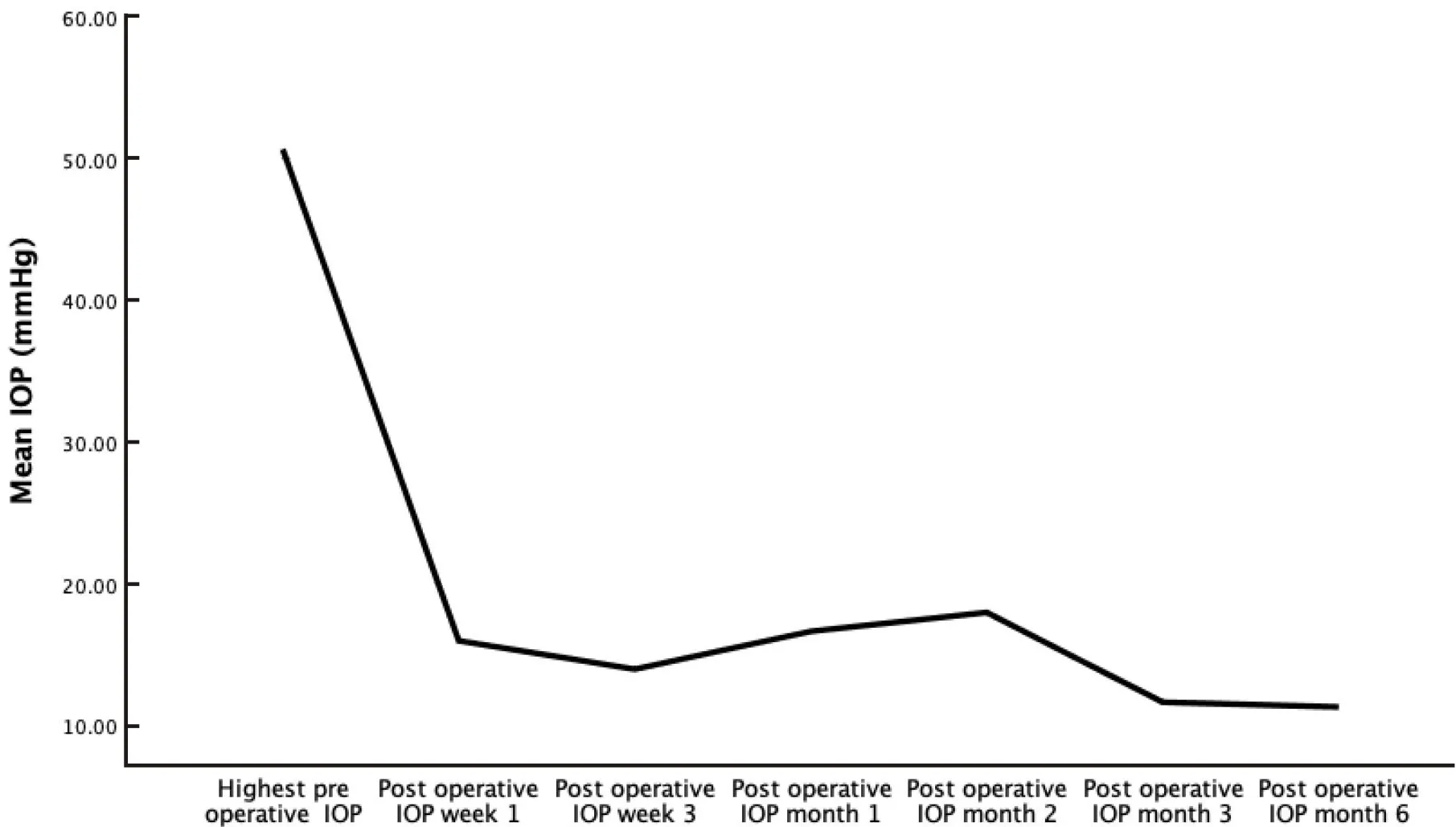
Figure 8 Mean intraocular pressure in the follow-up period after Paul glaucoma implant surgery.

Table 2 Postoperative anti-glaucoma medications after Paul glaucoma implant surgery
DISCUSSION
The PGI is a non-valved tube with a shorter wingspan and smaller end plant compared to the Baerveldt glaucoma implant (BGI). The PGI implant has the smallest tube lumen compared to Ahmed glaucoma valve and BGI and with occlusion using 6-0 or 7-0 Prolene prevents early hypotony[7].
IOP reduction of PGI is comparable to Ahmedvs. Baerveldt study with the same population. From the previous study, Baerveldt tube (60%) has shown higher IOP reduction than the Ahmed tube (58%)[16]. However, the limitations of this study were the short follow-up period and the small number of participants to assess the effectiveness of the PGI tube in the long term.
Compared to the previous PGI study, shallow anterior chamber, hypotony, and tube occlusions were the highest complications found[7-8,11-12]. In our study, we have 1 case of hypotony in the early follow-up period and self-resolve without posing any serious complications. To prevent hypotony, we used the 26 gauge needle with a single stab and carefully avoid peritubular leak intraoperatively. We also experienced good outcomes using Prolene 7/0 stenting to prevent early hypotony.
From our perspectives, the smaller tube diameter of PGI compared to Ahmed increased our concern for the risk of tube occlusion. From the previous study, occlusions of iris, vitreous, fibrin, or pigments were documented. However, in our cases, we did not experience any tube occlusions. Posterior chamber placement of the tube was found in two patients, one in secondary glaucoma to the Aphakic lens and one in secondary glaucoma to iridocorneal endothelial syndrome (ICE). No vitreous occlusions were found in both cases. However, both cases were partially success, and the decrease of IOP was not as low as we expected. Further analysis of follow-up data was needed regarding the tube placement position and IOP lowering effect. In three cases of silicon-filled eyes, there were also no silicone occlusions.
In 56% of the cases of Ahmed tube implant, the occurrence of hypertensive phase was noted. The most common occurrence of the hypertensive phase after the Ahmed tube implant occurred 1mo after the surgery and remained stable 6mo later. In most cases, the eyes experiencing the hypertensive phase did not show significant improvement in IOP control and continued to require the same number of glaucoma medications as before the procedure. The hypertensive phase was also found in cases after Baerveldt tube implant less common than Ahmed tube implant[17-18]. The occurrence of the hypertensive phase was less frequent after Baerveldt tube implantation compared to Ahmed tube implantation. Preoperative IOP and previous glaucoma surgeries are factors that increase the risk of developing a hypertensive phase. The presence of a hypertensive phase can indicate the need for long-term use of anti-glaucoma medication. This phase is believed to occur due to the formation of dense fibrous tissue over the plate, which may alter the pattern of newly formed collagens and other extracellular matrixes. The sudden cessation of medication after the Baerveldt implantation may also contribute to a rebound in IOP elevation. Prolonged preoperative use of medication can suppress IOP for an extended period. The development of a hypertensive phase after surgery suggests a higher likelihood of needing anti-glaucoma medication[19].
Another concern of glaucoma tube implant is corneal decompensation in the long term. Central and peripheral endothelial cell loss after glaucoma tube implantation in the anterior chamber has been associated with decreased endothelial cells 37% to 50% in 5 years[19-21]. The smaller lumen of PGI theoretically will also benefit in cases of secondary glaucoma post-keratoplasty. The smaller tube lumen eventually will reduce the endothelial cell loss compared to bigger lumen tube implants[7-8,11-12]. Anterior tube placement is considered safe for cases with low endothelial cell density cells[22]. One patient with secondary glaucoma post-keratoplasty did not experience a significant reduction in the density of corneal endothelial cells after 6mo of being treated with a PGI tube implant.
Tube exposure was a common complication after glaucoma tube implant. However, it can be reduced by several methods, include proper placement of the implant, sufficient tissue coverage of the device by the sclera, and careful wound closure[23-24]. Reoperations in our study were due to tube exposure and in both cases used the pericardium. We experienced better outcomes with scleral graft closure.
The limitations of our study include the short follow-up period, a limited range of cases, and the absence of a comparison with previous models used for treating similar glaucoma cases. Due to these limitations, our findings may not fully capture the long-term safety and efficacy of the PGI in the Indonesian population. To address these limitations and gain a more comprehensive understanding of the PGI’s efficacy, further studies with longer follow-up durations are necessary. These extended studies would enable a more thorough assessment of the safety and effectiveness of the PGI in various glaucoma cases within the Indonesian population. Additionally, it is essential to conduct comparative studies that compare the results of our current study with those of previous glaucoma tube shunt studies. By doing so, we can identify potential differences in outcomes and better understand the advantages and disadvantages of using the PGI compared to other existing treatment options. Conducting further studies would provide valuable insights for clinicians and researchers, enabling them to make more informed treatment decisions and improve glaucoma management for patients with diverse needs and conditions. Overall, a comprehensive and evidence-based approach to studying glaucoma tube shunts can significantly contribute to advancing glaucoma treatment and patient care.
CONCLUSIONS
PGI is a novel glaucoma tube shunt and is effective for lowering IOP. The complete success of the PGI tube in this study was 59%, with no serious postoperative complications found in our cases. One case of hypotony resolved in the early postoperative period. The surgical method of using a 7/0 Prolene stent is safe and effective in preventing early hypotony. Further studies with longer duration of follow-ups are required to assess the safety and efficacy of the PGI in the Indonesian population.

