
MODELING AND SIMULATION OF E1784K MUTATION AND SODIUM IONIC CHANNEL DISEASES
2011-10-08 12:10:04ZhangWenYuanYongfengKongShuhan
Zhang Wen,Yuan Yongfeng,Kong Shuhan
(1.School of Life Science,East China Normal University,Shanghai,200062,P.R.China;2.School of Computer Scienceand Technology,Harbin Institute of Technology,Harbin,100051,P.R.China)
INTRODUCTION
E1784K is a mutation in SCN5A gene which is the control gene of sodium ionic channel.In clinical reports,electrocardiograms(ECGs)of patients with the E1784K mutation exhibit the characteristics of abnormal prolongation of QT interval and T segment elevation in the right precordial leads,which indicates a phenotypic overlap between long QT syndrome(LQT3)and Brugada syndrome(BrS).Makita et al[1]reported the E1784K mutation resulted in a shift of transient sodium current(I NaT)inactivation curvein the hyperpolarising direction and an increase in density of late sodium current(INaL).However,it is still unclear whether the E1784K mutation-induced alternations in sodium channel functions are sufficient to illustrate the overlap between LQT3 and Br S.This problem is hard to validate by physiological experiment method[2-3]. Accordingly,in the present study,a computational model based on detailed experimental data is developed to investigate the problem as a hypothesis research method.
1 DEVELOPMENT OF COMPUTATIONAL MODEL
1.1 Altered kinetics of sodium ion channel under E1784K mutation condition
In the present study,the Ten Tusscher-Noble-Noble-Panfilov(TNNP)model which is the popular human ventricular cell computational model developed by Ten Tuss cher et al[4]is used.Since the TNNPmodel does not include I NaL model,a new ventricular I NaL model is developed according to the I NaL model[5]and the experimental data[6]on maximal conductance of I NaL in three types of canine ventricular cells,and then the model is incorporated into the TNNPcell computational model.Definition of the model is described as

where G NaL is the maximal conductance of I NaL channel and values of G Na L for epicardial(EPI),midmyocaridal(M)and endocardial(ENDO)cell types are 0.006 5,0.011 0 and 0.007 5 n S/p F respectively[6],mLis the activation-gate variance,hLthe inactivation-gate variance,Vmthe action potential(AP)of transmural membrane,E Na the equilibrium potential of sodium channel currents and computed by extra-cellular sodium concentration[Na+]oand intra-cellular sodium concentration[Na+]idepending on the equation of Nernst(Eq.(2)).
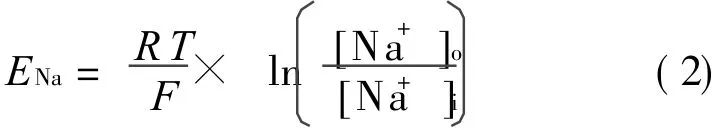
where R is the gas constant,F the Faraday constant and T the absolute temperature. [Na+]o equals 140 mmol/L and[Na+]i5 mmol/L.
According to recent experimental data under the E1784K mutation condition,Makita et al[1]found that the activation and inactivation curves of I NaT under the E1784K mutation condition were shifted to positive and negative directions compared with those under the control condition respectively.
The exponential regress equation(Eq.(3))is used to simulate effects of the E1784K mutation on I NaT.
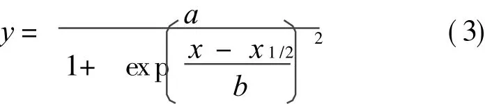
where x1/2represents m(activation-gate variance)or h(inactivation-gatevariance)in I NaT kinetics of TNNPmodel.Depending on Makita′s experimental observations[1],thevalues of m1/2 are changed from-56.86 mV to-36.98 mV,and h1/2from-71.55 mV to-101.9 mV.The values of b are changed from - 5.71 (control)to - 7.67(E1784K)for activation curve and from 5.97(control)to 6.62(E1784k)for inactivation curve respectively.Simulation results under both control and E1784K mutation conditions are shown in Fig.1.

Fig.1 Activation and inactivation curves of I NaT under control and E1784K mutation conditions
1.2 Kinetics of calcium and potassium ion channels under Br Scondition
The previous experimental studies[7-8]reported that the remarkable ST segment elevation in the right precordial ECG leads of Br S patients was associated with the depression of AP plateau from right ventricular outflow tract to ventricular epicardium,which was found to be related with an increase of the transient outward current I To and a decrease of the L-type calcium current I CaL.In order to simulate the effects of BrS-induced AP changes of epicardium cell,the Br Smodel developed by Ref.[9]is used in this paper.
(1)Simulations of ITokinetics changes under Br Scondition
G To,the maximum conductivity of I To,is increased by 27% and activation curve of its activation-gate variance r is shifted from 20 mV to 2.5 mV.
(2)Simulations of I CaL kinetics changes under Br Scondition
G CaL,the maximum conductivity of I CaL,is reduced by 35%.
1.3 Ventricular tissue model
One-dimensional(1-D)model of transmural ventricular strand is composed of 100 computational cells(TNNPmodel)in series.
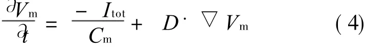
where t is the time,Cmthe cell capacitance per unit surface area,D the diffusion coefficient which represents conduction speed,I tot the total current across the membrane cell.
The length of each cell is 150μm,while transmural human ventricular cell is about 50—100μm.The length of transmural ventricular strand in stimulation is 15 mm whereas the real strand length of human is 4—14 mm.Following the results of Ref.[10],the length ratio of EPI,M and ENDO is 25∶ 35∶ 40.Except the EPI-M border,D is set to 0.054 mm2/ms.The conduction velocity of the simulated excitation wave is 0.48 m/s,and it belongs to the excitation conduction velocity scope of human heart chamber[10].Due to the protective activities of ventricular tissue,thereis a five-fold decreasein conduction velocity at the EPI-MIDDLE border.Accordingly,the value of D at this border is changed to 0.011 mm2/ms[6].
A 2-D transmural ventricular tissue sheet is modeled by expanding 1-D transmural strands.1-D strand direction(x-direction)is recognized as transmural membrane direction and 800 strands are arranged along y-direction in parallel.Both spatial resolutions of x-and y-directions are 0.15 mm,so the volume of simulated ventricular tissue is 15×120 mm2.
1.4 Computation of pseudo-ECG
Based on the 1-D strand model,a virtual electrode is set at 2 cm outside from the EPI end to simulate ECG(Fig.2)using the method in Ref.[10].According to source-field[11],the potential at virtual electrode is

Fig.2 Computational model of pseudo-ECG based on 1-Dventricular strand
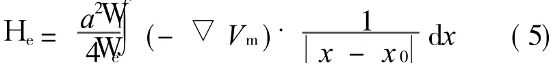
where H e is the electric potential field of whole strand,W i and W e are the intra-and extra-cellular conductivities respectively,a is the radius of the cell,x the local site point of strand,x0 the position of virtual electrode and|x-x 0|the distance from the local site to the electrode.The potential at virtual electrode is computed as an integral of electric field consisted of all cells.Consistent with Ref.[9],the values of Wi,Weand a are set to 0.000 74,0.001 26μs/cm and 0.001 1 cm.
2 SIMULATION RESULTSOF COMPUTATIONAL MODEL
2.1 Simulation results of altered sodium current under E1784K mutation condition
The current-voltage(I-V)curves of I NaT obtained from simulation and experimental results are normalized and compared(Fig.3).As illustrated,the curve shape and changing tendency of simulation data are nearly consistent with tho se of experimental data except the curve scope from-100 mV to-60 mV.In this range,the simula-
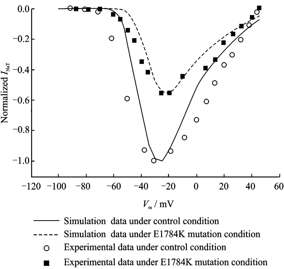
Fig.3 Current-voltage(I-V)relations for I NaT under control and E1784K mutation conditions
tion results are larger than experimental data.It is the main factor that theexperimental conditions(such as temperature and instruments)for developed computational models are different from those in clone cell experiments.Furthermore,there are differences in physiological features between clone and original cells,for example,the I NaT peak of clone cell is about five folds smaller than that of original cell.According to the shape and changing tendency of curves and physiological characters of INaT,the error between simulation and experimental results is within the acceptable range,which indicates the effectiveness of the above sodium ion channel model.
2.2 Comparison of action potentials
The effects of altered I Na T and I NaL under E1784K mutation condition on the cell APareillustrated in Fig.4.Compared with the control condition,the E1784K mutation condition results in a decrease in peak current density of INaT(Fig.4(a))and a persistent increase in I NaL current(Fig.4(b)).The altered I NaT and I NaL decrease the AP amplitude and prolong the AP duration(APD)of three ventricular cell types under E1784K mutation condition(Fig.4(d))in comparison with those under the control condition(Fig.4(c)),and APDs are listed in Table 1.
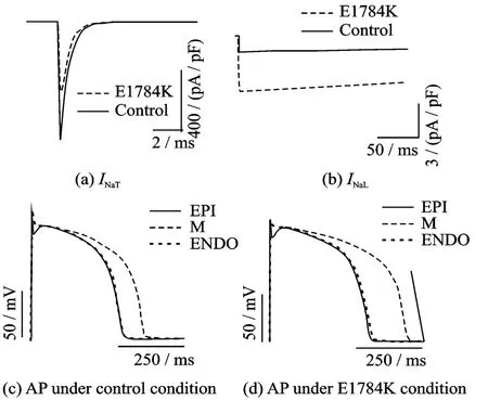
Fig.4 Effects of I NaT and I NaL on AP
Table 1 Ef fects of control and E 1784K mutation condi
tions on APs of three ventricular cell types ms

APD90 Control condition E1784K condition EPI 283.64 310.56 M 352.00 429.04 ENDO 285.30 316.68
2.3 Comparison of ef fective refractory period
As shown in Fig.5,the computed ERPs under both control and E1784K mutation conditions are plotted against basic cycle length(BCL)for three cell types.Compared with the control condition,the E1784K mutation condition increases ERPs of three cell types in whole scope of BCL.As the augmented ERP of M cell is the largest,the heterogeneity among the different cell types is increased.
2.4 Simulation results of pseudo-ECGs
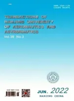
Simulation results of pseudo-ECGs in 1-D strand model under control and E1784K mutation conditions are illustrated in Fig.6.Time-varying APs of each cell in 1-D strand are illustrated in the upper parts of the figure,and the different colors represent the different potentials.Time runs x-direction from left to right,while space of 1-D strand runs y-direction from the bottom(ENDO)to the top(EPI).In the bottom parts of the figure,time-varying pseudo-ECGs simulated by 1-D strand model are shown.Compared with
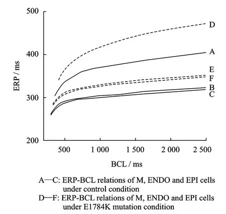
Fig.5 ERP-BCL relations of three cell types under control and E1784K mutation conditions
the control condition(Fig.6(a)),the pseudo-QT interval is prolonged and the amplitude and width of T-wave are increased under the E1784K mutation condition(Fig.6(b))because of the slow depolarization and repolarization of cells, the lengthened cell repolariza tion duration and the increased time difference of repolariza tion between cells induced by the E1784K mutation.
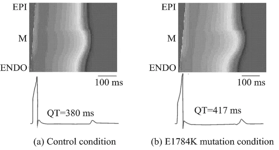
Fig.6 Simulations of pseudo-ECGs under two conditions
3 ANALYSISOF STIMULATION RESULTS
3.1 Analysis of physiological mechanism on stimulation results
The above simulation results indicate that the E1784K mutation decreases the peak current density of INaTbut increases INaLcurrent,APs and ERP.Furthermore,the dispersions of APs and ERP between cells are also enlarged. These changes result in the prolonged pseudo-QT interval and increase amplitude and width of T-wave.The prolonged QT interval and increased T-wave are typical ECG features of LQT3.But the ECG hallmark of BrS(elevated ST segment)has not been observed. Therefore,in this simulation study,the phenotypic overlap between LQT3 and Br Sdoes not appear.These results suggest that the E1784K mutation-induced changes in sodium channel currents are insufficient to produce the phenotypic overlap between LQT3 and BrS.But why does the overlap appear in patients with the E1784K mutation?
The previous experimental research[12]found that the marked ST segment elevation in ECG of Br Swas associated with the depression of the AP plateau in epicardium of right ventricular outflow,which was due to an increase of I To current and a decrease of I CaL current.This paper hypothesizes that function reconstructions of I To and I CaL currents under sodium channel dysfunctions caused by the E1784K mutation may be the major reason for producing the overlap between LQT3 and Br S.In order to validate the hypothesis,the two additional factors(increased I To and decreased I CaL)are reconsidered for the underlying mechanism of the E1784K mutation in this simulation.
Besides the effects of altered I To and I CaL,the altered kinetics of sodium current induced by the E1784K mutation also effects on APs of EPIcells and pseudo-ECGs.As shown in Fig.7,under the E1784K mutation condition,thereis not an obvious change on AP waveform of EPI cell except the markedly prolonged AP plateau.The QT-interval of pseudo-ECGs is prolonged and the amplitude and width of T-wave are increased.But the elevated ST segment is not observed.
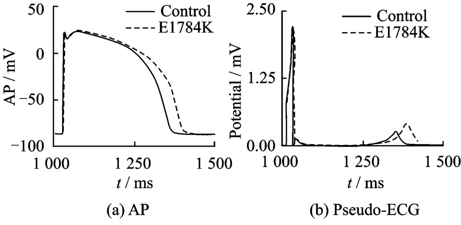
Fig.7 APs and pseudo-ECGs of EPI cell under control and E1784K mutation conditions
APs and pseudo-ECGs of EPI cell are simulated via increasing I To current using the methods in Section 1.2(for EPIcell only,the I Tovalues of M and ENDO cells stay unchanged).Fig.8 shows the waveforms of APs and pseudo-ECGs under control,I To(BrsⅠ )and″E1784K+I To(BrsⅠ )″conditions.Compared with the control condition,a faster repolarization,lower AP plateau,shorter APD arise under the ITocondition.But they just show features of BrSⅠ in ECG(the dome likee levat ion of ST segment without the prolonged QT interval)and the features of LQT3 are not observed.The AP plateau and APDs are augmented under″E1784K+I To″condition compared with those under I To condition,but these two parameters are much shorter than those under control condition.And the ST segment are elevated like a dome(feature of Br S)while pseudo-QT interval are prolonged(featureof LQT3),thus exhibiting the overlap between LQT3 and Br S.Ⅱ )″conditions,respectively.Compared with the control condition,there are a faster repolarization,lower AP plateau,shorter APD and elevated ST segment like a saddle after simulation of decreased I CaL.But the prolonged QT interval is not observed.The features of Br SⅡin ECGjust appear under the″E1784K+ I CaL″condition,and there is a mild decrease in AP plateau but an increase in APD compared with those under control condition.Also under this condition,features of both LQT3 ECG(prolonged pseudo-QTinterval)and BrS ECG(elevated ST segment in pseudo-ECGs)are observed,thus indicating the overlap between LQT3 and Br S.

Fig.8 APs and pseudo-ECGs of EPI cell under control,I To(Br SⅠ )and″E1784K+ I To(Br SⅠ )″conditions

Fig.9 APs and pseudo-ECGs of EPI cell under control,I CaL(Br SⅡ )and″E1784K+ I CaL(Br SⅡ )″conditions
3.2 Simulation Limitations
There are some limitations in the model:(1)The TNNPmodel has notincluded I NaL,and there are only canine experimental data of INaL;(2)Due to the lack of experimental data about the reconstruction of I To or I CaL induced by altered sodium channel,we directly use the results in Ref.[9]and assume that the altered sodium channel induced by the E1784K mutation results in an increase of I To and a decrease of ICaL.Accordingly,the precise reconstruction mechanisms of IToor I CaL induced by altered sodium channel cannot be obtained from the present work.However,these limitations do not influence the principal conclusions of this paper,that is,the E1784K mutation-induced sodium channel changes is insufficient to produce the phenotypic overlap between
APs and pseudo-ECGs of EPI cell are simulated via decreasing I Ca L current using the stimulation methods of BrS(I CaL values of M and ENDO cells stay unchanged)[12].Fig.9 shows the waveforms of APs and pseudo-ECGs under control,decreased I CaL(BrSⅡ )and″E1784K+ I CaL(Br S LQT3 and BrS,which may arise from a combined action of the mutation-induced changes in sodium channel currents and possible increased IToor decreased I CaL as seen in BrS.
4 CONCLUSION
To study the problem wether the E1784K mutation-induced sodium ionic channel alterations account for the overlap at tissue level,the simulation is implemented by the detailed computational model incorporated with experimental data in Ref.[1].The simulation results indicate that E1784K mutation-induced sodium channel alterations can explain increased APD and prolonged QT interval in the clinical ECGs,but cannot account for elevated ST segment observed in ECGs of patients with the E1784K gene mutation.The additional stimulations of IToand ICaLshow features of Br SⅠ and BrSⅡ without the LQT3 features respectively.The phenotypic overlap between LQT3 and Br Sphenomenon is only reproduced by the integration of I Na and I To or I Na and ICaL.Hence,the underlying cause of the phenotypic overlap of LQT3 and BrS may be a combined action of the E1784K mutation-induced changes in sodium channel currents with an increase of I To or a decrease of I CaL.In conclusion,the principal result in this paper suggests that the E1784K mutation-induced sodium current dysfunction is insufficient to produce the phenotypic overlap between LQT3 and Br S.The experimental study or clinical therapy about the E1784K mutation should not just concentrate on thedirect cause of the E1784K regulating-sodium channel but consider the complicated reconstruction effects of other channels such as I To and I CaL currents caused by the E1784K mutation.
[1] Makita N,Behr E,Shimizu W,et al.The E1784K mutation in SCN5A is associated with mixed clinical phenotype of type 3 long QT syndrome[J].The Journal of Clinical Investigation,2008,118(6):2219-2229.
[2] Zimmer T,Surber R.SCN5A channelopathies—An update on mutations and mechanisms[J].Progress in Biophysics and Molecular Biology,2008,98(2/3):120-136.
[3] Kapplinger JD,Tester D J,Alders M,et al.An international compendium of mutations in the SCN5A-encoded cardiac sodium channel in patients referred for Brugada syndrome genetic testing[J].Heart Rhythm,2010,7(1):33-46.
[4] Ten Tusscher K H,Noble D,Noble P J,et al.A model for human ventricular tissue[J].American Journal of Physiology-Heart and Circulatory Physiology,2004,286(4):H1573-1589.
[5] Xia L,Zhang Y,Zhang H,et al.Simulation of Brugada syndrome using cellular and three-dimensional whole-heart modeling approaches[J].Physiological Measurement,2006,27(11):1125-1142.
[6] Yan G X,Shimizu W,Antzelevitch C.Characteristics and distribution of M cells in arterially perfused canine left ventricular wedge preparations[J].Circulation,1998,98:1921-1927.
[7] Yan GX,Antzelevitch C.Cellular basis for the Brugada syndrome and other mechanisms of arrhythmogenesis associated with ST-segment elevation[J].Circulation,1999,100:1660-1666.
[8] Fish JM,Antzelevitch C.Cellular mechanism and arrhythmogenic potential of T-wave alternans in the Brugada syndrome[J].Journal of Cardiovascular Electrophysiology,2008,19:301-308.
[9] Ten Tusscher K H,Panfilov A V.Cell model for efficient simulation of wave propagation in human ventricular tissue under normal and pathological conditions[J].Physics in Medicine and Biology,2006,51(23):6141-6156.
[10]Zhang H,Hancox J C.In silico study of action potential and QT interval shortening due to loss of inactivation of thecardiac rapid delayed rectifier potassium current[J].Biochemical and Biophysical Research Communications,2004,322(2):693-699.
[11]Jaakko M,Robert P.Bioelectromagnetism:Principles and Applications of Bioelectric and Biomagnetic Fields[M].New York:Oxford University Press,1995:148-158.
[12]Satoshi N,Kengo F K,Hiroshi M,et al.Longer repolarization in the epicardium at the right ventricular outflow tract causes type 1 electrocardiogram in patients with Brugada syndrome[J].Journal of the American College of Cardiology,2008,51(12):1154-1161.
 Transactions of Nanjing University of Aeronautics and Astronautics2011年4期
- Transactions of Nanjing University of Aeronautics and Astronautics的其它文章
- VOLUMETRIC-SWEPT DISPLAY SYSTEM BASED ON HELIX ROTATING SCREEN AND DMD
- OPTIMIZATION DESIGN METHOD FOR INPUT IMPEDANCE MATCHING NETWORK OF LOWNOISE AMPLIFIER
- DIRECT SELF-REPAIRING CONTROL FOR HELICOPTER VIA QUANTUM CONTROL AND ADAPTIVE COMPENSATOR
- CHARACTERISTICSOF FAN STALLING BASED ON CORRELATED DIMENSIONS
- HYBRID SCHEME FOR COMPRESSIBLE TURBULENT FLOW AROUND CURVED SURFACE BODY
- PRE-CORROSION FATIGUE NOTCH FACTOR