Reconstruction of 3D Skin Model and Its Application in Evaluation the Efficacy of Cosmetic Raw Materials and Active Intredients
Song Xiaojie, Zhou Chunxia,Yao Qifeng,Wu Yue
R&D Center, JALA Corporation, China
In vitro three dimensional (3D) skin model was reconstructed based on technology and theory of cell biology and engineering. Normal human dermal fibroblasts and keratinocytes were used to construct three dimensional skin model, which has a very intact 3D anatomical structure that is highly similar to normal human skin (NHS). With the development of tissue engineering technology, as well as the huge demand of animal substitution methods for pharmaceutical or cosmetics industry, the skill and technology of reconstructed skin model in vitro has been greatly promoted. Now, 3D skin model has not only been applied in clinical treatment for burns, but also applied in safety and efficacy assessments.
Dermal-Epidermal Junction (DEJ) is located between epidermal and dermal, which consists of Laminin 5,Collagen IV (Col IV), Collagen VII (Col VII) and other important proteins. DEJ has many functions, including adhesion of epidermis tightly to dermis, and mediating exchange of substance between the epidermis and dermis. Aging and adverse environmental factors, such as ultraviolet exposure, can induce overlap and fracture in DEJ, and then reduce adhesion between epidermis and dermis, then further reduce nutrient delivery from dermis to epidermis, which accelerates skin aging.[1~3]Current studies[4~6]suggested that the condition of DEJ directly influences the status of whole skin, so maintaining a healthy DEJ status is the key to prevent skin aging. Upregulation of the key components of DEJ can renovate DEJ and strengthen the junction of the epidermis and the dermis, and further improve exchange of substance between the epidermis and dermis and maintain the survival and metabolism of epidermal keratinocyte, make DEJ structure strong and stable, prevent the skin from getting old.[6]Laminin 5 is an important protein at DEJ,and form a network with Col IV, which is the important support structure in DEJ.[7]Col VII and Laminin subunit form semicircle anchoring filaments, which pass through lamina lucida and lamina densa to form a “U” structure,and insert directly into dermal papillary layer, and tightly connect with collagen bundles composed of Collagen I(Col I), and then bind the basement membrane tightly to the dermis, and further make DEJ structure strong. Many studies supported that improved expression of Laminin 5 and Col VII plays an important role in maintaining DEJ stability and resisting aging and structure damage induced by UV-exposure.[10,11]Therefore, we can evaluate effect of cosmetics raw materials on anti-aging through detecting the key protein Laminin 5 and Col VII at DEJ.
Himalaya cedar extract is made from Himalayan cedar wood oil and glacial water. Medication of cedar has a long history.[12]And it is noted that NMR (nuclear magnetic resonance) half width of glacial water normally less than 90 Hz, it means glacial water is a kind of small molecular clusters. Previous studies[13,14]found that glacial water promoted absorption and penetration of bioactive substances. The anti-aging effect of Himalaya cedar extract consisting of Himalayan cedar wood oil and glacial water was evaluated on cellular level, while no relevant research was performed on 3D skin model.
By constructing 3D oriental skin model using normal human dermal fibroblasts and keratinocytes derived from skin of Chinese, we investigated the effect of Himalaya cedar extract on skin, particularly on DEJ in vitro, this study can provide further theoretical basis and technical support for application of Himalaya cedar extract in cosmetics.
Materials
Skin tissue samples were collected under the requirements of the Ethics Committee, and used for scientific research only. Cell culture medium: KSFM,DMEM, PBS, Biochemical (BC) grade, Invitrogen; fetal bovine serum, BC grade, Thermo; Trypsin, Hematoxylin staining solution, fuchsin basic, phosphotungstic acid,light green staining solution, EDTA, MTT, Triton X-100, Sigma; Mimedisc?, BASF; anhydrous ethanol,methylcyclohexane, acetone, Sinopharm Group Co. Ltd; K 10 antibody, Dako; β-catenin antibody, Col VII antibody,Santa Cruz; Involucrin antibody, Thermo; Laminin 5 antibody, Millipore; Elastin antibody, NOVOTEC;AlexaFluro 568, Invitrogen; DAPI, Roche;

Instruments
1,300 Series Class II A2 Biological Safety Cabinet,CO2incubator, Thermo Heraeus Multifuge X1R Centrifuge, Thermo Fisher(American); CKX53 inverted microscope, OLYMPUS(Japan); Zeiss Axio Scope.A1 fluorescence microscope, Zeiss(Germany);Fluorescence/Luminescence microplate reader,PerkinElemer(American).
Methods
Reconstructed skin in vitro
Normal human dermal fibroblasts (NHDF) and normal human epidermal keratinocytes (NHEK) were isolated from skin tissue according to method described by Wu et al.[15]NHDF-P6 were then thawed, cultured, and finally harvested, then seeded into the Mimedisc?scaffold and incubated for 21 days to form a dermal equivalent.Then NHEK-P2 cultured by feeder layer were thawed and seeded onto the surface of dermal equivalent, followed by immersion and air-liquid interphase culture stage, to finally form a 3D sponge matrix skin model until 35 days.
The structure of 3D skin model by Masson staining
Samples of 3D skin model were fixed in 10% neutral formalin, and dehydration, embedded in paraffin followed by sectioning, then dewaxed by methylcyclohexane and rehydration. Stained in hematoxylin solution for 10 min,washed in running water for three times, then stained with fuchsin basic for 10 min, discarded the staining solution directly, then differentiated in phosphotungstic acid for three times (5 min/time), followed by addition of light green staining solution and incubation for 10 min,later removed the dye, and washed by distilled water for 10 s, subsequently dehydration in anhydrous ethanol and methylcyclohexane, and finally mounted slides.

Evaluation the key proteins in three layers of 3D skin model
Frozen slides of normal skin tissues and 3D skin model were used in our research. Immunofluorescence was carried out to determine expressions of keratin 10 (K 10), Col VII, β-catenin, Involucrin in episermis,Laminin 5 and Col VII in DEJ, Elastin in dermis.Briefly, cold acetone was used to fix slides for 10 min at -20℃, followed by rehydration with the use of PBS,5% BSA was used to block slides for 60 minutes at room temperature. Slides were then incubated with above primary antibodies overnight at 4℃, followed by incubation with corresponding secondary Alexa Fluro 488, Alexa Fluro 568 antibodies and DAPI at room temperature protecting from light for 60 minutes. Finally, mounted the slides,then observed and photographed by Zeiss Axio Scope.A1 fluorescence microscope.
Evaluation the function of Himalaya cedar extract on 3D skin model
During the whole period of 3D skin model reconstruction,medium supplement with or without 2% Himalaya cedar extract were used to incubate 3D skin model from three days after seeding fibroblasts on Mimedisc?(D3) to D35.3D skin models untreated with 2% Himalaya cedar extract were presented untreated group (NT), served as negative control, 3D skin models treated with 2% Himalaya cedar extract were presented Sample group.
Analysis the effect of Himalaya cedar extract on thickness of 3D skin model
Medium supplement with or without 2% Himalaya cedar extract were used to incubate 3D skin model. After 35 days incubation, Masson’s Trichrome staining was performed to observe structure of 3D skin model. Three Masson staining images of sample group and untreated group were used to analyze thickness of epidermal, each image was taken 10 random points to record the thickness of the epidermis, then summarized the data of this three images from three different observers, and finally average of thickness was used to analyzed.
Analysis the effect of Himalaya cedar extract on key proteins expressions in 3D skin model
Frozen slides of 3D skin model treated with or without 2% Himalaya cedar extract were used for our study, Cold acetone was used to fix slides for 10 minutes at -20℃,followed by rehydration with the use of PBS, 5% BSA was used to block slides for 60 minutes at room temperature.Slides were then incubated with above primary antibodies(Laminin 5 or Col VII) overnight at 4℃, followed by incubation with corresponding secondary Alexa Fluro568 antibodies and DAPI in darkness at room temperature for 60 minutes. Finally, mounted the slides by mounting medium, then observed and photographed by Zeiss Axio Scope.A1 fluorescence microscope.
Statistic analysis
Microsoft Excel was used to analyze experimental results, comparison between two group was analyzed using t test. P value less than 0.05 was considered to be statistically significance, which was presented as “*”.Image-Pro Plus 7.0 was used to analyze images.
After immunofluorescence, all images of sample or untreated groups were taken under same exposure and background at the same time. Image pro plus was used to analyze intensity of all images to obtain two sets of data, t test was used to analyze the data. And P value less than 0.05 was considered to be statistically significance, which was presented as “*”. Result of non-treated(NT) group was used to normalized result of sample group, that is /NT in figures.
Result and dicussion
Stucture of 3D skin model
Masson staining results of normal skin tissue and 3D skin model were shown in Figure 1. We found that morphology of 3D skin model was similar with that of normal skin tissue by performing Masson staining. 3D skin model exhibited an intact, completely differentiated and organized stratified epidermis, well structure of DEJ and dermis containing alive fibroblasts. Epidermis was observed with characteristic stratum basal, stratum spinous, stratum granular and stratum corneum; The alive fibroblasts and the extracellular matrix (ECM),such as collagen, which is stained green can be observed in the dermis. The reconstructed 3D skin model in vitro witch used Epidermal keratinocyte and dermal fibroblast derived from Chinese people incubating for 35 days has well and complete structure.
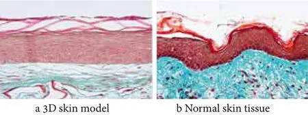
Figure 1. Masson staining of 3D skin equivalent and normal human skin (X25)
Characterization of key proteins in various layers of 3D skin model
The comparison of key proteins expressed in human skin between normal skin tissue and 3D skin model was performed using immunofluorescence. K10, β-catenin,Involucrin in epidermis; Laminin 5 and Col VII in DEJ,Elastin in dermis were examined. As shown in Figure 2, all key proteins were expressed in 3D skin model, and their locations were consistent with normal skin tissue, which further confirmed that the 3D skin model constructed in our lab simulated the structure of normal human skin very well, which can be used to test the efficacy of cosmetics.
Effect of Himalaya cedar extract on structure of 3D skin model
Masson staining was performed to analyze the thickness of epidermis, the results were summarized in Table 1, 2% Himalaya cedar extract efficiently increased the thickness of epidermis in 3D skin model compared with non-treated(NT) , the thickness of epidermis increased by 20%. The thickness of skin epidermis is associated with the viability and regeneration of keratinocytes, and can reflect the overall vitality and health of the skin. Cell viability and regeneration ability of keratinocytes were gradually reduced during the aging of skin, which obviously reduced thickness of epidermis and induced decline in skin barrier and resistance, and further decreased overall vitality and health of the skin.[19]Therefore,2% Himalaya cedar extract significantly increased thickness of epidermis in 3D skin model, indicating that 2% Himalaya cedar extract increased the cell viability and regeneration ability of keratinocytes, and has a potential role in delaying skin senility and improving overall vitality and health of the skin.

Figure 2. Immunofluorescent staining of relevant proteins in 3D skin equivalent and normal human skin (X25)

Table 1. The epidermal thickness of skin equivalent
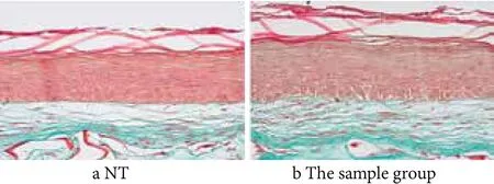
Figure 3. Masson staining of 3D skin equivalent (X25)
Effect of Himalaya cedar extract on expression of Laminin5 and Col VII in DEJ
To further explore the underlying mechanism of Himalaya cedar extract, we further detected the effect of Himalaya cedar extract on expressions of key proteins in DEJ, such as Laminin5, Col VII using immunofluorescence. Expression of Laminin 5 in 3D skin model treated or untreated with Himalaya cedar extract was exhibited in Figure 4. As shown in Figure 4, 2% Himalaya cedar extract evidently increased expression of Laminin 5 in 3D skin model in comparison with that in 3D skin model untreated with Himalaya cedar extract. Laminin 5 level was increased by 20% through Fluorescent quantitative analysis,suggesting that 2% Himalaya cedar extract obviously enhanced Laminin 5 expression.
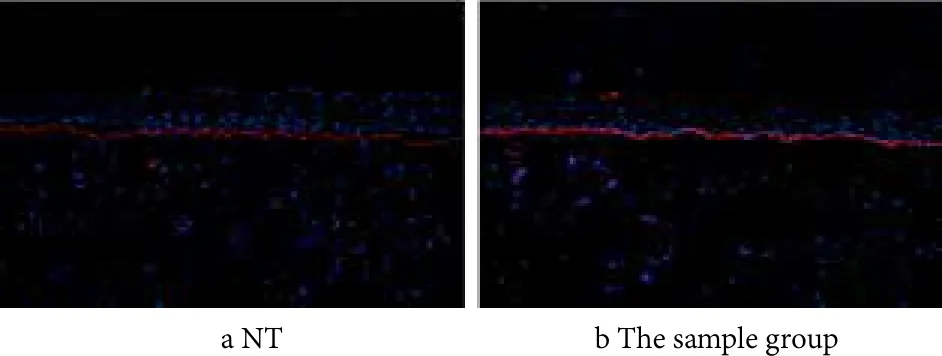
Figure 4. Immunofluorescent staining of Laminin 5 (X10)
Expression of Col VII in 3D skin model treated or untreated with Himalaya cedar extract was exhibited in Figure 5. As shown in Figure 5, 2% Himalaya cedar extract evidently up-regulated expression of Col VII in 3D skin model in comparison with that in 3D skin model untreated with Himalaya cedar extract. Col VII level was increased by 47% by fluorescent quantitative analysis,suggesting that 2% Himalaya cedar extract obviously promoted Col VII expression.
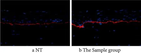
Figure 5. Immunofluorescent staining of Collagen VII (X25)
Above all, Himalaya cedar extract improved the expressions of Laminin 5 and Col VII, which play important role in promoting the structural integrity of DEJ,and further strengthened DEJ and influenced the exchange of substances between DEJ, improved cell viability and regeneration ability of keratinocytes, and then delayed skin senility. In addition, structure of 3D skin model reconstructed in vitro was very similar with that in normal human skin, it can use this 3D skin model instead of normal human skin to detect the effect of active ingredients or to evaluate the influence of stimulation on expressions of key proteins, therefore can instead of human skin to evaluate efficacy of cosmetic actives. All results collectively suggested that Himalaya cedar extract apparently enhanced key proteins in DEJ expressions, and strengthened DEJ structure, which provided theoretical basis of the application for delaying skin senility in cosmetics.
Conclusion
3D skin model with sponge matrix constructed in vitro well simulated the structure of normal human skin with completely differentiated and organized stratified epidermis, dermis and DEJ. The key proteins were all expressed in 3D skin model, and their locations were consistent with normal skin tissue.
The effect of Himalaya cedar extract on DEJ was determined using 3D skin model, and 2% Himalaya cedar extract can evidently improve whole function of skin (thickness of epidermis was increased by 20%),furthermore significantly enhanced expressions of key proteins in DEJ. The expression of Col VII increased by 47%, Laminin 5 expression increased by 20%.
[1] Li Xianyu;Gu MingyN; Keiko Muta;et al. Study of Chinese women skin gelatinase as an etiologic photoaging factor and development of cosmetic containing is inactivator. Proceedings of the 10th Seminar on Cosmetics-China,2014.
[2] Miralles F. UV irradiation induces the muring urokinase-type plasminogen activator gene via the c-Jun N-terrminal kinase signaling pathway:requirement of an API enhancer element.Mol Cell Biol 1998, 18(8),4537-4547.
[3] Ogura Y; Matsunaga Y; Nishiyama T; et al. Plasmin induces degradation and dysfunction of laminin 332 (laminin 5) and impaired assembly of basement membrane at the dermalepidermal junction. Br J Dermatol 2008, 159(1),49-60.
[4] Chanls. Human skin basement membrane in health and in autoimmune diseases. Frontiers in Bioscience 1997(2),343-352.
[5] Zhu Xuejun. The research status of skin basement membrane belt.. : Chinese Journal of Dermatology 2001, 34(4), 312-313.
[6] Zhang Lili; Li Xianyu; Mu Rong; et al. The structure and functions of skin basement membrane. Chinese Journal of Aesthetic Medicine 2016, 25(10), 113-117.
[7] Zhou Pengfei;Liu Hui;Lu Wancheng; et al. Basement membrane and skin care. Flavour Fragrance Cosmetics 2013,6(3), 58-60.
[8] Michael Hertl; Rüdiger Eming; Christian Veldman. T cell control in autoimmune bullous skin disorders . The Journal of Clinical Investigation 2006, 116, 1159-1166.
[9] Alexander Nystr?m; Daniela Velati; Venugopal R. Mittapalli ,et al.Collagen VII plays a dual role in wound healing. The Journal of Clinical Investigation 2013,123(8),3498-3509.
[10] Paula Benny; Cedric Badowski; E.Birgitte Lane; et al. Making more matrix: Enhancing the deposition of dermal-epidermal junction components in vitro and accelerating organotypic skin culture development, using macromolecular crowding.Tissue Engineering: Part A 2015, 21(1/2), 183-192.
[11] Eijiro Tokuyama; Yusuke Nagai; Ken Takahashi; et al.Mechanical stretch on human skin equivalents increases the epidermal thickness and develops the basement membrane.PLOS ONE 2015,11, 1-12.
[12] Zhang Junmin; Shi Xiaofeng; Fan Bin. Research progress on chemical constituents and pharmacological activities of Cedrus. Chinese Traditional Patent Medicine 2009, 31(6),928-932.
[13] Lu Jiang.. The quality and function of small molecule water.Medicine and Health Care 2011(10), 71.
[14] Li Fuxin. Water, medicine or poison? China market press 2007, 27-37.
[15] Wu Jinjin;Zhu Tangyou. Skin Tissue Engineering. People's military medical publishing house 2009, 72-103.
[16] Dezutterdambuyant C; Black A; Bechetoille N; et al. Evolutive skin reconstructions: from the dermal collagen-glycosaminoglycanchitosane substrate to an immunocompetent reconstructed skin. Bio-medical Materials and Engineering 2006, 16(4),85-94.
[17] Damour O; Black A, Pascal P; et al. Transplantation and Changing Management of Organ Failure. Springer 2000,49-54.
[18] Black A. F.; C. Bouez; E Perrier;et al. Optimization and characterization of an engineered human skin equivalent.Tissue Engineering 2005, 11(5/6), 723-733.
[19] Dos Santos, Morgan, Metral, Elodie, Boher, Aur′elie, et al.In vitro 3-D model based on extending time of culture for studying chronological epidermis aging. Matrix Biology 2015,9(47), 85-97.
 China Detergent & Cosmetics2018年1期
- China Detergent & Cosmetics的其它文章
- An In Vitro Biological Model for Evaluation of the Anti-Dandruff Performance of Hair Care Products
- Models for Assessing Pollution Damage of Asian Hair
- Technical Approaches and in-vitro Evaluation Methods for Scalp Care
- Regulation of Talcum Powder in Cosmetics at Home and Abroad
- Technical Standard System Construction Plan for Surfactants and Detergents in the“13th Five-year Plan” Period
- A Summary on Chinese Daily Chemical Journals
