Apelin-13 inhibits apoptosis and excessive autophagy in cerebral ischemia/reperfusion injury
Zi-Qi Shao, Shan-Shan Dou, Jun-Ge Zhu, Hui-Qing Wang, Chun-Mei Wang,Bao-Hua Cheng, , Bo Bai,
Abstract Apelin-13 is a novel endogenous ligand for an angiotensin-like orphan G-protein coupled receptor, and it may be neuroprotective against cerebral ischemia injury. However, the precise mechanisms of the effects of apelin-13 remain to be elucidated. To investigate the effects of apelin-13 on apoptosis and autophagy in models of cerebral ischemia/reperfusion injury, a rat model was established by middle cerebral artery occlusion. Apelin-13 (50 μg/kg) was injected into the right ventricle as a treatment. In addition, an SH-SY5Y cell model was established by oxygen-glucose deprivation/reperfusion, with cells first cultured in sugar-free medium with 95% N2 and 5% CO2 for 4 hours and then cultured in a normal environment with sugar-containing medium for 5 hours. This SH-SY5Y cell model was treated with 10-7 M apelin-13 for 5 hours. Results showed that apelin-13 protected against cerebral ischemia/reperfusion injury. Apelin-13 treatment alleviated neuronal apoptosis by increasing the ratio of Bcl-2/Bax and significantly decreasing cleaved caspase-3 expression. In addition, apelin-13 significantly inhibited excessive autophagy by regulating the expression of LC3B, p62, and Beclin1. Furthermore, the expression of Bcl-2 and the phosphatidylinositol-3-kinase (PI3K)/Akt/mammalian target of rapamycin (mTOR) pathway was markedly increased. Both LY294002(20 μM) and rapamycin (500 nM), which are inhibitors of the PI3K/Akt/mTOR pathway, significantly attenuated the inhibition of autophagy and apoptosis caused by apelin-13. In conclusion, the findings of the present study suggest that Bcl-2 upregulation and mTOR signaling pathway activation lead to the inhibition of apoptosis and excessive autophagy. These effects are involved in apelin-13-induced neuroprotection against cerebral ischemia/reperfusion injury, both in vivo and in vitro. The study was approved by the Animal Ethical and Welfare Committee of Jining Medical University, China (approval No. 2018-JS-001) in February 2018.
Key Words: central nervous system; brain; brain injury; factor; pathways; apoptosis; autophagy; neuroprotection; regeneration Chinese Library Classification No. R453; R741; R363
Introduction
Stroke is an acute disease caused by diseases of the vasculature that transports blood to the brain. It is one of three major disease-related causes of death worldwide.Ischemic stroke accounts for approximately 87% of all stroke patients (Benjamin et al., 2019). The causes of ischemic neuronal death are complex, and the pathological mechanisms are also complex and diverse, including energy failure, excitotoxicity, neuroinflammation, apoptosis, and oxidative stress (Eltzschig and Eckle, 2011; Hatakeyama et al.,2020). Recently, many studies have reported that autophagy plays an important role in the occurrence and development of ischemic stroke (Papadakis et al., 2013; Chen et al., 2014;Wang et al., 2018). Autophagy is an important process in the evolution and conservation of intracellular materials in eukaryotes (Parzych and Klionsky, 2014; He et al., 2019).Physiological autophagy can protect cells themselves, thus contributing to the maintenance of cellular homeostasis.However, excessive autophagy may cause autophagic cell death or even accelerate disease progression (Guo et al.,2018). The regulation of autophagy may therefore be a novel drug target for ischemic stroke.
The Bcl-2 family of proteins can combine with the proapoptotic proteins Bax or Bak1 to form heterodimers that regulate apoptosis (Suzuki et al., 2000). However, Bcl-2 also regulates autophagy by interacting with Beclin1, which plays a vital role in autophagosome formation (Pattingre et al.,2005; Vega-Rubín-de-Celis, 2019). The rapamycin-sensitive mammalian target of rapamycin (mTOR) complex 1 signaling pathway is the converging point of multiple signaling pathways(Jung et al., 2010; Rabanal-Ruiz et al., 2017). It affects cell autophagy via upstream and downstream signals, and is the most studied signaling pathway. The phosphatidylinositol-3-kinase (PI3K)/Akt/mTOR pathway is an important pathway of mTOR complex 1, and previous studies have reported that the PI3K/Akt/mTOR pathway also participates in apoptosis (Cantley,2002; Wang et al., 2017, 2020; Zhang et al., 2020). These findings suggest that regulating Bcl-2 and activating the PI3K/Akt/mTOR pathway may be important and novel strategies for targeting apoptosis and autophagy in ischemic stroke.
Apelin is a novel endogenous ligand for APJ (O’Dowd et al., 1993; Tatemoto et al., 1998). In recent years, studies have reported that the apelin/APJ system is involved in the pathophysiological processes of cardiovascular system diseases, digestive system diseases, metabolic diseases,and tumor angiogenesis (Yang et al., 2016; Hou et al., 2017;Castan-Laurell et al., 2019; Huang et al., 2019; Kuba et al.,2019; Yan et al., 2020). Apelin-13 has the strongest affinity to APJ (Boal et al., 2016). Many reports have suggested that apelin-13 protects against neuronal damage (Yang et al.,2014; Wu et al., 2017; Jiang et al., 2018; Zhu et al., 2019; Luo et al., 2020). Apelin-13 may also be protective in myocardial ischemia/reperfusion (I/R), which has a similar mechanism to cerebral I/R (Yang et al., 2015; Chen et al., 2016; Gunes et al.,2018), suggesting that apelin-13 may have similar protective effects in ischemic stroke. Nevertheless, the exact mechanisms of apelin-13 remain to be elucidated in ischemic stroke. Here,we investigated the neuroprotective effects of apelin-13 on PI3K/Akt/mTOR- and Bcl-2-regulated apoptosis and autophagy in models of cerebral I/R injury.
Materials and Methods
Animals
Forty specific-pathogen-free male Spragu-Dawley rats, aged 8-9 weeks (weighing 260-280 g), were purchased from Pengyue Experimental Animal Co., Ltd., Jinan, China (license No. SCXK (Lu) 2019-0003). All rats were provided with sufficient water and suitable food, and were maintained in a temperature-controlled room at approximately 25 ± 1°C. The experiments were conducted in accordance with the National Experimental Animal Feeding Guidelines. The experimental procedures were approved by the Animal Ethical and Welfare Committee of Jining Medical University, China (approval No.2018-JS-001) in February 2018.
Sprague Dawley rats were divided into sham, middle cerebral artery occlusion (MCAO), apelin-13, and MCAO + apelin-13 groups (n= 10 per group) according to simple randomization using a random number table in SPSS 11.0 (SPSS Inc., Chicago,IL, USA). Rats in the sham and apelin-13 groups were treated the same as those in the MCAO group, but the monofilament was not advanced into the common carotid artery. For the drug treatment, rats in the sham and MCAO groups were injected with vehicle, while rats in the apelin-13 and MCAO +apelin-13 groups were injected with apelin-13. A flow chart of thein vivostudy design is shown in Figure 1.
MCAO model establishment
Before the surgery, all animals had access to water, but were fasted for 12 hours to prevent intestinal obstruction after anesthesia. The rats were weighed, and 10% chloral hydrate(300 mg/kg) was then administered by intraperitoneal injection as the anesthesia (Kleinschnitz et al., 2011;Langhauser et al., 2012). Anesthetized rats were immobilized,their fur was removed, and the area around the surgical site was disinfected. The right common carotid artery, right external carotid artery, and internal carotid artery were exposed, and a 3.0 nylon monofilament was then inserted through the right common carotid artery into the internal carotid artery. The monofilament was advanced 18-22 mm beyond the bifurcation to block the middle cerebral artery.The suture was removed after 2 hours.
Intracerebroventricular injection
At the onset of reperfusion, a burr hole was made at the position of the right lateral ventricle (stereotactic coordinates from the bregma: anterior-posterior: -0.8 mm; mediallateral: 1.6 mm (Paxinos et al., 1980)) using a Dremel drill(Foredom, Bethel, CT, USA). Next, 10 μL apelin-13 (50 μg/kg;Phoenix Pharmaceuticals, St. Joseph, MO, USA) or 10 μL vehicle (0.9% NaCl) in a 10 μL microsyringe (Shanghai Anting Microsyringe Factory, Shanghai, China) was injected at a depth of 3.8 mm, at 2 μL/minute. To minimize drug leakage,the needle was left in place for at least 3 minutes after the injection.
Neurological score
After 24 hours of reperfusion, the neurological score of each rat was evaluated by two blinded investigators using the Longa Score Scale, as follows: 0, no neurological deficit; 1, failure to fully extend the left forepaw; 2, circling to the left; 3, falling to the left; and 4, no spontaneous walking (Longa et al., 1989).
2,3,5-Triphenyl-2H-tetrazolium chloride staining
After 24 hours of reperfusion, the rats were killed by overdose chloral hydrate anesthesia. Brains were carefully removed,incubated on ice for 25 minutes, and cut into five 2-mm coronal slices. After staining with 1% 2,3,5-triphenyl-2H-tetrazolium chloride (TTC) (Sigma, St. Louis, MO, USA) at 37°C in the dark for 15-20 minutes, the brain sections were fixed with 4%paraformaldehyde for 24 hours at room temperature. The infarct areas were measured by two blinded investigators using ImageJ software (Media Cybernetics, Bethesda, MD, USA).
Determination of lactate dehydrogenase
The injured hippocampus of the rats was homogenized in saline solution (0.9% NaCl) using an ultrasonic crusher(Jiangsu Tron Intelligent Technology, Nanjing, China),and were then centrifuged at 400 ×gfor 10 minutes. An lactate dehydrogenase (LDH) kit from Nanjing Jiancheng Bioengineering Institute (Nanjing, China) was used, and LDH levels in the supernatant were measured following the manufacturer’s instructions at 450 nm on a microplate reader(BioRad, Hercules, CA, USA).
Slice preparation
After 24 hours of reperfusion, the rats’ chests were opened after anesthesia and injected with 4% paraformaldehyde from the tip of the heart. Brains were fixed in 4% paraformaldehyde for 2 days at 4°C and then left to sink in 30% sucrose solution.After completely sinking in the solution, 30 μm sections were cut using a microtome (Thermo Scientific, Inc., New York, NY,USA).
Nissl staining
The brain sections were mounted on adhesive slides and covered with Nissl staining solution (Beyotime, Shanghai,China) for 10 minutes at 37°C in the dark. The slides were then immersed in 95% alcohol for approximately 5 seconds.After dehydration through an alcohol gradient, the injured hippocampus sections were observed using an inverted fluorescence microscope (IX 71; Olympus, Tokyo, Japan)(Shiffman et al., 2018).
Double immunofluorescent staining
Injured hippocampus sections, mounted on adhesive slides, were first permeabilized using 0.3% Triton X-100 for approximately 35 minutes. They were then blocked with goat serum for 60 minutes. Next, sections were incubated for 24 hours at 4°C with a mixture of the following primary antibodies: rabbit or mouse anti-NeuN (a marker of neurons;1:1000; Cat# ab104225 and ab104224; Abcam, Cambridge,UK) and mouse anti-p62 (a marker of autophagy; 1:500;Cat# ab91526; Abcam) or mouse anti-caspase-3 (a marker of apoptosis; 1:500; Cat# 9662; Cell Signaling Technology,Danvers, MA, USA) or rabbit anti-microtubule-associated protein 1 light chain 3 (LC3B; a marker of autophagy; 1:500;Cat# NB100-2220; Novus, Littleton, CO, USA). The slices were then incubated at room temperature with Cy5- and fluorescein isothiocyanate-conjugated rabbit/mouse IgG (both 1:50; Cat# BA1031 and BA1105; Boster Biological Technology,Wuhan, China) in the dark for 2 hours. Immunofluorescence was observed using an inverted fluorescence microscope(Olympus IX 71), and immunofluorescent density was quantified using ImageJ software.
Cell culture and oxygen-glucose deprivation/reperfusion treatment
Human neuroblastoma SH-SY5Y cells were obtained from the Cell Resource Center, Chinese Academy of Sciences, Shanghai Academy of Life Sciences (Shanghai, China) and cultured in a humidified atmosphere incubator with 5% CO2at 37°C.Dulbecco’s modified Eagle medium (GibcoTM, Thermo Fisher Scientific, Waltham, MA, USA) with 100 U/mL streptomycin,100 U/mL penicillin, and 10% fetal bovine serum (Solarbio,Beijing, China) was used to culture the cells.
For the oxygen-glucose deprivation (OGD) injury, SH-SY5Y cells at about 70% density were cultured in glucose-free Dulbecco’s modified Eagle’s medium and were incubated with 95% N2and 5% CO2in a tri-gas incubator (Thermo Fisher Scientific). After 4 hours of OGD treatment, the cells were cultured in glucosecontaining medium and exposed to the original environment for an additional 5 hours. In addition, cells were treated with apelin-13 (10-7M) for 5 hours at the onset of reperfusion.Cells were also treated with the inhibitors of the PI3K/Akt/mTOR pathway, LY294002 (20 μM) and rapamycin (500 nM;MedChemExpress, Monmouth Junction, NJ, USA), 30 minutes before apelin-13 treatment. A flow chart of thein vitrostudy design is shown in Figure 1.
Cell Counting Kit-8 assay
SH-SY5Y cells in the different groups were cultured in 96-well plates. Cell Counting Kit-8 reagent (10 μL, Dojindo, Shanghai,China) was added to the cells and they were returned to the incubator for 2 hours. To calculate cell viability, a microplate reader at 450 nm was used to measure the optical density of the cells.
Hoechst 33342 staining
Cells in the different groups were cultured in 12-well plates.After OGD/R treatment, the SH-SY5Y cells were stained with 1 mL Hoechst 33342 (Solarbio) at 4°C for 30 minutes in the dark. Hoechst 33342 staining solution becomes embedded in the broken DNA of apoptotic cells and emits strong blue fluorescence (Crowley et al., 2016). Images of the stained cells were obtained using an inverted fluorescence microscope,and the numbers of apoptotic cells were counted using ImageJ software.
Acridine orange staining
Autophagic vacuoles can be stained red by acidic dyes such as acridine orange (Thomé et al., 2016). The different groups of cells cultured in 12-well plates were stained with 1 mL acridine orange (5 mg/mL, Solarbio) for 30 minutes at 37°C. Images of the stained cells were obtained using an inverted fluorescence microscope.
Western blot assay
Proteins extracted from hippocampal tissue or cells were separated using 8-12% sodium dodecyl sulfatepolyacrylamide gel electrophoresis, until the designated positions were reached. The proteins were then transferred to polyvinylidene difluoride membrane at 4°C for different periods of time. After blocking at room temperature for 1.5 hours with 5% non-fat milk powder in Tris-buffered saline with Tween-20, the membranes were incubated at 4°C overnight with the following primary antibodies against autophagy and apoptosis markers and other relevant pathway proteins: mouse anti-Bcl-2 (1:1000; Cat# 15071; Cell Signaling Technology), rabbit anti-Bax (1:1000; Cat# 2772; Cell Signaling Technology), rabbit anti-phospho(p)-Akt (1:1000; Cat# 4060;Cell Signaling Technology), mouse anti-Akt (1:1000; Cat# 2920;Cell Signaling Technology), rabbit anti-mTOR (1:1000; Cat#2983; Cell Signaling Technology), rabbit anti-p-mTOR (1:1000;Cat# ab84400; Cell Signaling Technology), rabbit anti-PI3K(1:1000; Cat# 4292; Cell Signaling Technology), rabbit antip-PI3K (1:1000; Bioworld, Nanjing, China), mouse anti-p62(1:1000; Cat# ab91526; Abcam), rabbit anti-Beclin1 (1:1000;Cat# ab62557; Abcam), rabbit anti-caspase-3 (1:1000; Cat#BS4605; Proteintech, Wuhan, China), rabbit anti-LC3B (1:1000;Cat# NB100-2220; NOVUS), and mouse anti-β-actin (1:2500;Cat# TA-09; Zhongshan Golden Bridge Biotechnology, Beijing China). Next, the membranes were incubated for 60 minutes at room temperature with peroxidase-conjugated anti-mouse or anti-rabbit secondary antibodies (1:5000, Cat# ZB-2305 and ZB-2301; Zhongshan Golden Bridge Biotechnology). The blots were revealed using an enhanced chemiluminescent kit(Liankebio, Hangzhou, China), and optical density values were quantified using ImageJ software.
Statistical analysis
Normally distributed data are represented as the mean ±standard error of mean. Statistical analysis was conducted using one-way analysis of variance followed bypost hocTukey’s test. Non-normally distributed data (the neurological scores) were analyzed using the non-parametric Mann-WhitneyUtest. All data were analyzed using GraphPad Prism 5.0 (GraphPad Software, Inc., La Jolla, CA, USA).P< 0.05 was considered statistically significant.
Results
Apelin-13 is neuroprotective against cerebral I/R injury
Intracerebroventricular administration of apelin-13 significantly improved neurological scores on the Longa Score Scale (0-4)in the MCAO rat model (P< 0.01; Figure 2A). Furthermore,infarct sizes (observed in TTC staining) were significantly decreased by the intracerebroventricular administration of apelin-13 (P< 0.001; Figure 2B and C). Nissl staining and the LDH assay revealed that neuronal death in the hippocampus of the MCAO rat model was also significantly inhibited by intracerebroventricular administration of apelin-13 (P< 0.05;Figure 2D-F). Additionally, in the SH-SY5Y OGD/R model, cell viability was increased by apelin-13 treatment (P< 0.001;Figure 2G). As shown in Table 1, the rat mortality rate was not sufficient to affect the experiment’s progress, and only successfully operated rats were used in the experiment.Together, these results indicate that apelin-13 is neuroprotective against cerebral I/R injury bothin vivoandin vitro.
Apelin-13 alleviates neuronal apoptosis in cerebral I/R injury
Apoptosis in the hippocampus was significantly inhibited by the intracerebroventricular administration of apelin-13, as exhibited by decreased caspase-3 expression in the MCAO rat model (P< 0.05; Figure 3A and B). Similarly, in the SH-SY5Y OGD/R model, the ratio of apoptotic cells was significantly decreased with apelin-13 treatment compared with the untreated group, as measured by Hoechst 33342 staining(P< 0.001; Figure 3C). These results suggest that apelin-13 significantly alleviates neuronal apoptosis in cerebral I/R injury models.
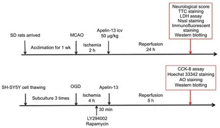
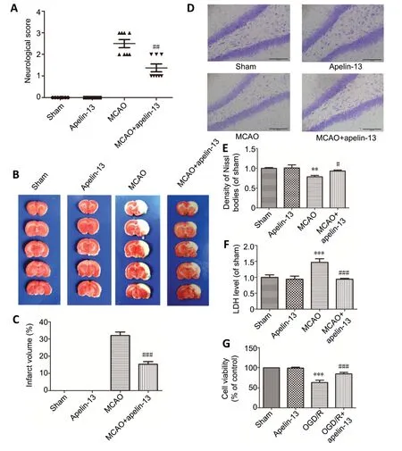
Figure 2 |Apelin-13 is neuroprotective against cerebral ischemia/reperfusion models.

Table 1 |Quantitative analysis of the experimental animals
Apelin-13 alleviates autophagy in cerebral I/R injury
In the MCAO rat model, autophagy in the hippocampus was significantly increased, with upregulated expression of LC3B-II (P< 0.05; Figure 4A and B) and downregulated expression of p62 (P< 0.05; Figure 4C and D). These effects were markedly reversed by the intracerebroventricular administration of apelin-13. Acridine orange staining in SHSY5Y OGD/R cells revealed that the increase in autophagic vacuoles was significantly inhibited by apelin-13 treatment(Figure 4E). Moreover, both the upregulated expression of LC3B-II (P< 0.01; Figure 4F) and downregulated expression of p62 (P< 0.05; Figure 4G) caused by OGD/R in these cells were significantly attenuated by apelin-13 treatment. Therefore,apelin-13 treatment alleviated autophagy both in the hippocampus of the MCAO rat model and in SH-SY5Y OGD/R cells.
Bcl-2 is involved in the apelin-13-induced inhibition of apoptosis and autophagy
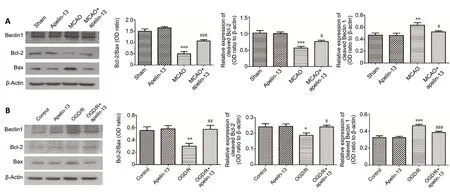
To explore whether Bcl-2 contributes to the effects of apelin-13 on apoptotic inhibition, we evaluated the ratio of Bcl-2/Bax in the MCAO rat model and SH-SY5Y OGD/R cells. Compared with MCAO group, the Bcl-2/Bax ratio was significantly increased in the MCAO + apelin-13 group (P<0.001; Figure 5A). In addition, the expressions of Beclin1 and Bcl-2 were investigated to clarify the role of Bcl-2 in autophagy. Beclin1 expression was increased in the MCAO rat model and SH-SY5Y OGD/R cells, whereas Bcl-2 expression was decreased. Apelin-13 treatment was able to reverse these effects (P< 0.05; Figure 5B). Together, these results suggest that apelin-13 treatment increases the expression of Bcl-2,thus inhibiting beclin1-dependent autophagy. These results therefore indicate that Bcl-2 upregulation participates in the apoptotic and autophagic inhibition of apelin-13.
The PI3K/Akt/mTOR pathway is involved in the inhibitory effects of apelin-13 on apoptosis and autophagy
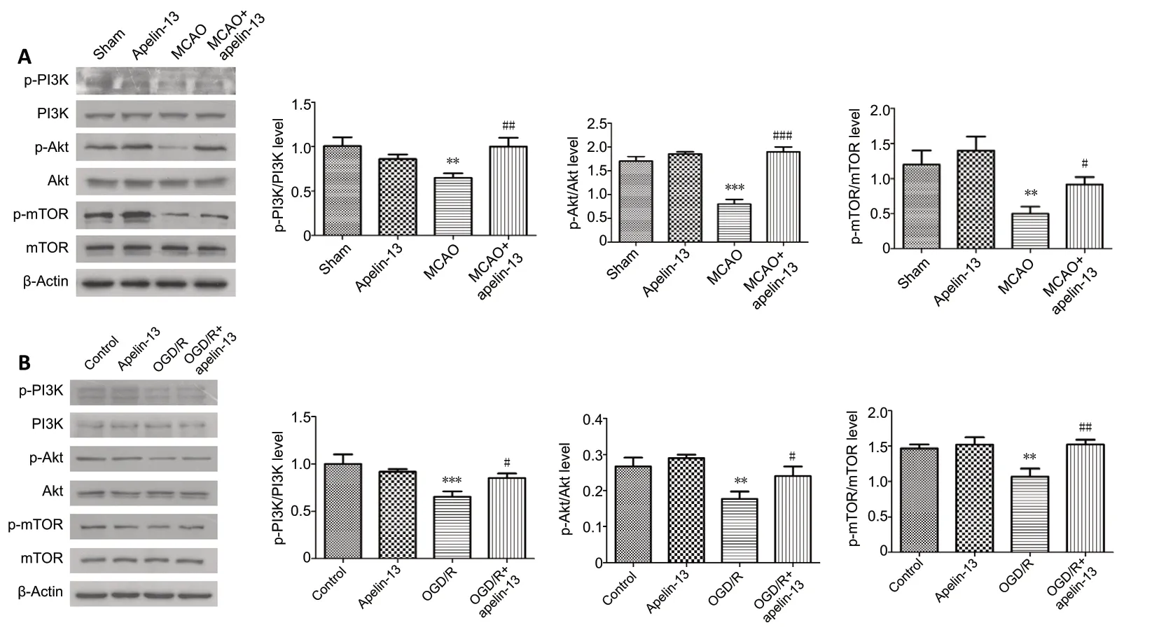
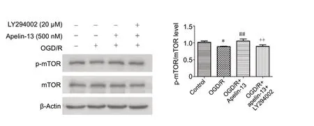
In the MCAO rat model and SH-SY5Y OGD/R cells, the PI3K/Akt/mTOR pathway was significantly inhibited. However,apelin-13 treatment reversed this inhibition by upregulating p-mTOR/mTOR, p-Akt/Akt, and p-PI3K/PI3K (P< 0.05;Figure 6A and B). To clarify the role of this pathway in the neuroprotective effects of apelin-13 on apoptosis and autophagy in SH-SY5Y OGD/R cells, 20 μM LY294002 and 500 nM rapamycin treatment were used. Treatment with LY294002, which inhibits the PI3K/Akt signaling pathway,significantly alleviated the inhibitory effects of apelin-13 on apoptosis and autophagy, such as the increase in p-mTOR/mTOR (P< 0.01; Figures 7 and 8). Likewise, treatment with rapamycin, which inhibits mTOR, also alleviated the inhibitory effects of apelin-13 on autophagy (P< 0.01; Figure 8A and B).The apelin-13-induced inhibition of apoptosis in SH-SY5Y OGD/R cells was significantly alleviated by LY294002 and rapamycin treatment (P< 0.001; Figure 8C). Together, these results suggest that the PI3K/Akt/mTOR pathway participates in the inhibitory effects of apelin-13 on apoptosis and autophagy.

Figure 3 |Apelin-13 alleviates neuronal apoptosis in cerebral ischemia/reperfusion models.
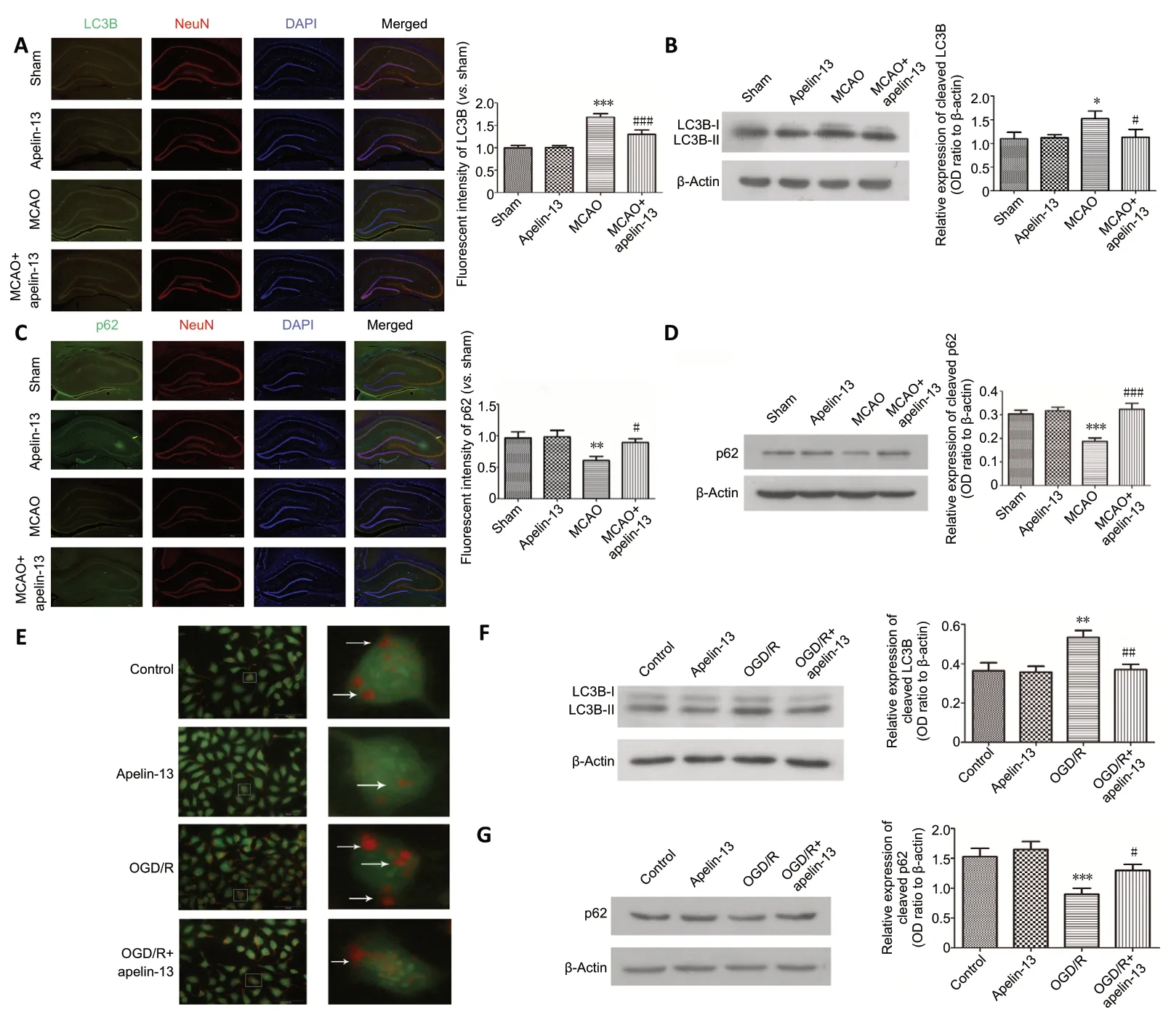
Figure 4 |Apelin-13 alleviates autophagy in cerebral ischemia/reperfusion models in vivo and in vitro.
Discussion
Ischemic stroke is a disease that seriously endangers human life and health worldwide (Benjamin et al., 2019).The protection of neurons against insult and death plays a significant role in post-stroke recovery. Previous studies have reported that the apelin/APJ system has protective effects and can resist excitotoxic damage, oxidative stress damage, and serum deprivation-induced apoptosis (O’Donnell et al., 2007;Zeng et al., 2010; Kasai et al., 2011). Similar to the results of our previous studies (Xin et al., 2015; Wu et al., 2018),in the present study, we further confirmed that apelin-13 can protect against cerebral I/R injury. We also investigated some proposed mechanisms of the neuroprotective effect of apelin-13 against cerebral I/R injury, in bothin vivoandin vitromodels.
It has been reported that neuronal death in cerebral ischemia can be divided into two morphologically distinct types:necrosis and apoptosis (Puyal and Clarke, 2009; Park et al.,2019). To explore the neuroprotective effect of apelin-13 on necrosis, we detected dehydrogenase activity in braintissue using TTC staining and the LDH assay. We revealed that apelin-13 could effectively reduce necrosis levels. Apoptosis is a programmed death process, and therefore plays a more important role in ischemic stroke (Fricker et al., 2018).Caspase-3 is an important regulatory factor during apoptosis,and cleaved caspase-3 is the activated form of this protein.In the present study, cleaved caspase-3 expression was significantly increased in the MCAO rat model, and the rate of apoptosis was also significantly increased in SH-SY5Y OGD/R cells. However, these changes were alleviated by apelin-13 treatment, which suggests that apoptotic inhibition may play a vital role in the neuroprotective effects of apelin-13.
Accumulating evidence indicates that autophagy, a third type of neuronal death, plays a major role in ischemic stroke (Shi et al., 2012; Wang et al., 2018; Yan et al., 2019). However,few studies have reported whether or not apelin-13 exerts its protective effects by inhibiting autophagy in ischemic stroke.In the autophagosome extension stage of autophagy, the LC3 precursor is processed by Atg4 into cytosolic-soluble LC3B-I.LC3B-I is then covalently linked to phosphatidylethanolamine by Atg7 and Atg3-II-phosphatidylethanolamine activity, to become lipid-soluble LC3 (LC3B-II). LC3B-II can bind to newly formed membranes until the formation of autolysosomes.Thus, LC3B-II is often used as a marker of autophagy formation (Rodríguez-Arribas et al., 2017), and is currently the only known marker that is localized on autophagy vesicle membranes (Chifenti et al., 2013). The autophagosome membrane receptor protein SQSTM1/p62 combines substrates (misfolded proteins or protein aggregates).It can then degrade p62 and other misfolded proteins,thus removing damaged organelles and releasing ATP and nutrients for reuse. Furthermore, p62 can also be negatively correlated with autophagic activity, reflecting the strength of autophagolysosomal lysozyme activity and autophagic flux.Thus, p62 can be used as a molecular marker of autophagy(Min et al., 2018). Autophagy is something of a double-edged sword: both too high and too low levels of autophagy cause damage (Chen et al., 2014). Autophagy can protect cells from metabolic stress and oxidative damage, and participates in the maintenance of cell homeostasis as well as the synthesis,degradation, and recycling of cell products (Vilimanovich et al., 2015). However, although physiological levels of autophagy are beneficial to neuronal survival, excessive levels may lead to neuronal death. In the current study, LC3B-II expression was markedly decreased by apelin-13 treatment in both thein vivoandin vitromodels, whereas p62 expression was increased. These results indicate that apelin-13 may protect against ischemic stroke injury by inhibiting excessive levels of autophagy.
Apoptosis and autophagy are two important cellular processes for the maintenance of cellular homeostasis. Moreover, the relationship between apoptosis and autophagy is complex and diverse (Mukhopadhyay et al., 2014). Under some conditions,autophagy inhibits apoptosis, which is a cell survival pathway.However, autophagy itself can also induce cell death, and can also work with apoptosis and induce cell death as a backup mechanism in response to apoptotic defects (Gump and Thorburn, 2011). In this previous study, the simultaneous upregulation of apoptosis and autophagy triggered cell death in ischemic stroke. Apoptosis and autophagy are interrelated and mutually regulate one another; thus, they must share common signaling pathways and regulatory proteins (Gump and Thorburn, 2011; Mukhopadhyay et al., 2014). Bcl-2-related proteins play an important role in apoptosis. Bcl-2 anti-apoptotic proteins have also been reported to markedly attenuate Beclin1-dependent autophagy (Pattingre et al.,2005). The present results demonstrated that apelin-13 treatment increases the ratio of Bcl-2/Bax to inhibit neuronal apoptosis, and increases the expression of Bcl-2 to inhibit Beclin1-dependent autophagy. Furthermore, the PI3K/Akt/mTOR pathway is an important autophagic pathway, and many studies have reported that this pathway may be a common pathway for apoptosis and autophagy (Wang et al., 2017,2020). In myocardial I/R, it has been previously reported that the PI3K/Akt/mTOR signaling pathway is regulated by apelin-13(Jiao et al., 2013). Moreover, multiple reports have suggested that a range of drugs can upregulate mTOR phosphorylation via the PI3K/Akt pathway to inhibit autophagy in ischemic stroke (Luo et al., 2014; Huang et al., 2018). In the present study, we demonstrated that apelin-13 treatment upregulated PI3K/Akt/mTOR-related phosphorylated protein levels in models of cerebral I/R injury. The neuroprotective effects of apelin-13 on autophagy were weakened by LY294002 and rapamycin treatment, as well as by apoptosis.
Our research has some limitations. First, we only used the PI3K/Akt/mTOR inhibitorsin vitro, and did not use themin vivo. In addition, we did not inhibit the expression of Bcl-2 to further verify its mechanism. Furthermore, although apelin-13 was able to inhibit both apoptosis and excessive autophagy, we did not probe the relationship between apoptosis and autophagy in the current study. Further studies are therefore required to determine these unresolved issues.The neurological test that was performed here is more related to motor impairments, which are unlikely to depend heavily on the hippocampus, and we will study the effect of apelin-13 treatment on the hippocampus in the future.
In summary, apelin-13-induced neuroprotection against cerebral I/R injury involved the inhibition of apoptosis and excessive autophagy by Bcl-2 and the mTOR pathway. These findings offer a novel direction for exploring the roles and mechanisms of apelin-13. Because of the limitations of current thrombolytic therapies for ischemic stroke, neuroprotection has become a main research focus. Our results provide a new potential therapeutic approach for the clinical treatment of ischemic stroke.
Author contributions:Study design: BHC, BB; experimental implementation: ZQS, SSD, JGZ; data analysis: ZQS, HQW, CMW;manuscript writing: ZQS, BHC. All authors read and approved the final manuscript.
Conflicts of interest:The authors declare no competing financial interests.
Financial support:The study was supported by the National Natural Science Foundation of China, Nos. 81870948 (to BB), 81671276 (to BHC), 81501018 (to CMW); the Natural Science Foundation of Shandong Province of China, No. ZR2014HL040 (to BHC); Program Supporting Foundation for Teachers’ Research of Jining Medical University of China,No. JYFC2018KJ003 (to SSD). The funding sources had no role in study conception and design, data analysis or interpretation, paper writing or deciding to submit this paper for publication.
Institutional review board statement:The study was approved by the Animal Ethical and Welfare Committee of Jining Medical University, China(approval No. 2018-JS-001) in February 2018.
Copyright license agreement:The Copyright License Agreement has been signed by all authors before publication.
Data sharing statement:Datasets analyzed during the current study are available from the corresponding author on reasonable request.
Plagiarism check:Checked twice by iThenticate.
Peer review:Externally peer reviewed.
Open access statement:This is an open access journal, and articles are distributed under the terms of the Creative Commons Attribution-NonCommercial-ShareAlike 4.0 License, which allows others to remix,tweak, and build upon the work non-commercially, as long as appropriate credit is given and the new creations are licensed under the identical terms.
 中國(guó)神經(jīng)再生研究(英文版)2021年6期
- 中國(guó)神經(jīng)再生研究(英文版)的其它文章
- Entacapone promotes hippocampal neurogenesis in mice
- Electroacupuncture improves learning and memory functions in a rat cerebral ischemia/reperfusion injury model through PI3K/Akt signaling pathway activation
- Normobaric oxygen therapy attenuates hyperglycolysis in ischemic stroke
- MicroRNA-670 aggravates cerebral ischemia/reperfusion injury via the Yap pathway
- Corticospinal excitability during motor imagery is diminished by continuous repetition-induced fatigue
- TP53-induced glycolysis and apoptosis regulator alleviates hypoxia/ischemia-induced microglial pyroptosis and ischemic brain damage
